

 PDF
PDF ePub
ePub Citation
 Print
Print


The gluteus medius is the main abductor and internal rotator of the hip and is an essential stabilizer of the hip during the stance phase of gait. Tears within the gluteus medius and minimus tendon were first described by Bunker et al.1) in 1997. They called them ‘rotator cuff tears of the hip’ due to its morphologic similarities with the anatomy of soft tissue around the shoulder. Their exact etiology and incidence remain unclear, but similar to rotator cuff tears in the shoulder, tears of the hip abductor tendons seem to occur through a degenerative and progressive process2). To our knowledge, a few cases of acute tear in the gluteus medius or minimus3456) have been reported, but none involving acute tears isolated solely within the gluteus medius in a young male.
CASE REPORT
A 20 year-old male patient visited the hospital due to acute lateral hip pain that occurred during a foot volleyball game. The patient suddenly felt a sharp pain while performing a forcible kicking motion consisting of hip abduction and internal rotation movements. On physical examination, the pain was aggravated at the hip joint on external rotation with abduction putting the gluteus musculature under tension. The range of motion of the right hip was diminished due to pain predominantly during internal rotation, and the tenderness was present in the right buttock area. There was no swelling, erythema or ecchymosis around the hip. Trendelenburg sign was slightly positive, while exhibiting weakness during a manual strength test of resisted right hip abduction with the knee extended and flexed to minimize the contribution of the iliotibial band.
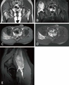
Although a simple radiograph showed no remarkable findings, clinical suspicion of an acute disruption of the gluteus musculature compelled a magnetic resonance imaging (MRI) scan. The MRI in turn revealed a nearly total rupture of the right gluteus medius tendon at the femoral attachment point, along with a partial tear of the gluteus medius muscle and an associated intramuscular hematoma (Fig. 1).
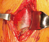
We performed operative treatment consisting of open repair and hematoma evacuation. Under the spinal anesthesia, a lateral approach was used on the left lateral decubitus position. A lateral skin incision centered over the greater trochanter was used and the iliotibial band was split longitudinally, permitting the visualization of the lateral surface of the greater trochanter. Upon entry into the peritrochanteric space, large hematoma and seroma were discovered. However, contrary to pre-operative MRI reports, intra-operative findings unsuspectedly revealed that the superficial fascia and sheath of the gluteus medius remained intact without retraction or defect (Fig. 2). Therefore, only hematoma evacuation was performed for pain relief. Postoperatively, the patient underwent non weight bearing ambulation for 6 weeks after surgery before allowing gradual progression full weight bearing. No active hip abduction or passive hip internal rotation was allowed during those 6 weeks. At 8 weeks post-surgery, he underwent full weight bearing and was pain free and working on active strengthening of his hip abductors. The patient was informed that this case would be submitted for publication.
DISCUSSION
Although there are significant differences between the hip and shoulder joint, their structural and functional similarities established by previous reports allowed for the introduction of the concept of rotator cuff pathology to the hip joint1). However, despite a growing interest in the pathoanatomy and treatment of abductor tears of the hip, the mechanism of injury still remains unclear5). Historically, tears of the gluteus medius and minimus have been thought to be attritional, and associated with chronic peritrochanteric pain5). Many authors speculate that, similar to the shoulder, an analogous progression of stepwise degeneration occurs within the muscle-tendon unit and at its bony interface78). These changes are believed to ultimately cause insertional failure as early tendinitis and tendinopathy lead to partial undersurface tearing and eventually to complete avulsion of the abductor attachment5).
Although the exact incidence of gluteus medius and minimus tears is unknown in both the general and athletic populations6), the incidence peaks between the fouth and sixth decades, with a female:male ratio of 4:189). This supports that the hip rotator cuff tears most commonly occur through a degenerative and progressive process similar to the shoulder.
According to previous studies, the treatment of choice for symptomatic tears of the gluteus musculature is typically conservative management such as rest, activity modification, physiotherapy, medication, and corticosteroid injections. Surgical repair should be considered only in cases refractory to conservative treatment10). The indications for surgical repair presented by other authors are symptomatic tears, functional failures, failure to respond to non-operative treatments combined with a pathologic MRI etc35). However, standardization of such courses of treatments has not yet reached a consensus and they were mostly applied to the chronic tears of middle-aged patients with degenerative processes. Therefore, such treatment methods may not have been suitable for our case. Considering the patient was at a young and active age, was in need of a rapid recovery of function and rapid pain relief through hematoma decompression, we have decided to proceed with surgical treatment. However, because intra-operative findings revealed a partial tear of the gluteus medius in which the fascia and sheath were intact, the plan for tendon repair was put on hold and only hematoma evacuation was performed. At 8 weeks post-surgery, even without repair, the patient could get a satisfactory recovery.
Although our case only pertains to incomplete partial tears and may be limited in covering grounds for complete tears, in regards to treating young patients with acute gluteus medius tears, we should consider the possibility that conservative treatment can be sufficient for full recovery.
 XML Download