

 PDF
PDF ePub
ePub Citation
 Print
Print


Bisophosphonate agents effectively prevent fractures caused by osteoporosis and also produces excellent increases in bone mineral density (BMD) and several studies have demonstrated their outcomes1). However, with respect to long-term bisphosphonate treatment, many researchers have reported decreases in the effects of bisphosphonate over time and many potential risks associated with bone weakness due to the prolonged suppression of bone turnover, such as the risks of atypical fractures in the femur234). To date, most reports on atypical fractures due to long-term bisphosphonate use have described fractures in the subtrochanter or the diaphysis of the femur4), and no reports have described acetabular insufficiency fractures associated with prolonged bisphosphonate treatment. In this report, we described two cases of insufficiency fracture of the acetabulum following long-term treatment with alendronate.
CASE REPORT
1. Case 1
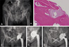
A 77-year-old female visited the clinic due to aggravating bilateral hip pain for several days without any history of recent trauma. Her history included hysterectomy for cervical cancer approximately 17 years ago and she had no radiation therapy for the cancer. She had started taking 70 mg of oral alendronate once per week 6 years ago due to a diagnosis of osteoporosis. Fractures of the right ramus of the pubis, the right wing of sacrum, the left medial acetabular wall and the left femoral head were diagnosed via hip X-rays (Fig. 1A). The lateral cortex of the lateral femur was observed to be thickened in comparing with right side, but it was not significant and authors could not have confidence that the finding was correlated with the significant change from the alendronate use (Fig. 1A). The BMD measured in the 2nd lumbar vertebra region was 0.821 g/cm2. The T-score was -1.0. The BMD measured in the right femoral neck was 0.748 g/cm2. T-score was -1.8. Alkaline phosphatase (ALP) was 109 IU/L (normal range, 50-128 IU/L), osteocalcin was 4.4 ng/mL (normal range, 3.2-12.2 ng/mL) and cross-linked telopeptide of collagen type I (CTx) was 0.495 ng/mL (normal range, 0.01-1.00 ng/mL). All markers were within normal ranges. Surgery was performed on the right sacrum with a cortical screw and an external fixator, and the left acetabulum and the femoral head were treated using total hip arthroplasty (THA) (Fig. 1C). A cementless press-fit cup and stem were used for THA. Alendronate treatment was stopped, and treatment with human recombinant parathyroid hormone was started. The potential risk of metastatic malignancy from the patient's prior cervical cancer was excluded via bone biopsy at three points of acetabular dome and one point of fracture site. Bone biopsy results revealed no osteocytes in the lacuna and neither osteoclasts nor osteoblasts on the surface of the bone tissue (Fig. 1B). One year after THA was performed, the acetabular cup had protruded into the pelvic cavity, causing a large deficit in the medial wall (Fig. 1D). Revision arthroplasty was performed with a cancellous bone allograft supported by wire mesh (Fig. 1E).
2. Case 2
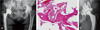
A 61-year-old female visited the clinic due to pain in the left inguinal area. The patient had no history of trauma, and her pain, which started 5 to 6 months earlier, was worsening. She had previously been diagnosed with rheumatoid arthritis (RA) and had received THA for her right hip 5 years ago. Her RA involved no sites other than both hips, and we planned THA for the left hip with recovery of the right hip. Systemic manifestations due to RA were unremarkable and were controlled with nonsteroidal anti-inflammatory drugs. Rheumatoid factor was normal at 11.9 IU/mL (normal range, <14 IU/mL). The patient's osteoporosis had been treated with 70 mg alendronate for 6 years. At the clinic, a fracture in the left medial acetabular wall was observed in X-ray images (Fig. 2A). The BMD measured in the 2nd lumbar vertebra region was 1.081 g/cm2. The T-score was -1.0. The BMD measured in the left femoral neck was 0.975 g/cm2. T-score was -1.6. ALP and CTx were within normal ranges (ALP, 83 IU/L; CTx, 0.140 ng/mL), whereas osteocalcin levels were abnormally low (0.3 ng/mL). Bone biopsy were taken at two points of acetabular dome by bone biopsy needle. The biopsies revealed no osteocytes in the lacuna and neither osteoblasts nor osteoclasts on the surface of the bone tissue (Fig. 2B).Internal fixation of the fracture and the implantation of autologous cancellous bone on the medial wall were followed by THA with a Ganz reinforcement ring (Fig. 2C).
DISCUSSION
Researchers have reported low incidence rates of insufficiency fractures of the hip, most of which involve the pubis, sacrum, and/or femur5). For whatever reason, insufficiency fractures involving the acetabulum are extremely uncommon. Cases of insufficiency fractures in the acetabulum have been reported in patients with RA who were treated with high-dose corticosteroids6) and in a patient with severe osteoporosis7). We determined that the insufficiency fractures described in the current report were caused by the long-term use of alendronate for the following reasons; First, the patients in these cases had experienced pain without any trauma. Second, we could not identify any ordinary cause of an insufficiency fracture, such as severe osteoporosis, advanced RA, radiation therapy, reconstructive surgery or a metabolic disease5). BMD for the involved patients produced higher T-scores than the diagnostic threshold for osteoporosis. The second patient had previously been diagnosed with RA but had no history of high-dose corticosteroid treatment. RA is a predisposing cause of insufficiency fracture because high-dose corticosteroid treatment is used to treat patients with advanced RA6). Third, both of the described patients had used alendronate for more than 5 years, and biopsies revealed signs of an extreme reduction in bone metabolism, such as the loss of osteocytes in the lacunae and a sparse number of osteoblasts and osteoclasts48). These findings for atypical fractures due to bisphosphonate use are indicative of a phenomenon known as “frozen” bone2).
Several cases of atypical insufficiency fractures of the femur following bisphosphonate use have been reported34). Currently, the dominant opinion is that bisphosphonate treatment increases the risk of insufficiency fractures by inhibiting bone turnover129). There are two possible reasons that the subtrochanter and the diaphysis of the femur are the most common regions where such insufficiency fractures occur. First, these areas have high levels of cortical bone. If bone turnover declines, the strength of cortical bone is significantly weakened, and the risk of fracture increases2). This explanation is also applicable to the acetabulum. Second, the lateral part of the femur is the site where tensile strength is concentrated, and the associated stress causes the area to become fragile in response to ordinary stress8). This explanation is not applicable to the acetabulum. However, a significant degree of weight stress can concentrate at the acetabulum because it is part of a weight-stress pathway down to the legs. Therefore, the acetabulum is also at risk for insufficiency fracture following bisphosphonate use. In this case, however, the fracture happened at the medial wall instead of acetabular dome area, where weight is largely concentrated on. This is assumed that the ratio of the cortical bone to the medial wall is relatively high and that the site is relatively thin.
Insufficiency fractures in the acetabulum require greater care to manage than fractures in the subtrochanter or the diaphysis because acetabulum fractures involve the joint. Thus, further treatment beyond osteosynthesis is generally needed for acetabulum fractures; in the cases described in this report, we conducted THA. Certain authors have reported the use of THA in cases involving an insufficiency fracture of the acetabulum67). Our cases, however, differed from cases described in prior reports because the cases in this study involved fractures in “frozen” bone due to alendronate use. The first case demonstrated that conventional THA with a cementless cup could potentially risk early failure in an acetabulum with “frozen” bone (Fig. 1B). The cementless cups that are commonly used today are fixed on the acetabulum via press-fitting at an early time point; over time, the cup undergoes prolonged fixation via the on-growth or in-growth of acetabular bone on the surface of the cup10). In the first case, we achieved secure initial fixation but failed to attain prolonged fixation at a year after surgery. However, it is assumed that this failure may be the complication from the instability of the cementless cup due to complicated fracture, especially the fracture involved posterior column of pelvis. On the other hand, the authors noted the long-term use of alendronate as the potential cause of the fractures of this report. It was suspected that changes in bone quality in “frozen” bone may have inhibited the acetabular bone's on-growth or in-growth process. We believe that it is necessary to perform preparative procedures to secure the acetabulum, such as cup reaming and bone grafting, and to consider other types of implants for the acetabulum, such as a revision cup and reinforcement rings.
In this study, the authors first considered that these cases would have potential for rapid destructive coxarthrosis (RDC). However, RDC has a limit to figure out as the definite cause and the cause of RDC is not clear. The patient's history and biopsy findings in this case showed the possibility of fracture due to prolonged use of alendronate. Particular care should be devoted to the management of this type of fracture because there exist many potential risks of failure associated with conventional treatment and a strong possibility of disability in patients who undergo THA.
 XML Download