

 PDF
PDF ePub
ePub Citation
 Print
Print


INTRODUCTION
The use of acetabular cup revision is on the rise as demands for total hip arthroplasty, improved life expectancies and the need for individual activity increase1). Despite decreased wear rates and advancement in bearing surfaces, acetabular revision is inevitable in cases of subsequent cup loosening caused by bone loss resulted from acetabular osteolysis due to wear particulate debris from bearing surfaces. In addition, acetabular revision is commonly performed in cases of hip instability caused by malpositioning of the acetabular cup.
CLASSIFICATION OF ACETABULAR BONE DEFICIENCY
For an acetabular cup revision to be successful, bone growth should be achieved with stable cup placement and fixation within the remaining supportive bone of the acetabulum. Since the amount of remaining supportive acetabular bone stock in the patient plays an important role in the success of revision, proper classification of the degree of acetabular bone defect is necessary preoperatively. The popular classification systems for acetabular bone defect include the American Academy of Orthopedic Surgeons (AAOS) and Paprosky classification systems. The AAOS classification system was first described in 1989 by D'Antonio et al.2), which divides bone defects into segmental and cavitary patterns. Although this system categorizes the presence of pelvic discontinuity separately, it has limitations in reflecting the location and size of acetabular defect. For this reason, the Paprosky classification was developed to help select an appropriate revision cup according to acetabular defect type, size and location3). In Paprosky type 2 defects, the acetabular walls are compromised and migration of the acetabular component is less than 3 cm. In Paprosky type 3 defects, the acetabular wall and columns are compromised and migration of the acetabular component is more than 3 cm. Since there are challenges in choosing an appropriate acetabular cup in Paprosky type 3 defects, careful selection of the acetabular component is mandatory (Table 1).
TREATMENT APPROACHES
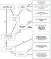
Several surgical techniques for acetabular cup revision are available including acetabular liner exchange, high hip center, oblong cup, trabecular metal cup with augment, bipolar cup, bulk structural graft, cemented cup, uncemented cup including jumbo cup, acetabular reinforcement device (cage), trabecular metal cup cage and others. The optimal treatment option can be chosen depending on circumstances (Fig. 1)4).
Acetabular liner exchange is a revision surgery option best suited for cases involving large periacetabular osteolysis but maintained acetabular cup stability. With an intact liner locking mechanism, chip bone graft can be performed through screw holes after screw removal. When the liner locking mechanism fails, a liner is fixed with cement after screw removal. Disadvantages of this technique are insufficient bone grafting and higher rates of postoperative dislocation5).
Acetabular cup removal is primarily indicated for cases where there are difficulties in achieving acetabular stability despite impaction chip bone graft through screw holes of the acetabular cup due to a high degree of bone loss around the acetabular component. Additionally, this technique can be applied when performing secure fixation of the acetabular polyethylene liner due to severe damage to the liner locking mechanism. Moreover, acetabular cup removal is conducted when severe damage and deformity are present in the metal shell of the acetabular cup as the femoral head penetrates through the acetabular polyethylene liner. After complete exposure of the border following removal of soft tissues and osteophytes surrounding the acetabular component, the interface between the acetabular cup and the acetabular bone is isolated circularly using the curved osteotome. Additionally, using the Explant Acetabular Cup Removal System (Zimmer, Warsaw, IN, USA), the space between the cup and the acetabular bone can be effectively separated by rotating the osteotome around the acetabular cup.
The common treatment options for acetabular cup revision are described below. Acetabular cup revision can be performed using uncemented cups for type 2A and type 2B, and uncemented cup with medial chip bone graft for type 2C. In type 3A defects, the jumbo cup is chosen for spherical shape and high hip center, and trabecular metal cup, cemented cup, oblong cup and bulk structural graft are chosen for oblong shape.
In type 3B defects without discontinuity, cage with chip bone graft, trabecular metal cup with augment and bulk structural graft are used, in type 3B defects with discontinuity, cage or trabecular metal cup with internal plate, trabecular metal cup cage and acetabular transplant can be selected.
The most important factor determining the likely success of acetabular cup revision is obtaining primary stability through stable placement and fixation of the acetabular cup on the remaining host acetabular bone stock and tight compact chip bone graft.
In cases when jumbo cups are used, achieving intrinsic cup stability is essential for successful surgery through wedge fit by maintaining posterior bone stock, antero-superior acetabulum and ischial area, and grafting with chip bone as small as possible is desirable67).
When intrinsic cup stability is challenging with a jumbo cup, it is necessary to choose a cage or a trabecular metal cup with augment alternatively.
When using cages to promote stability of cage strong impaction of chip bone graft, a sufficient number of screws (as many as 3 or 4), and secure fixation of screws on good bone stock are mandatory8910).
In cases where trabecular metal augments are used, the cup size should be determined to obtain relative stability by performing expanded reaming until two points of fixation are achieved. Initially, the augment should be securely fixed on the host bone in the acetabular bone defect with multiple screws. Subsequently, intrinsic stability of cup is obtained by reaming for an optimum contact between the metal shell cup and the augment, as well as the host bone surface, and overall primary cup stability should be achieved through additional screw fixation11121314).
In recent years, to improve the relatively high failure rate of the antiprotrusio cage at long-term follow up, acetabular cup revision using a cup cage construct has been suggested. In this combined procedure using cage and trabecular metal shell, fixation is established for partial stability through the trabecular metal shell, then be reinforced using a cage for secure initial stability, and favorable short-term clinical results have been reported15).
Custom triflange implants are used for patients with massive acetabular bone loss and pelvic discontinuity. By manufacturing and inserting implants after obtaining reconstruction images using 3-dimensional computed tomography, favorable short-term clinical outcomes have been reported16).
With recent advances in implants, a variety of acetabular reconstruction options have been introduced. Selecting the optimal treatment option can be chosen according to patient factors, including the degree of acetabular defect. However, choosing the most effective treatment modality should also be based on the long-term outcomes of well-designed clinical studies of recently developed implants.
CONCLUSION
The keys to achieving successful acetabular cup revision include: i) accurate evaluation of bone defect preoperativey and intraoperatively; ii) choosing the proper method of acetabular revision according to the evaluation of acetabular bone deficiency; and iii) applying the proper technique to achieve primary implant stability (e.g., precise grafting technique, and stable implant fixation).

 XML Download