

 PDF
PDF ePub
ePub Citation
 Print
Print


Pelvic clamps, which can be used for emergent stabilization to control such hemorrhages, can be life-saving1). These clamps are easily applied and control bleeding through a tamponade effect on the pelvis. However, several complications have been reported during or after pelvic C-clamp application, including secondary clamp displacement, clamp loosening and pin perforation into the pelvis2). These complications are mainly caused by primary pin misplacement, likely as a result of blind procedure in an emergency situation with unclear surface landmarks from swelling or distortion. We describe a case of superior gluteal artery pseudoaneurysm that developed after emergent blind AO pelvic C-clamp application.
CASE REPORT
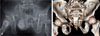
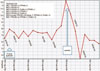
A 66-year-old man presented to the emergency room three hours after a motor vehicle accident. Initial X-ray and pelvic computed tomography (CT) images revealed diastasis of the symphysis pubis and right sacroiliac joint. In addition, there were fractures of the superior and inferior rami, and the right hemipelvis was externally rotated (Fig. 1). Associated injuries were multiple rib fractures and hemothorax. On arrival, mental status was drowsy. The patient's blood pressure on admission was 60/40 mmHg, with a pulse rate of 150 beats/minute. The laboratory data showed a low hemoglobin level (7.0 g/dL). These vital signs are representative of hypovolemic shock. The patient was intubated and further resuscitated with lactated Ringer's solution and packed red blood cells. The vital signs were summarized in graph in chronological order (Fig. 2).
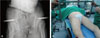
Given the situation's urgency, pelvic ring stabilization was obtained blindly using a pelvic C-clamp (Fig. 3) in the emergency room. The patient's vital signs stabilized after external pelvic fixation. However, a radiograph revealed that the pelvic C-clamps were misplaced at the left superoposterior bony surface of the acetabulum. The patient recovered from his state of hemorrhagic/hypovolemic shock. Extubation was done the day after admission. The metal status changed to ‘alert’. Surgery was scheduled on five days after the trauma. The pin site oozing was increased on four days after the trauma, the pins of pelvic C-clamp became more loosened. We removed pelvic C-clamp on scheduled operation day so that we checked abdominal contrast CT as recommendations of abdominal trauma department, because the huge appearance of our pelvic C-clamp was too big patient with that to enter our CT machine.
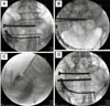
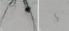
Under the general anesthesia including intubation, an ilioinguinal approach was used on supine position to perform open reduction and internal fixation of the diastasis symphysis pubis and pubic rami fracture. The diastasis of right sacroiliac joint was also reduced and fixed using two sacroiliac screws that were tightly locked over the sacrum (Fig. 4). Exsanguinate bleeding from the pelvic floor persisted, but we thought of bleeding from the fracture site and finished the operation. Intraoperative bleeding estimated by anesthesiologist was about 2,000 mL. Operation time was three and half hours. On the first postoperative day, the patient's hemoglobin and blood pressure began to drop. Angiography was performed of the common iliac artery, internal and external right iliac arteries, and their branches. Angiography revealed a pseudoaneurysm of the left superior gluteal artery. The aneurysm was immediately embolized (Fig. 5). However, the patient's vital signs did not recover and he eventually expired.
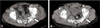
On case review, we could find a pseudoaneurysm of left superior gluteal artery that we missed, on abdominal contrast CT just before surgery, just after removal of pins of pelvic C-clamp. It is a finding that was not found in pelvic CT checked in emergency department before pin insertion (Fig. 6). It might be the cause of exsanguinate bleeding from the pelvic floor seen during the surgery.
DISCUSSION
The major cause of death within one day of a pelvic ring disruption is exsanguination from hemorrhage34). A 38% mortality rate was reported in hypotensive patients, compared with only 3% mortality in hemodynamically stable patients5). Early diagnosis and hemorrhage control are crucial with regard to patient survival. Acute reduction and external flxation of the disrupted pelvis controls retroperitoneal bleeding by decreasing the pelvic volume and inducing a tamponade-like effect. In such a critical situation, external devices can be easily applied. The pelvic C-clamp is an effective option with a simple external frame; it is particularly useful in type C injuries6).
However, it is not always easy to apply the pelvic C-clamp. It is properly placed on the lateral cortex of the ilium, where a palpable groove is formed by angulations of the iliac wing7). Unfortunately, in the setting of trauma, it is sometimes difficult to locate the anatomic landmarks around the pelvis because of pelvic ring distortions, swelling or soft tissue hematomas. Several complications may occur when a pelvic C-clamp is applied in an emergent situation. Several articles have described the potential complications, including secondary clamp displacement, clamp loosening, pin perforation into the pelvis, incomplete closure of the anterior pelvic ring, bacterial contamination at the sacroiliac joint screw fixation, and nerve/vessel injuries78). According to Pohlemann et al.9), perforation may occur when the clamp attachment points are too far anterior. In contrast, when they are too far posterior, secondary fragment displacement may occur9). Finally, when the attachment points are too distal, with displacement into the greater sciatic notch, vascular structures may be compromised9).
A pseudoaneurysm results from a ruptured arterial wall. When its wall is compromised, the artery bleeds into the surrounding soft tissue, forming a sac that directly communicates with the arterial lumen8). Pseudoaneurysms involving the superior gluteal artery are rare. Although the exact incidence is not known, fewer than 40 cases have been reported. Regardless, pseudoaneurysms are considered emergencies because they are liable to rupture at any time, with life-threatening hemorrhage10).
In a critical situation, there is no choice but to apply the pelvic C-clamp blindly. However, it is safer to apply a pelvic clamp under an image intensifier than it is to do so blindly. In the case that we presented, the pins were misplaced during an emergent application of a pelvic clamp. Regardless, although it is rare, a vascular injury must be considered in the case of emergent pelvic C-clamp application because of its potential severity. In particular, it is worth considering that a vascular complication may arise after pin removal from a pelvic C-clamp application. If we are aware of this potential, it may be helpful to confirm the contrast CT before surgery after removal the C-clamp.
 XML Download