

 PDF
PDF ePub
ePub Citation
 Print
Print


INTRODUCTION
Subtrochanteric femoral fractures are commonly caused by high-energy injuries in young patients, low-energy injuries in osteoporotic patients1), and rarely low-energy injuries in long-term bisphosphonate users.
Severe comminuted subtrochanteric fractures have many post-operative complications such as malunions, nonunions, and metal failures related to biomechanical characteristics. Subtrochanteric fractures happen from more stress force on the medial cortex and tensile force on the lateral cortex and have a relatively high ratio of cortical bone to cancellous bone, which has relatively less blood supply2345).
Good results of the cephalo-medullary nails have been reported in proximal femoral fractures recently16789). Based on the length of nails and shape of screws fixed in a femoral head for proximal fragment fixation, various designs of the implant were used7910). There are many excellent clinical and radiological results of proximal femoral nail anti-rotation in the treatment of intertrochanteric femoral fractures which uses helical blade type screws111213). A long intramedullary (IM) nail has a biomechanical advantage over short IM nail theoretically. It has a longer working length than the short IM nail, and it can protect the remnants of the femur shaft below the fracture site141516). Up to now, we have some reports of subtrochanteric fractures including simple and complex type. However, there was no investigation of long IM nail focused in the severe comminuted subtrochanteric fractures (Seinsheimer's classification type IV or V).
MATERIALS AND METHODS
Between March 2010 and March 2013, 21 patients with subtrochanteric femoral fractures were treated by long proximal femoral nail antirotation II (PFNA II; DePuy Synthes, Billerica, MA, USA). According to Seinsheimer's classification, five patients were type IV and 16 patients were type V. Ten cases were AO classification 32C1.1, 6 cases were 32C2.1 and 5 cases were 32C3.117). A senior surgeon did all operations. The study subjects consisted of 13 men and eight women with an average age of 64.8 years (range, 43-85 years). The follow-up period was 30.9 (12-50) months. Of the causes of injury, 11 were of a traffic accident (52.4%), 5 were a fall from a height more than 3 m (23.8%), and 5 cases were of a simple fall (23.8%). The study was approved by Konyang University Hospital's institutional review board.
1. Operation Techniques
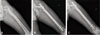
All patients were placed supine position on the fracture table during the operation. Patients had about an 8-cm skin incision proximally from the apex of greater trochanter of the femur and a guide pin inserted from the trochanteric apex, passing through the fracture site. The proximal reamer was then used before inserting the nail into the medullary space. Additional reaming was done in patients with small isthmic diameters of the IM canal less than that of IM nail to insert nail as long as possible. A 340 mm-length nail was used in 18 cases, and a 380-mm nail was used in 3 cases. All patients had a helical blade screw and two distal interlocking screws were used for firm fixation (Fig. 1). In 10 cases, we could get sufficient reduction with closed methods. But in 11 cases a minimal semi-open reduction technique18). In case of semi-open reduction, we made a 3 cm lateral incision at the level of the lesser trochanter about 1-2 cm posterior to the longitudinal axis of the femur. The tip of the Hohman retractor was placed at the distal fragment which was displaced to anterior and we elevated the handle of the retractor toward the anterior aspect of the thigh using a curved tip placed at the lesser trochanter as a fulcrum. To maintain the reduction bone clamp was applied. In 4 of 11 cases, we used the Dall-Miles cable (Stryker, Kalamanzoo, MI, USA) for maintaining the reduction.
2. Evaluation
Clinical and radiographic results were assessed for bridging callus and outcome measures were applied at a final follow-up of a minimum of 1 year. Radiological union was defined as the presence of a bridging callus in three cortices and were analyzed in the follow-up radiographs19). For evaluation of reduction state, the degree of neck-shaft angle was measured on immediate postoperative and last follow-up radiographs20). The sliding length of helical blade screw was measured for the amount of collapse of the fracture site21). To confirm the position of helical blade screw in the femoral head, tip-apex distance (TAD) was calculated22). Evaluation of leg length discrepancy (LLD) was calculated by comparing the length of contralateral uninjured femur and length of fractured femur shaft last follow-up radiographs. For clinical evaluation, modified Koval index was assessed at pre-operation, and last follow-up23).
RESULTS
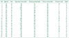
The individual demographic data of all patients are presented in Table 1.
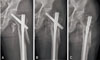
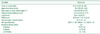
All of the fractures healed at union time of 24.2±3.8 weeks (range, 18-30 weeks). The decrease in neck-shaft angle was 4.5°±3.5°(range, 0.4°-9.5°). The sliding length of helical blade screw was 4.2±3.3 mm (range, 0.3-11.7 mm). LLD was 3.0±1.8 mm (range, 0-7 mm). Mean TAD was 23.0±9.1 mm (range, 11.7-44.4 mm) and TAD above 25.0 mm was measured in 7 cases (33.3%) (Table 2). There was no cut-through or cut out of the helical blade screw or metal failure. However, in the 2 cases who experienced prolonged discomfort and pain at the trochanteric area were induced by excessive sliding or protrusion of helical blade screw, so the helical blade screw was removed after bone union (Fig. 2).
On pre-operative evaluation, modified Koval index score of all the patients was 5 points. On the evaluation of modified Koval index score at final follow-up, the score of 15 patients (71.4%) was 5 points, 4 patients (19.0%) was 4 points, 1 patient (4.8%) was 3 points because she got an another surgery due to intertrochanteric femoral fractures on contralateral side, so she was able to do only independent household ambulation; last 1 patient (4.8%) responded 0 point due to her underlying disease and general condition but was not associated with the surgery. Operation time was 96±25 minutes (range, 55-140 minutes) (Table 2). None of the patients had malunion, skin problems, or infection. Two patients complained postoperative trochanteric area pain due to helical blade irritation even though the union was achieved. The sliding length was 11.7 mm and 11.5 mm, respectively. The pain was relieved after removal of the blade (Fig. 2).
DISCUSSION
Nowadays, IM nails are used more often than extramedullary devices in the treatment of proximal femoral fractures because IM nails have more biological and mechanical advantages; which includes better preservation of blood supply at fracture sites surrounding soft tissue, and better endurability of weight and stresses than extramedullary devices1324252627). We attempted to reduce and fix the fracture site in the manner of a closed reduction as much as possible, but in the cases of severe comminuted or reducible proximal fragment, we reduced the fracture site with the Hohmann retractor or Dall-Miles cable using minimal skin incisions to mitigate the damage of the soft tissues including periosteum. There was no case that shows complications of non-union or infection. We should avoid widespread soft tissue dissection and wiring that can lead to nonunion or infection by reducing blood supply at the extramedullary area of the fracture site3527).
We can employ the concept of working lengths of plates to IM nails. In the comminuted fractures, enough working length provides less stress to the implant and more strain than a rigid fixation. In that perspective, long IM nails can contribute less stress to the nail, which could cause the metal failure of unstable fracture than a short one16). In addition, patients with osteoporosis theoretically, have more advantage with the long nail than the short nail to protect remaining bone below the nail if fall occurs afterwards because the short nail tip could elevate stress concentration on the bowing site of the femur1415). However, short nails could be inserted into the IM space easily even in patients with remarkable ante curvature of the femur shaft. Also, we can easily fix the distal interlocking screw of short or standard PFNA-II using insertion handle and aiming arm. Some authors have mentioned in their research aimed at AO/OTA Type 31-A3 fractures that long IM nail did not show statistically significant clinical and radiological outcomes2829). They reported that using a long nail was no more helpful for old-aged patients with poor general conditions as it could increase operation time and fluoroscopic radiation time28). The intertrochanteric area is metaphyseal bone which has enough blood supply and has sufficient contact surface for the union. The previously reported good or equal results of short nail applied to the intertrochanteric femur should not be associated with comminuted subtrochanteric fractures, which is more unstable and difficult to get enough contact surface. Application of long-PFNA II's has a risk of penetration of anterior cortex during the insertion because it has 1,500 mm of the radius of curvature which is not enough for a severe bowing femur30). However, in this study, comminuted subtrochanteric fractures haves fewer concerns about the penetration by nail tip because the comminution could provide a minimal correction of the bowing, which leads to minimal LLD that does not affect the clinical results. LLD of the complications is reported with various incidence ranged from 17 to 34% and Borens et al.10) say that LLD under 2.0 cm does no matter clinically. In all our cases, there was no patient of LLD over 2.0 cm, and no one complained of discomfort in everyday life though it was derived from low expectation due to severely comminuted fractures. Overall reduction was satisfactory. In this study, the mean of decrease in neck shaft angle and helical blade sliding was 4.5° and 4.2 mm, respectively. Multi-fragmentary subtrochanteric femoral fracture has a lack of medial buttress. Comminution leads the acceptable blade sliding which finally leads bone unions. We assume that the spontaneous varus change of neck shaft angle arisen from the lack of medial buttress. However, the clinical results were acceptable.
In 2 cases of this study, patients complained of trochanteric pain. All of them had reverse obliquity fracture line at the helical blade screw insertion site in the lateral cortex. We cautiously think that relatively excessive sliding was caused and facilitated by weakened helical blade support at the lateral cortex, especially iatrogenic comminution of the lateral cortex during the operation31). It is also known that trochanteric pain after surgery can be caused by injury to the insertion site of gluteus medius muscle during reaming for nail insertion32), and the possibility of post-operative trochanteric pain should be explained to patients before surgery.
In terms of bone union, Borens et al.10) reported 17.2 weeks of mean union time with long gamma nail. Kim et al.6) reported 18.5 weeks with an IM nail and he reported that a relatively long union period derives from largely a displaced fracture site or comminution of medial cortical bone. In our study, mean union period was 24.3 weeks (range, 17.6-30.5 weeks) and we could consider severe comminution and displaced fragments as the cause of relatively long union periods.
Limitations of this study are as follows. First, this study is not a comparative study with that of other fixation methods especially short length nail. Second, this study has a small number of cases and short term follow up period. Third, this study has a retrospective and nonrandomized features. It is essential to expand cases of study, and we need a long-term and prospective evaluation for reduce the influence of variations including ages and injury levels as well.
 XML Download