

 PDF
PDF ePub
ePub Citation
 Print
Print


INTRODUCTION
Many biomechanical studies show the advantages of the spiral blade in the proximal femoral nail antirotation (PFNA; Synthes, Solothurn, Switzerland) system for intertrochanteric femoral fractures12345). Although it is known PFNA system provides high union rates with low major complication rates, geometric discrepancies exist between the proximal femur and PFNA system. This geometric mismatch is associated with lateral cortical impingement, which causes lateral cortical fracture and intraoperative loss of reduction when inserting the PFNA5678). PFNA II devices have been introduced as an improved PFNA design to overcome these problems. The PFNA II design modifications include the flat lateral shape of the proximal portion and a decrease in the mediolateral bending angle from 6° to 5°. Some pilot studies have demonstrated that the flat lateral shape of PFNA II could reduce the chances of lateral cortical impingement when inserting nails and may lessen the chances for an intraoperative lateral wall fracture and intraoperative loss of reduction8910). However, few reports have analyzed the geometric discrepancies between the proximal femur of Koreans and PFNA and/or PFNA II or the causes of lateral cortical impingement.
Thus, we answered the following questions in this study: Are there significant geometric discrepancies between the proximal femur of Koreans and PFNA system? What kinds of anatomical discrepancies lead to lateral cortical impingement? Are problems, such as lateral cortical impingement and intraoperative loss of reduction, eliminated after using PFNA II? Are intertrochanteric femoral fractures in the Korean population still a problem when using PFNA II?
MATERIALS AND METHODS
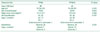
Between May 2008 and October 2011, 173 consecutive patients were admitted to our hospital with an acute proximal femoral fracture. The following 73 patients were excluded from the study: 51 elderly patients with a displaced femoral fracture treated with hip arthroplasty, 12 young patients with a femoral neck fracture treated with screws fixation, and 10 patients with subtrochanteric fermoal fractures. We retrospectively reviewed this left a cohort of 100 patients with intertrochanteric fractures treated with proximal femoral nail (Table 1). The first 38 patients were treated with PFNA between May 2008 and July 2009. This nail was replaced by the PFNA II for the next 62 cases between July 2009 and October 2011. The mean age of the patients at surgery was 72 years in the PFNA group and 74 years in the PFNA II group. Each fracture was classified according to the AO/OTA classification11). Minimum follow-up was 32 months (mean, 36 months; range, 32-70 months). No significant differences in sex, body mass index or fracture classification were observed between the two groups. Eligible patients' information had been reviewed and Institutional Review Board approval of the present study was given prior to commencing the study by the human subjects committee of Pusan National University Yangsan Hospital (IRB No. 05-2014-002).
In principle, all patients were operated by single surgeon in the same technique using PFNA system. All operations were performed with the patient lying supine on a fracture table. When closed reduction was unacceptable in some cases, limited open reduction was performed using various reduction instruments, as necessary. We regard the Lauenstein lateral view as the most important view for determining the position of the spiral blade. Many surgeons prefer to place the affected extremity in excessive adduction, the so-called “banana position”, but this makes it difficult to visualize the exact Lauenstein lateral view intraoperatively because of a space limitation between the affected extremity and the image intensifier tube. Thus, we adducted the patient's torso as much as possible to facilitate the inserting nail, rather than using excess adduction on the affected extremity.
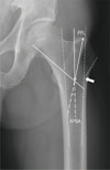
We evaluated the preoperative true plain anteroposterior (AP) radiograph of contralateral unaffected hip to establish the anatomical measurement of the proximal femur. Standardised true AP radiographs, which present controlled rotation and tilt, should be obtained on supine position with the foot internally rotated 15° to show best view of the proximal femur. Proximal femoral length (PFL), AP bending angle (APBA), and the femoral neck shaft angle were measured on true plain AP radiographs of the hip based on the technique of Tyagi et al.8) (Fig. 1). These values were measured independently and at the different times by three observer and was reassessed twice at intervals of two weeks on the images of all 100 patients. The femoral shaft axis was defined for the bisecting line between inner cortical extension lines of the medial and lateral cortex. Then another line was drawn at 130° to the femoral shaft axis from the inferior margin of the femoral neck that coincided with the insertion angle of the lag screw12) and a crossing point was made. PFL was the distance between the tip of greater trochanter and this crossing point. The APBA was formed by the femoral shaft axis and a line extending from the PFL. The angle made between a line extending from the lateral cortex and that of the slope of the lateral cortex of the proximal femur and the point where these two extended line meet was defined as the impingement point. The measured geometric values of the proximal femur were compared with the PFNA and PFNA II dimensions.
The postoperative assessment was performed using postoperative plain AP and lateral radiographs for the quality of the reduction, implant position, and the presence of lateral cortical impingement between the nail and lateral cortex of the proximal femur. Anatomical reduction was considered to have occurred if displacement was less than 5 mm, implant position was not varus, and the angulation was less than 20° on the lateral view13). Intraoperative loss of reduction and/or lateral wall fractures were reviewed when we identified impingement on the lateral cortex. We determined the position of the spiral blade tip in the femoral head based on the Cleveland index14) on both AP and lateral views.
All patients were followed up at 1, 3, and 6 months and yearly with physical examination of the affected hip joint. Plain AP and lateral radiographs were obtained at each visit and reviewed for fracture union or implant failure. Fracture union was determined radiographically as the appearance of a bridging callus on three or four cortices on AP and lateral views and clinically as a lack of pain around the affected site. The proximal protruding length of the nail tip and the sliding distance of the spiral blade were measured on last follow-up radiographs. Operating time, time to union, and union rate were estimated. In addition, we recorded any intraoperative and postoperative complications related to the implant.
All quantitative and qualitative variables are represented by mean value and range. The Kolmogorov-Smirnov test was used for normality analysis of quantitative data. Continuous variables were analyzed using Mann-Whitney test for two independent samples. The chi-square test (or Fisher's exact test where appropriate) and the linear by linear association were used for analysis of categorical data. All tests were two-tailed. Intraclass correlation coefficient with 95% confidence interval was calculated to check for intra- and inter-observer reliability, where one indicates perfect correlation and zero indicates poor correlation. All analyses were carried out using the statistical package PASW Statistics version 18.0 (IBM Co., Armonk, NY, USA). A P-value of <0.05 was used as the significance threshold.
RESULTS
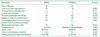
The results of the proximal femur morphological dimensions of the preoperative contralateral unaffected hip in the PFNA and PFNA II cases are summarized in Table 2. The geometry of the proximal femur revealed large differences between patients. The proximal dimensions of PFNA and PFNA II were determined for one fixed size. In addition, PFL was shorter than the corresponding measurements for both proximal femoral nails. Mean APBA was greater than 6°, the largest mediolateral banding angle of both proximal femoral nails. These results show a geometric mismatch between the proximal femur and PFNA system in the Korean population with short stature.
1. Comparative Results between the Two Groups (Table 3)
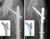
Patients treated with PFNA were more likely to have lateral cortical impingement and intraoperative loss of reduction. Thirteen cases of lateral cortical impingement were observed in the PFNA group, whereas no lateral cortical impingement was observed in the PFNA II group (Fig. 2)15) (P=0.000). Six cases of intraoperative loss of reduction were observed in the PFNA group and 1 case in PFNA II group (P=0.011). Three cases of lateral wall fracture during nail insertion were observed in the PFNA group and 2 cases were detected in the PFNA II group (P=0.365).
Overall, proximal protrusion of the nail tip was seen in 71 patients (71.0%), and the mean length of the proximal protruding nail tip was 4.7 mm (range, 0-15 mm). No difference was observed regarding proximal protrusion of the nail tip between the PFNA and PFNA II groups (PFNA: 29 patients [76.3%], 4.6 mm; PFNA II: 42 patients [67.7%], 3.9 mm) (P=0.378). Of the 71 patients with proximal protrusion of the nail tip, 21 were male (55.3%) and 50 were female (80.6%). Although proximal protrusion of the nail tip tended to be more common in females, no significant difference was observed between the PFNA and PFNA II groups. However, proximal protrusion of more than 10 mm was observed in 15 patients and all 15 cases occurred in females. These 15 patients suffered constant discomfort during activity, particularly during abduction at the follow up.
The evaluation of the quality of the reduction on postoperative radiographs showed that 27 patients (71.1%) in the PFNA group and 50 patients (80.6%) in the PFNA II group achieved anatomical reduction (overall, 77.0%; P=0.330). According to the Cleveland index on the AP and lateral views, the tip of the spiral blade was placed in the 5, 6, 8, and 9 zone of the femoral head in 34 patients (89.5%) in the PFNA group and 61 patients (98.4%) in PFNA II group, which had a low risk of complications (P=1.000).
Operating time was shorter in the PFNA II group than that in the PFNA group (29 minutes vs. 36 minutes; P=0.010). No differences were found for fixation-related complications, time to union, union rate and sliding distance of the spiral blade at last follow up between the two groups.
2. Implications for Lateral Cortical Impingement
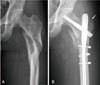
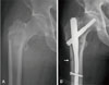
We re-analyzed the PFNA group according to the presence of impingement (Table 4). The difference in the occurrence of the intraoperative loss of reduction in the 13 cases with lateral cortical impingement and the 25 cases without lateral cortical impingement in the PFNA group was significant. Five cases of intraoperative loss of reduction were observed in the group with lateral cortical impingement, but only 1 case was found in the group without lateral cortical impingement (Fig. 3). In the cases of intraoperative loss of reduction during nail insertion, nail was reinserted after additional reduction using various reduction instruments or further reaming of entry point, as necessary. The intraoperative loss of reduction during nail insertion was correlated with lateral cortical impingement (P=0.012). However, no difference in the frequency of intraoperative lateral wall fractures was observed between the two groups, with or without lateral cortical impingement in the PFNA group (P=0.265) (Fig. 4).
We also investigated whether the short proximal femur in Korean patients is associated with lateral cortical impingement. The mean difference between the proximal length of the PFNA and PFL of unaffected proximal femur in the PFNA group was 7.65 mm. The mean difference was 9.3 mm in the group with lateral cortical impingement and 6.8 mm in the group without lateral cortical impingement (P=0.032).
DISCUSSION
This was a retrospective study in which two different intramedullary fixation devices for treating intertrochanteric fractures in Koreans were compared for perioperative radiographic outcomes. Although the relatively small sample size may have lead to weaker statistical power, there were few reports regarding the geometric discrepancies between the proximal femur of Korean and PFNA or PFNA II on plain radiographs. Another limitation of this study was that the different follow up times for the groups have not been accounted for in the analysis used
The PFNA system was first designed by AO/ASIF in 2003 and is considered a suitable implant for unstable intertrochanteric femoral fractures. Although it is known PFNA system provides high union rates with low major complication rates, some reports have shown penetration and cutout of the helical blade161718). Macheras et al.6) found a geometric discrepancy between the proximal femur and PFNA, which could lead to lateral cortical impingement that cause intraoperative loss of reduction and a lateral wall fracture. Geometric discrepancies in morphological incompatibility, the bending angle on a coronal section, angle of inclination of the lateral cortex and neck shaft angle, and height of the greater trochanter are greater in Asians of short stature71019). The PFNA II was designed to overcome these concerns. Therefore, we were interested in determining whether the PFNA II is a perfectly designed implant without problems, even in the Korean population.
We analyzed proximal femoral geometry in the Korean population using true hip plain AP radiographs of the contralateral unaffected side showing the largest proximal femur diameter according to the measuring technique of Tyagi et al.8) who used a coronal computed tomography (CT) section from a randomly selected cases without any obvious pathology. Although a CT scan distinguish cortical from cancellous bone, a CT scan exposes the patient to a greater dose of radiation, and is more expensive, time consuming compared to plain radiographs, and is not always routinely available. Thus, we used the true hip plain AP radiographs of contralateral unaffected side for the measurements as a modification of their technique.
We found significant anatomical discrepancies between the proximal femur of Koreans and the proximal end of the PFNA. This geometric mismatch could lead to lateral impingement or fracture displacement during insertion. Tyagi et al.8) reported significant differences between proximal femur bending angle and that of the nail, and also between the inclination angle of the lateral cortex to that of the nail. They explained that these discrepancies contribute to difficulties during insertion and impingement between the nail and lateral cortex in patients with subtrochanteric fractures. However, lateral cortical impingement did not always occur when using the PFNA, and no statistical difference was observed between the proximal femur angle and lateral cortical impingement in our results. We found a significant correlation between the short proximal femur of Korean patients and lateral cortical impingement in the PFNA group. This is important, as lateral cortical impingement may occur due to the angle differences of the angle as well as the short proximal femur of patients. In other words, lateral cortical impingement may occur depending on nail depth when PFNA is used on the short proximal femur of Koreans.
Lateral cortical impingement during nail insertion is associated with intraoperative loss of reduction or a lateral wall fracture67820). Macheras et al.6) reported that impingement in the PFNA cases resulted in fragmentation of the lateral wall and the fragmentation resulted in loss of reduction into varus. However, lateral wall fractures in patients with intertrochanteric fractures were not correlated with lateral cortical impingement in the PFNA cases in our study. One study revealed that an intraoperative lateral wall fracture occurs more frequently in AO/OTA 31A2.2 and 31A2.3 than in 31A1 and 31A2121). Hsu et al.22) noted that lateral wall thickness is a reliable predictor of post-operative lateral wall fracture and Boopalan et al.20) found that the combined forces of proximal canal reaming and reaming at the base of the lateral wall with a blade reamer result in a higher incidence of lateral wall fractures. These results suggest that various factors are involved in the occurrence of intraoperative lateral wall fractures. However, we found there difference in intraoperative loss of reduction between the PFNA and PFNA II groups. Intraoperative loss of reduction during nail insertion in the PFNA group was correlated with the presence of lateral cortical impingement in our study. Overall, PFNA II was expected to prevent impingement and loss of reduction during nail insertion in patients with intertrochanteric femoral fractures compared with those receiving PFNA.
However, the PFNA II design still has problems. Proximal protrusion of the nail tip was seen in 71 patients (71.0%), and no difference in proximal protrusion of the nail tip between the PFNA and PFNA II groups. In particular, proximal protrusion of more than 10 mm was observed in 15 patients, and all 15 cases occurred in females. Thus, it is problematic that proximal end of PFNA II is long compared with the proximal femur of Korean patients, particularly females. Therefore the short proximal end of the nail should be modified for the geometry of the Korean population. In addition, it is important and necessary to positively carry out preoperative planning using such as templates, along with the development of new devices appropriate to patients' shape. The operating time was slightly shorter in the PFNA II group than that in the PFNA group. We also consider this result as a consequence of the PFNA II device, which can reduce the incidence of lateral cortical impingement, intraoperative loss of reduction, and the learning curve.
CONCLUSION
PFNA system is believed to be a significant advancement in the surgical treatment of intertrochanteric femoral fractures. However, geometric discrepancies between the proximal femur of the Korean population and PFNA lead to lateral cortical impingement and intraoperative loss of reduction. Our results demonstrate that the flat lateral surface of PFNA II helps avoid lateral cortical impingement, leading to better fixation of intertrochanteric femoral fractures. However, the longer proximal portion of the PFNA II compared with the PFL in Koreans remains a problem. We expect that these numerical values can be used to design a new device with a shorter proximal nail, which would be more appropriate for the Korean population.


 XML Download