

 PDF
PDF ePub
ePub Citation
 Print
Print


Femoral stress fractures are caused by repetitive application of force, often by overuse, such as repeated jumping up and down or running long distance. Most femoral stress fractures occur in the neck, condylar area and proximal shaft, rarely supracondylar area, middle shaft, distal shaft and head1). We report a case of stress fracture that occurred in trochanteric area and the result of fixation with intramedullary nailing.
CASE REPORT
A 53-year-old lady presented to our outpatient department with a pain on the right hip and limping gait. Her gait was antalgic, with moderate decrease in weight bearing on the right lower extremity. On the physical examination, mild swelling and localized tenderness were found at the greater trochanteric area. The range of motion (ROM) was symmetrical and within normal limits in the lower extremities, but the patient reported hip pain on either abduction or internal rotation of right hip joint.
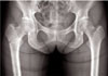
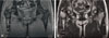
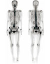
Anteroposterior and lateral radiographs showed transverse fracture below the greater trochanter and above the lessor trochanter (Fig. 1). For differential diagnosis we performed magnetic resonance imaging (MRI) and Tc-99 m bone scan. MRI revealed the intertrochanteric fracture with surrounding marrow edema (Fig. 2), whole body bone scan showed a focal hot uptake on the fracture site (Fig. 3). There was no evidence of metabolic and endocrinologic disorders on laboratory tests.
She had a past history of L4-5 fusion due to spondylolisthesis, but there was no history of any other comorbidities. She was a window cleaner working at single-story buildings, not at high floors of skyscraper. She worked three days a week, and the frequency of jumping ups and downs around the window frames was ten to twelve times a day. She recalled that the onset of her symptoms was approximately three weeks ago, which was two years after the initiation of her working. She denied any type of trauma, and she described her primary symptom as a dull ache in the right hip. Approximately two weeks after the onset of her symptoms, she experienced severe pain after work and difficulty with normal ambulation. She encountered menopause four years ago, but the bone mineral density showed no osteoporosis. Dual energy X-ray absorptiometry showed a lumbar spine T score of +0.2 and a contralateral trochanter T score of –0.9. Body mass index (weight/ height2) was 22.5 kg/m2 (52/1.522).
We decided a fixation of the fracture because the trochanteric fracture may lead to adverse consequence such as displacement and prolonged morbidity. We performed hip nailing with Asian gamma nail under the spinal anesthesia.
The patient was hospitalized for 7 days after surgery. The day after surgery, she was permitted to ambulate with crutches and touch-down weight bearing on the right extremity. Following discharge she was referred for out patient physical therapy consisting of ROM exercise and partial weight bearing ambulation with crutches. The patient was also instructed in isometric exercises for the gluteal, quadriceps, hamstring, and hip abductor muscles. Six weeks after surgery, the patient was ambulatory without an assistive device or weight bearing restriction.
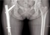
Anteroposterior and lateral radiographs obtained two months after surgery (about three months after onset of symptom) revealed bony union with good alignment (Fig. 4).
The last follow-up visit one year after surgery, the patient was fully back to work without pain or discomfort in right lower extremity.
DISCUSSION
Stress fractures of the lower extremity most commonly involve the tibia and metatarsal bones. Femoral stress fractures are rare, representing only about 5% of all stress fractures2345678). Volpin reported that most of femoral stress fractures were located in diaphysis4), and fewer than 10% of stress injuries involved the femoral neck. However, a recent study have suggested that three most common sites were the femoral neck (50%), the condylar area (24%), and the proximal shaft (18%) in Finnish military conscripts1). Other sites were the supracondylar area, middle and distal third of the shaft and femoral head. There were no trochanteric injury1). Most stress fractures occur in military recruits and athletes. However, stress fractures in labors are very rare5). To our knowledge, our case was the first of trochanteric stress fracture, happened by labor-related stresses.
The differential diagnosis for hip pain is wide and pathological fractures (congenital defects, general systemic disease, metastatic bone tumors), pseudo-fractures (Paget's disease, osteomalacia, rickets), osteitis due to tuberculosis or syphilis, simple fracture due to violence, and osteomyelitis must be distinguished3). A physician must first rule out those causes which are potentially dangerous.
We diagnosed the fracture first by plain radiographs, but ordered MRI and bone scan for the confirmation and ruling out other pathologic conditions. MRI revealed the fracture with surrounding edema and bone scan showed a focal hot uptake on the trochanteric area. No evidence of other pathologic condition was seen on both studies.
The risk factors that can cause stress fractures include excessive use, nutritional deficiencies, and endocrine disorders. In addition, stress fractures may arise from long standing rheumatoid arthritis, osteoporosis, steroid therapy, joint stiffness, or the correction of angular deformity9). In current case, there was no risk factor except amenorrhea.
At junction of the proximal and middle thirds of the femoral shaft lies the insertion of adductor muscles, which may cause proximal femoral shaft fracture1). The underlying mechanism of current case is unclear. However, authors suggest that the trochanteric stress fracture might have been caused by strong and repetitive stresses on trochanteric region at the insertion point of abductor muscles, and focal bending stresses beyond the ability of the bone to tolerate have resulted in fracture.
Because the patient had a difficulty on weight bearing and the fracture had a risk of displacement, we performed internal fixation with hip nailing. For the purpose of early ambulation and fast recovery to work, internal fixation should be considered as initial treatment for patient with trochanteric stress fracture, even for non-displaced fracture10).
 XML Download