

 PDF
PDF ePub
ePub Citation
 Print
Print


Pubic symphyseal diastasis (open-book injury) commonly presents in anteroposterior compression (APC) type injuries described by Burgess et al.1). In APC type II patterns, widening of the symphysis of more than 2.5 cm has been correlated with rupture of anterior sacrospinous ligament and thought to be required operative fixation like a symphyseal plating with or without sacroiliac (SI) joint fixation2345). The APC type III patterns is characterized by vertically unstable injury associated with a posterior disruption of SI complex, which requires both anterior and posterior fixation146). Because the classification of APC injury type is based on using static radiographs, stress radiographs are known as a useful adjunct in classifying type of APC pelvic injuries17).
A concern prone to be mistaken APC type II for APC type I without stress radiographs have been demonstrated in the literature. In a recent article, the intraoperative stress examination has led to a change in the treatment plan in more than 25% of patients on 22 patients presumed APC type I (symphyseal diastasis <2.5 cm) injuries7). However, to our knowledge there is few report demonstrating a concern that APC type II may be mistaken for APC type III in the literature. We report a case which should require a supplementary SI screws insertion because after plating of diastasis pubic symphysis, even though preoperative radiograph and computed tomography (CT) revealed pure diastasis of pubic symphysis without any findings suggesting vertical instability and disruption of posterior SI complex which act as a hinge if injury type was APC II.
CASE REPORT
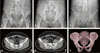
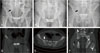
A 28-year-old man was admitted to our hospital via emergency department. He stated he was pressed between reversing 10-ton truck and wall at a construction site. Initial assessments upon admission revealed stable hemodynamics and alert mental status. He complained of pubic pain. Urologic problems by initial trauma were excluded by urologist except subtle scrotal swelling. Plain radiographs and three-dimensional CT revealed an APC type II pelvic ring injury with 3.0 cm sized diastasis of pubic symphysis (Fig. 1A-C). On a following CT, no widening was observed at anterior aspect of right SI joint (Fig. 1D-F). We scheduled an operation for open reduction and internal fixation with plate on diastasis of pubic symphysis at 4 days after injury. Surgery was performed under image intensifler guidance, in the supine position through a Pfannenstiel approach. After reduction of symphysis pubis was tried but failed, using a Weber pointed reduction clamp placed anteriorly at same level on pubic body, satisfactory reduction was obtained using a Farabeau clamp after temporary two screw insertion on superior border of both pubis, and confirmed by fluoroscopy. Due to a difficulty of reduction, double plating was done at anterior and superior border of pubis (Fig. 2A-C). After plating, SI joint abnormality such as widening was excluded by fluoroscopy.
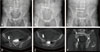
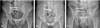
On postoperative radiograph checked on one day after operation, right SI joint widening was observed (Fig. 2A-C). Following CT was rechecked for confirming such a finding (Fig. 2D-F). We planned a supplementary operation using percutaneous SI screws at the second day after initial operation. The patient was positioned supine with applying radiolucent padding under his sacrum not to compress buttock muscle. Under an image intensifier, we inserted half-threaded cannulated screw with a washer into S1 vertebral body placed from the lateral surface of the iliac wing for achieving the reduction of widen SI joint. Then second fully-threaded screw was inserted into S2 body as a positional screw. The patient recovered well and was discharged from the hospital on the seventh day after second operation. On postoperative radiograph (Fig. 3A-C) and CT (Fig. 3D-F), satisfactory reduction of right SI diastasis was confirmed. Partially weight bearing on the injured side and fully weight bearing on the contralateral side were permitted after removal of drainage. One year after injury he had no anterior pain and his follow-up radiograph showed no screw breakage, fixation failure or heterotopic ossification (Fig. 4A-C).
The patient was asked if data concerning the case could be submitted for publication, and he consented.
DISCUSSION
Pubic symphysis is known as a non-synovial amphiarthrodial joint that is formed at the junction of the two innominate bones anteriorly. The joint comprises a fibro-cartilaginous disc that is stabilized by the anterior capsular and ligamentous structures. The typical mechanism of injury for an open-book pelvic ring injury is due to a force directed from anterior to posterior, or an external rotation or abduction force applied to either or both legs8).
In APC type II patterns, widening of the symphysis of more than 2.5 cm has been correlated with rupture of anterior sacrospinous ligament and thought to be required operative fixation like a symphyseal plating with or without SI joint fixation2345). The APC type III patterns is characterized by vertically unstable injury associated with a posterior disruption of SI complex, which requires both anterior and posterior fixation146). In general, diastasis of pubic symphysis without any evidence of vertical instability is thought to have intact posterior SI complex. Thus only anterior plating is enough to reduce disrupted pelvic ring due to anatomical properties of posterior pelvic ring similar to suspension bridge. Until now, retrospective studies suggest that posterior ring fixations may not be necessary in APC type II injury pattern injury8910).
Static radiographs may lead to underestimation of the actual injury, even if no pelvic manipulation has occurred. Stress X-rays were performed on a series of 22 patients with a symphyseal diastasis between 1 and 2.5 cm to determine the extent of pelvic ring instability7) Despite an injury, radiographic measurement of an average diastasis of 1.8 cm, the average diastasis with applied stress was 2.5 cm. Such findings of intraoperative stress examination have led to a change of the treatment plan in more than 25% of patients7). In the present a case, though initial radiographs revealed 3.0-cm pubic symphyseal diastasis, CT revealed 1.2-cm diastasis and no widening of SI joint. We assumed that unintended significant variable recoil may occur even if any pelvic wrapping was not tried. Such unintended recoil could happen by the patient transportation process using bed sheet for checking CT or the convex table setting during checking CT, which is different from flat table setting during checking plain radiograph. These may mask more serious posterior ring lesions than we think.
Even if initial radiograph showed SI diastasis, following preoperative CT showed minimal SI diastasis. Thus we think there may be injured anterior SI complex and intact posterior SI complex which could be treated by only anterior plating. However, though anterior plating was done, postoperative radiograph and CT revealed widening of SI joint. We presumed that there would be concealed injury of posterior SI ligamentous complex which should act as a hinge if injury type was APC II and it could make consequent seesaw effect by forced reduction of widened pubic symphysis with double anterior plating.
When CT was checked for scrutinizing associated posterior ring lesions that have occurred after diastasis of pubic symphysis was measured more than 2.5 cm by initial radiograph, it should be scrutinized not only associated SI joint widening but also whether diastasis of pubic symphysis on CT was equal to that of initial radiograph. Making a comparison between initial radiographs and following CT, if a smaller measurement of pubic symphyseal widening was observed on following CT, it should be considered a possibility of underestimation of a SI joint injury or APC type III injury. Even when pelvic open-book injury was thought that only anterior plating is required without SI joint fixation, intraoperative stress test for excluding concealed posterior ring disruption should be considered.
 XML Download