

 PDF
PDF ePub
ePub Citation
 Print
Print


The factors that prevent concentric reduction of the hip in traumatic fracture-dislocation are usually a bony fragment and infrequently soft tissues such as a torn acetabular labrum, a ruptured capsule, or a ligamentum teres. In some cases, small osteochondral fragments are difficult to detect in preoperative plain radiographs or computed tomography (CT) scans. Hip pain can be the result of intra-articular pathology that can potentially be a source for early degeneration and progression to osteoarthritis. In place of open surgery, hip arthroscopy is used increasingly as a less invasive surgical procedure for the diagnosis and treatment of acetabular labrum123) or for the extraction of loose bodies45). We present three cases, arthroscopic surgery of incarcerated acetabular osseo-labral fragment following reduction of traumatic hip dislocation. This study was approved by the Institutional Review Board (Chungnam National University School of Medicine, CNUH 2013-06-011-001) and informed consent was waived from all patients.
CASE REPORTS
1. Case 1
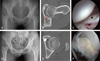
A 24-year-old woman presented to our hospital with complaint of intermittent left hip joint catching pain and snapping sensation for 3 years. Three years ago, she was admitted at our hospital with history of pedestrian traffic accident followed by fracture-dislocation of the left hip joint. After closed reduction, CT scans showed the fracture in anterior wall of the acetabulum. She underwent internal fixation for acetabular fracture using the fully threaded cannulated screw and was lost to follow-up after surgery. The plain anterior-posterior radiography showed asymmetry of the joint space and CT scans showed an intra-articular bony fragment on the left hip joints. She underwent hip arthroscopy for bony fragment removal. In arthroscopic findings, acetabular osseo-labral fragment was widely detached from the superior to posterior acetabular rim and incarcerated in the joint space. We made standard three portals (anterior, anterolateral, posterolateral) and additional distal anterolateral portal (1-2 cm distal to anterolateral portal). We performed reduction of inverted labral tear and refixation using two 2.7 mm absorbable suture anchor (Bioraptor; Smith & Nephew, Andover, MA, USA), and the bony fragment was removed. In addition, there were evidence of chronic cam type femoroacetabular impingement and osteochondral defect on femoral head. We performed femoral osteoplasty across the head-neck junction to protect secondary injury and micro-fracture in femoral head. After operation, concentric reduction and complete removal of bony fragment were confirmed using CT scans (Fig. 1). The visual analogue score (VAS) for pain improved from 7 preoperatively to 3 postoperatively and 1 at 3 months follow-up. At 2 year follow-up, modified Harris hip score (mHHS) was improved from 52 preoperatively to 92 postoperatively and hip outcome score of activity of daily living (HOS ADL) and sport related activity (HOS sport) were improved from 58 and 56 preoperatively to 93 and 91 postoperatively, respectively.
2. Case 2
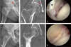
A 17-year-old man was referred to our hospital with a one year history of left hip joint pain, sometimes severe catching pain, and worsened with activity. Physical examination revealed a hip pain when moving leg and slightly limited range of motion was observed due to pain. A year ago, the patient felt groin pain following hip fracture-dislocation cause to passenger traffic accident. In medical history, he was treated only the closed reduction of hip joint and rest in a private clinic. The plain anterior-posterior radiography showed asymmetry of the medial joint space and CT scans showed an intra-articular bony fragment on the hip joint. The patient underwent hip arthroscopy for bony fragment removal. In arthroscopic findings, acetabular osseo-labral fragment was widely detached from the superior to posterior acetabular rim and incarcerated in the joint space. We performed reposition of inverted labral tear and refixation using two 2.7 mm absorbable suture anchor (Bioraptor) using standard 3 portals and additional distal anterolateral portal, and the bony fragment was removed. After operation, concentric reduction and complete removal of bony fragment were confirmed using CT scans (Fig. 2). At 2 year follow up, the symptoms of the patient were disappeared. mHHS was improved from 55 preoperatively to 93 postoperatively and HOS ADL and HOS sport were improved from 59 and 62 preoperatively to 92 and 93 postoperatively, respectively.
3. Case 3
A 54-year-old woman presented to our hospital with complaints of right hip pain and limited range of motion. She was complaint that the pain type was different from before: she felt catching pain when moving the leg. Two months ago, she was involved in traffic accident and sustained posterior dislocation of the right hip associated with a fracture of the inferior femoral-head. The hip was reduced spontaneously while the patient was being positioned for plain radiographs in local hospital. Post-reduction plain radiographs showed that the femoral-head fragment was aligned satisfactorily however bony fragment and asymmetry joint space were found. In medical history, she was recommended bed rest in a local hospital because of femoral-head fracture. The patient underwent hip arthroscopy for bony fragments removal. In arthroscopic findings, acetabular osseo-labral fragment was widely detached from the superior to posterior acetabular rim and incarcerated in the joint space. We performed labral debridement and excision of bony fragments because of the labral tear was irreducible and bony fragments were small (bony deficit across the posterior acetabulum was less than 10%) (Fig. 3). At 2 year follow up, the symptoms of the patient were improved but mild pain (VAS 1) still remained. Her mHHS was improved from 47 preoperatively to 85 postoperatively, and HOS ADL and HOS sport were improved from 51 and 49 preoperatively to 85 and 87 postoperatively.
DISCUSSION
A torn osseo-labral fragment rarely prevents consentric reduction of the hip in a traumatic dislocation and difficult to detect. Kim et al.6) reported the mechanism of incarcerated acetabular labrum following reduction of traumatic hip dislocation: An acetabular rim fracture or detachment of the labrum from the acetabular rim results from the posteriorly dislocating femoral head; when the hip is manually reduced, the torn labrum with or without the fractured fragment of the acetabular rim is pushed into the acetabulum, and it is trapped between the joints. And Mullis and Dahners5) found that 7 of 9 hip dislocation patients had loose bodies although no loose bodies were detected preoperatively and the joints were congruent on imaging. This illustrates that conventional radiographs and CT scans do not depict the extent of chondral or labral pathology associated with traumatic hip dislocation.
It is important to obtain complete early reduction to prevent the rapid development of degenerative arthritis. Even though the joint appears normal, we should thoroughly examine the inside of the joint for possible soft tissues incarceration. In our cases, we also missed the diagnosis radiologically at first time. However, the persistent patient's unusual hip catching pain, despite the reduction, caused concern about the possible soft tissues incarceration. It is thus important to recognize that a difference in the width of the joint space in postreduction plain radiographs suggests soft tissues incarceration.
Conventional treatment of hip fractures involves opening the joint to perform reduction and fixation and excision of fracture fragments. Recently, hip arthroscopy is used increasingly as a less invasive, safe and quickly able to regain mobility. Nonconcentric hip reduction or intra-articular loose bodies are ideal indications for hip arthroscopy 457). Hence, we also performed arthroscopic incarcerated fragment reduction and fixation and extraction in three cases.
This three case reports emphasize the nonconcentric hip reduction due to osseo-labral incarceration that are not shown clearly on plain radiograph and importance of unusual pain types; catching pain. We must consider that the hip arthroscopy is limited treatment but safe and effective as a subsequent procedure to treat incarcerated acetabular labrum following reduction of traumatic hip dislocation. It also constitutes for diagnosis of intra-articular pathologic lesions not evident on preoperative imaging studies.

 XML Download