Acetabular labrum normally attached to the margin of bony acetabular rim. Well known morphological variant of this structure can be seen in developmental dysplasia of hip (DDH) is an inverted labrum1). In DDH, peripheral margin of the acetabular labrum is interposed between bony acetabulum and femoral head. But there are a few reports about inverted labrum in the absence of dysplasia23). Patients with a labral tear usually had an osseous dysmorphism consistent with femoroacetabular impingement and hip dysplasia, of course there is no known bony abnormality45). Osteoarthritis caused by these bony abnormalities is well known. In this paper, we'd like to report a case had double contour sign of acetabular dome combined with intrusion of acetabular labrum and also describe its clinical importance, X-ray, computed tomogram (CT), magnetic resonance image and arthroscopic findings.
CASE REPORT
A 56 year old female patient visited our clinic with the left groin pain for 3 years. She didn't have any memorable trauma history and didn't like to exercise except intermittent riding a bike. She had received conservative treatment including activity restriction, non-steroidal antiinflammatory drug and physical therapy in the primary care hospital. But there was no improvement of symptoms, rather it was worsened. When she visited our hospital, she couldn't walk more than 30 minutes without pain.
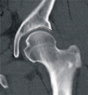
The nature of pain was deep aching, aggravated by pivoting action. Sometimes, she felt a sense of spontaneous subluxation and reduction of her hip joint. Impingement test was negative and Patrick test was positive. Anteroposterior and frog leg pelvis X-ray was checked (Fig. 1). Lateral center edge angle was 35°, acetabular inclination was 9°. No definite femoral head asphericity like pistol grip deformity and acetabular retroversion like cross over sign were observed. Usually, acetabular dome should be smooth one contour and the joint space should be even in the medial and lateral aspect in the absence of osteoarthritis. But weight bearing acetabular dome was slightly abnormal with double contour and widening of lateral joint space. For further evaluation, we checked CT and magnetic resonance arthrogram (MRA). In the CT scanning, we could confirm widening of the lateral one third of joint space and double contour of acetabular dome like gull wing, especially in the anterior half of the acetabulum (Fig. 2). In the MRA scanning, labral intrusion was observed between the anterosuperior acetabular dome and femoral head. But there was no definite labral tear (Fig. 3). We tried to do a hip arthroscopy. During the hip arthroscopy, anterosuperior labral tear and intrusion of labrum was observed (Fig. 4A). Labral tear and acetabular cartilage damage was minimal. Simple debridement and cauterization was performed (Fig 4B). Immediate after the operation, weight bearing was permitted in the tolerable pain range. Range of motion of the affected hip wasn't restricted.
She has pain free hip joint without progression of osteoarthritis at 2 years follow up.
DISCUSSION
Acetabular labrum acts as a seal, ensuring more constant fluid-film lubrication within the hip joint and limiting the rate of fluid expression from the articular cartilage layers of the joint, as indicated by a greater hydrostatic fluid pressurization within the intra-articular space when the labrum is intact6). Labral tear was known as a source of hip pain especially anterior groin pain7). Labral tear may be caused by traumatic event, but main cause of the tear is associated with osseous dysmorphism called femoroacetabular impingement or acetabular dyaplasia45). In the absence of trauma history and these bony abnormalities, the cause of acetabular labral tear, was thought to be idiopathic.
Harris et al.2) suggested that the intra-articular labrum was a developmental abnormality and postulated that this abnormality was the cause of the degenerative arthritis. Byrd and Jones3) also reported the labral inversion is a cause of osteoarthritis. But these two papers focused on the labrum itself and didn't mention the primary acetabular bony abnormality though Byrd and Jones3) talked about secondary bone change. Although several authors described this lesion as a labral inversion, we thought labral inversion is not a proper description. Pure labral inversion means whole labrum is caught in joint including peripheral margin. Rather, labral intrusion is better expression. In the presence of dysplasia and femoroacetabular impingement, labral tear may happen at the labro-cartilagenous junction. Labral tears alter the mechanical or biochemical environment within the joint, accelerating degenerative changes and progression of chondral pathology over time. This is well known pathophysiology of osteoarthritis in dysplatic hip and femoroacetabular impingement89).
In our case, there is no known osseous abnormality aforementioned. New osseous abnormality no one reported up to this time was observed so called double contour sign of acetabular dome that is confirmed at the X-ray, CT and MRA scanning. We postulated followings. In the hip with double contour sign at the acetabular dome, there is a labrum under the lateral subchondral bone instead of articular cartilage and the junction between the acetabular cartilage and the labrum may be subjected to higher mechanical demand but mechanically inferior compared to original acetabular hyaline cartilage. This vulnerable acetabular labrum may be susceptible to tear and cause anterior hip pain and exposure of subchondral bone. Also femoral head will move toward anterolateral aspect within the acetabulum. After then accelerated cartilage damage and early osteoarthritis will be happened. We thought this is one of unknown cause of idiopathic osteoarthritis of hip joint. To confirm this postulation, further study for patients who underwent total hip arthroplasty due to primary osteoarthritis may be needed. To treat this lesion, we recommend arthroscopic intervention including labral debridement, microfracture technique for exposed subchondral bone to preserve the native hip joint and delay the requirement for future total joint arthroplasty especially in young active patients.
Our patient is symptom free at 2 years follow up. But long-term follow up is needed whether the arthroscopic surgery can delay or eliminate needs for total joint replacement. This is our postulation for development of osteoarthritis in the near normal hip joint without femoroacetabular impingement and dysplasia. It should be verified by further study.


 PDF
PDF ePub
ePub Citation
 Print
Print


 XML Download