

 PDF
PDF ePub
ePub Citation
 Print
Print


INTRODUCTION
The early use of bisphosphonates are believed to interfere with bone remodeling of the callus to cortical bone and delay fracture healing in patients with osteoporosis by inhibiting osteoclast function123). For this reason, the optimal time to consider bisphosphonate therapy still remains controversial. The incidence of secondary fracture is very high within the first 1 year after the primary fracture, and 50% of the second fractures have been reported to occur within the first 12 months in men and 19 months in women1). It has been suggested that the early use of bisphosphonates during or after the first two weeks following surgical intervention may reduce fracture and mortality rates by increasing bone mineral density (BMD), therefore a decrease in the incidence of secondary fracture can be expected by administering bisphosphonates as early as possible after osteoporotic fracture1). Although a previous study has shown that the time of starting bisphosphonate treatment following surgery has no influence in bone union or healing, the exact effect of administration time is not completely understood2). Therefore, the authors aimed to verify the optimal time of bisphosphonate administration for management of osteoporotic intertrochanteric fractures and chracterize its effects on bone healing and complications.
MATERIALS AND METHODS
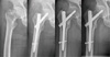
This multicenter study retrospectively analyzed data on 284 patients (284 hips) who underwent surgery due to osteoporotic intertrochanteric fracture from December 2002 to December 2012 in 3 different hospitals. The mean age of patients was 77.3 years (range, 58-92 years) and 52 were men and 232 women. The mean follow-up period was 68.4 months (range, 6.4-123.0 months). The implants used were dynamic hip screw (DHS; Depuy Synthes, Warsaw, IN, USA) in 20 hips, proximal femoral nail anti-rotation (PFNA; Depuy Synthes) in 119 hips, and Gamma-nail (Stryker, Mahwah, NJ, USA) in 145 hips. All patients had osteoporotic fractures with BMD T-scores of less than -2.5, and had no history of anti-osteoporotic drug use preoperatively. From the first postoperative day, range of motion and non-weight bearing ambulation was initiated, and weight bearing was allowed starting three months after surgery. As shown in Table 1, administered bisphosphonates were zolendronate (n=56), ibandronate (n=89), risedronate (n=81) and alendronate (n=58). In addition, patients were given a daily dose of 1,500 mg calcium and 800 IU vitamin D. To identify the effect of time to administration of bisphosphonate on callus formation at the fracture site, patients were divided into three groups with varying times to bisphosphonate administration post surgery: 1 week (group A; n=102), 1 month (group B; n=89) and 3 months (group C; n=93) (Table 2). No differences were found in age, gender, body mass index, BMD and fracture types according to Evans classification between the three groups (Table 3). Oral and intravenous administration routes were determined by patients' choice and the distribution of administration time are shown in Table 4. Koval scores and change of Koval scores 1 year after surgery were compared between the three groups for clinical evaluation. Bone union was judged as callus appearance across the fracture line on sagittal and coronal radiographs taken on the 4th, 8th, 12th, and 16th postoperative weeks, with no evidence of: i) prosthetic loosening, ii) blurring of the fracture line, and iii) pain during hip motion (Fig. 1). In addition, complications such as infection, mal-union or displacement were also examined. For statistical analyses, ANOVA was performed using IBM SPSS Statistics ver. 21.0 (IBM Co., Armonk, NY, USA).
RESULTS
Koval scores one year after surgery for groups A, B, and C were 2.44±1.74, 2.36±2.23, and 2.43±1.89, respectively. Although a considerable standard deviation was found in group B, there was no significant difference between the groups. Changes in Koval scores between pre- and post-fracture were 0.84±0.73, 0.519± 0.78, and 0.78±0.85 in groups A, B, and C, respectively, showing no significant difference between the groups (Table 5). The duration of union in groups A, B, and C was 4 weeks after surgery in 6 cases (5.9%), 8 (9.0%), and 4 (4.3%); 8 weeks in 27 (26.5%), 22 (24.7%), and 24 (25.8%); 12 weeks in 47 (46.1%), 41 (46.1%), and 45 (48.4%); 16 weeks in 18 (17.6%), 15 (16.9%), and 15 (16.1%); 20 weeks in 3 (2.9%), 1 (1.1%), and 5 (5.4%); and 24 weeks in 1 (1.0%), 2 (2.2%), and 0 (0.0%). For all three groups, union occurred most commonly 12 weeks after surgery (46.1%, 46.1%, and 48.4%, respectively). Union rates were similar in all three groups. A higher rate of early (1 month after starting bisphosphonate therapy) union was achieved in group B (9.0%) compared to groups A (5.9%) and C (4.3%). Also, union was achieved in 24.7% of group B cases 8 weeks after surgery, indicating a decreased union rate compared to groups A (26.5%) and C (25.8%). Finally, union was observed in 2.2% of group B cases 24 weeks after surgery, exhibiting a higher rate of delayed healing compared to that of groups A (1.0%) and C (0.0%). However, these differences did not reach to statistical significance. The mean duration for bone union was 12.4, 11.9 and 12.3 weeks after surgery for groups A, B, and C (P=0.883) (Fig. 2), respectively. These results were not statistically significant difference based on the time of bisphosphonate administration. There was no case with reoperation due to complications such as nonunion, infection and prosthetic loosening. Although displacement of lag screw was detected in 3, 5, and 7 cases for groups A, B, and C, respectively, these differences were not statistically significant (P>0.472).
DISCUSSION
The World Health Organization defines osteoporosis as a systemic skeletal disease characterized by reduction in bone mass and deterioration of the micro-architecture [3]. According to a cohort study by Shin et al.4) in 2010, the prevalence of lumbar spinal osteoporosis in adults aged 50 years or older were 24% in women and 12.9% in men. The rise in morbidity and mortality following hip fractures has been reported567), and one of the causes for increased morbidity and mortality is a new osteoporotic fracture occurring on the contralateral side of the hip or spine89). Secondary osteoporotic fracture affects approximately 4-10.4 in 100 fractured patients per year, and occurs predominantly within the first year after the primary fracture. Although differences among studies were observed, the use of bisphosphonates reduced the risk of fracture by 50% in the group at high risk of osteoporotic fracture compared to the group with no use. These findings suggest that bisphosphonate therapy is warranted in patients with osteoporotic fracture to prevent secondary fractures5). However, a screening test for osteoporosis is performed in only 12-24% of patients with hip fracture and prescribed in only 8.6-19.3%, therefore the importance of osteoporosis has been largely underscored1011). Prevention of secondary fracture is a critical factor in lowering mortality rates and increasing average life expectancy. Since compliance with osteoporosis therapy is an important parameter reducing the incidence of secondary fractures12), a prolonged duration from fracture to bisphosphonate therapy may induce attrition or withdrawal during follow-up. However, no definite standards have been established regarding the time of bisphosphonate administration following osteoporotic fracture.
There have been many confusing reports about the effect of bisphosphonate on bone healing. Multiple previous animal studies have revealed that bisphosphonate delays the bone healing1314). However, Bauss et al.15) and Munns et al.16) have suggested that the use of bisphosphonates had no influence on bone union, while a number of recent studies have described that bisphosphonate treatment improved fracture healing1718192021). The pharmacological action of bisphosphonates is understood to inhibit bone resorption by blocking osteoclasts from breaking down bone due to accumulation to bone surfaces. Thus, the initiation of bisphosphonate therapy has been recommended following fracture healing in the past due to concerns that bisphosphonates could interfere with bone remodeling22), delay bone union, and weaken primary stability of prosthetic components by affecting bony ingrowth in patients treated with cementless hip arthroplasty or internal fixation232425). Only a few studies have objectively investigated the impact of bisphosphonate use on bone healing in patients with osteoporotic fracture. Recent papers have rather advocated no deterioration of biomechanical properties and increased bone ingrowth26). According to Molvik and Khan27), there is no evidence that supports the notion that bisphosphonates result in nonunion or delay in union time in fracture healing in the femur, the distal end of the radius and proximal tibia. A double-blinded randomized controlled trial by Shane et al. was unable to demonstrated a correlation between start time of intravenous zoledronic acid administration and union time of femoral fracture28).
In a prospective multicenter study, Gong et al.29) administered risedronate to patients with femoral fracture in three different groups (1 week, 1 month, and 3 months following surgery). The average duration to bone union was 10.70, 12.90, and 12.30 weeks, respectively, and no statistically significant differences were observed. On the contrary, faster fracture union was seen in the early administration group, contradicting previous findings. Moreover, in a randomized controlled non-blinded study, Abrahamsen et al.30) administered alendronate to 24 patients after internal fixation with plate screw on distal radius fracture. The administration of alendronate was started 2 weeks and 3 months after surgery in the experimental and control groups, respectively. The mean duration to bone healing at their study was 6.70 and 6.80 weeks after operation, showing no statistical difference. It has not been proved and remains controversial whether bisphosphonates affect the incidence of postoperative nonunion in patients with femoral fracture. Recently, it has been suggested that surgical technique and implants are possible causative factors for nonunion. Weil et al.25) have attracted attention by advocating that delays in healing may be caused by a decrease in load transfer due to stress shielding31). For this reason, early ambulation has been recently recommended, and the wider use of load sharing devices has been suggested6).
The results of this study revealed that there was no difference in time to achieving union of intertrochanteric fractures, and the presence of complications according to the time of starting bisphosphonate treatment. Unlike other areas, the intertrochanteric region of the femur is characterized primarily by weight-bearing trabeculae, a large amount of cancellous bone and abundant blood supply that stimulate new bone formation32). Because of these anatomical characteristics of the intertrochanteric region, the early use of bisphosphonates appears to have insignificant effects on bone union despite decreased bone remodeling by reducing osteoclastic activity. Therefore, this would suggest that early administration of bisphosphonates is helpful in preventing secondary fractures, improving the patient's quality of life, and increasing life expectancy in patients with osteoporotic intertrochanteric fracture.
The limitations of the present investigation, as a multicenter study, are possible variations due to differences in surgeons, surgical outcomes and different implants and drugs, determination of the exact time of fracture healing due to the nature of intertrochanteric fracture, and the influence of subjective judgement. Further studies are warranted to resolve these limitations by performing a single-center study with larger sample size or a prospective study to compare the incidence of secondary fractures, mortality following primary fractures, and duration to death. In this way, more objective study results can be achieved by improving the limitations of the retrospective cohort design.
CONCLUSION
The initiating time of bisphosphonate administration following surgery does not affect the clinical outcomes or the occurrence of complications in patients with osteoporotic intertrochanteric fracture. Therefore, early administration of bisphosphonates would be helpful to prevent additional fractures and reduce mortality in patients with osteoporotic fracture.


 XML Download