

 PDF
PDF ePub
ePub Citation
 Print
Print


Atypical femoral fractures (AFFs), which may occur anywhere along the femur from just distal to the lesser trochanter to just proximal to the supracondylar flare, are stress or insufficient fractures induced by low energy trauma or no trauma and have specific X-ray findings. Bisphosphonate therapy appears to be associated with a high risk of AFFs. Although periprosthetic fractures have been excluded from the AFF definition established by the American Society for Bone and Mineral Research (ASBMR) in 20131), some authors have reported periprosthetic fractures showing specific features of atypical fractures around a well-fixed femoral stem234). We report 3 cases of periprosthetic femur fractures exhibiting specific radiographic features of AFFs after hip arthroplasty and long-term use of bisphosphonates, and provide a brief review of relevant literature.
CASE REPORT
1. Case 1
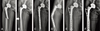
A 43-year-old woman visited our hospital due to a fracture of the right femur. She had a history of long-term steroid therapy (>20 years) for systemic lupus erythematosus, underwent hip arthroplasty in our hospital 10 years earlier due to bilateral avascular necrosis of the femoral heads, and had a history of risedronate use for the last 5.5 years. Her right femur fracture occurred due to a minor trauma after slipping in a puddle. Radiographs taken in the emergency center revealed specific features of an AFF classified as type B1 according to the Vancouver system5) with lateral cortical thickening and short oblique orientation around the right prosthesis without comminution (Fig. 1A). According to the results of retrospective radiographic analysis and history, intermittent pain in the right thigh persisted for the last 4 months, and radiographs obtained 4 months before a complete fracture showed localized periosteal thickening of the lateral cortex and a transverse lucent fracture line (Fig. 1B). Before the fracture, the femoral component remained stable with no prosthetic loosening or osteolysis.
Since a tapered-wedge stem was used, anterior plate fixation was performed with a single short plate, and then percutaneous cerclage wiring was performed without bone grafting (Fig. 1C). Three months after fixation surgery, she re-visited our hospital because of right thigh pain during ambulation with no associated trauma, and fixation failure was identified by radiography, which was due to non-union at the same site (Fig. 1D). Autogenous bone grafting and re-fixation with dual plates were performed (Fig. 1E), and then teriparatide was administered as subcutaneous injections in the abdomen (20 µg once daily for 6 months). Successful bony union was achieved 11 months postoperatively. She was followed-up for 2.5 years after re-fixation and ambulates independently without any complications (Fig. 1F).
2. Case 2
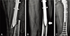
A 74-year-old woman visited our hospital due to a left femur fracture that occurred after minor trauma due to slipping down during ambulation. She received bipolar hemiarthroplasty 13 years earlier due to a left femoral neck fracture and revision left hip arthroplasty because of erosion of acetabular cartilage 11 years earlier. She also had a history of risedronate use for 6 years after revision left hip arthroplasty. Radiographs demonstrated specific findings of AFF classified as type B1 according to the Vancouver system5) with a short oblique pattern and no comminution around the prosthesis (Fig. 2A). According to the results of retrospective radiographic analysis and history, intermittent pain in the left thigh persisted for the last 2 years, and radiographs obtained 3 months before the fracture revealed localized periosteal thickening of the lateral cortex and a transverse lucent fracture line (Fig. 2B). No loosening or osteolysis of the femoral component was observed before the fracture. The fracture was treated by internal fixation with a long plate without bone grafting (Fig. 2C). Bisphosphonate treatment was stopped, and then teriparatide was administered as a subcutaneous injection in the abdomen (20 µg once daily for 6 months). The patient is currently (7 months after operation) under observation and does not have any complications.
3. Case 3
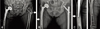
A 86-year-old woman visited our hospital after slipping down and complained of left thigh pain. She received bipolar hemiarthroplasty 9 years earlier due to a right femoral neck fracture, and had a history of risedronate and alendronate therapy for 9.3 years. Radiographs revealed specific findings of AFF with a short oblique pattern in the left femoral subtrochanteric region. Although lateral cortical thickening and periosteal reaction of the lateral cortex were observed in the right prosthesis, no transverse lucent fracture line was visible and the femoral component was well-fixed and stable (Fig. 3A). According to retrospective analysis, cortical thickening and periosteal reaction around the right periprosthetic area and localized periosteal thickening of the lateral cortex in the left subtrochanteric area were detected on the radiographs taken 1.3 years before the complete fracture of the left femur. The fracture of the left femur was managed using closed reduction and cephalomedullary nailing (Fig. 3B), and teriparatide was administered as a subcutaneous injection in the abdomen (20 µg once daily for 4 months). Although a bone scan revealed a slight positive finding in the right femur at that time, patient did not complain of any symptoms. Therefore, we decided to follow-up the patient closely.
A progressive radiolucent line was identified around the periprosthetic area during regular follow-up. Radiographs taken 6 months after surgery revealed a transverse lucent fracture line on the right femur with periosteal thickening of the lateral cortex. Definite positive bone scan findings were observed at that time, and the patient complained of pain in the right femur (Fig. 3C). As preventive therapy, minimally invasive plate fixation was performed without bone grafting (Fig. 3D). She is currently (8 months after last operation) under observation and does not have any complications.
DISCUSSION
The 2013 ASBMR diagnostic criteria for AFFs include (1) fractures associated with no trauma or low-energy trauma; (2) a fracture line originates at the lateral cortex and is substantially transverse in its orientation or becomes oblique; (3) complete or incomplete fractures (involving only the lateral cortex); (4) non-comminuted or minimally comminuted fractures; and (5) localized periosteal reaction or thickening along the lateral cortex at the fracture site1). The diagnosis of AFFs requires the presence of at least 4 of these major criteria. The minor features include a generalized increase in cortical thickness of the femoral diaphysis, prodromal symptoms such as thigh pain, a bilateral incomplete or complete femoral diaphysis fracture, and delayed fracture healing, but the AFF definition excludes periprosthetic and pathologic fractures1).
However, specific features of atypical fractures have been recently observed around the periprosthetic region after hip arthroplasty234). Niikura et al.2) performed open reduction and plate fixation in a patient with a history of hip arthroplasty and prolonged bisphosphonate therapy, who was diagnosed with a periprosthetic fracture with AFF features after low-energy trauma. Curtin and Fehring3) used conservative management of incomplete fractures with AFF features in three patients who complained of thigh pain after hip arthroplasty and had a history of long-term bisphosphonate use. Cross et al.4) reported conservative management and suspension of bisphosphonate treatment for a patient with a history of prolonged bisphosphonate therapy and thigh pain after localized periosteal thickening was detected around a well-fixed cemented femoral stem.
The common characteristics of these studies are a history of long-term bisphosphonate therapy and a stable and well-fixed femoral stem at the time of fracture diagnosis. All three patients in this case report also had prodromal symptoms including thigh pain after hip arthroplasty, a history of long-term bisphosphonate treatment (for 6.9 years on average), and a stable femoral component at the time of fracture diagnosis. In two cases, non-comminuted complete fractures extended medially through the lateral cortex. These fractures were managed with open reduction and plate fixation. In the third case, preventive plate fixation was performed after early detection of periosteal reaction and thickening of the lateral cortex around the femoral component.
The type of AFF was either incomplete or complete. Surgical treatment is unavoidable in patients with complete fractures, and multiple studies have demonstrated the possibility of delayed union or nonunion67). Since incomplete fractures associated with thigh pain have the potential to progress to complete fractures, it is important to perform internal fixation as a preventive measure through early detection7). To achieve this goal, radiographic exams and detailed history taking are warranted on a regular basis. The possible causes of thigh pain after hip arthroplasty include prosthetic loosening, periprosthetic fractures, and hip instability8). However, the possibility of thigh pain as a prodromal symptom of AFFs has to be taken into consideration, despite a stable and well-fixed femoral component. In particular, thorough radiographic exams and detailed history taking are crucial in patients with a history of long-term bisphosphonate use, and active interventions such as prophylactic internal fixation are required when symptoms persist.
 XML Download