

 PDF
PDF ePub
ePub Citation
 Print
Print


Vancouver system has been successfully used to classify and treat a periprosthetic fracture of the femur after hip arthroplasty. However, there is a concern about the possibilities that insufficiency fracture around the implant can occur. This concern is growing because of increased number of elderly patients with osteoporosis who received hip arthroplasty1).
To date, there have been a few reports about atypical fractures at the distal part of femoral stem2345). However, report of periprosthetic insufficiency fracture in subtrochanteric area were not frequent. Recently, we observed two cases of insufficiency fractures at the lateral cortex of subtrochanteric area around the radiographically loose cemented femoral component.
CASE REPORT
1. Patient 1
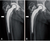
A 75-year-old woman underwent cemented total hip arthroplasty, and made regular follow up for 32 years without any clinical symptoms. Although the patient did not recall the exact date that she started medication, her family remembered that she has been taking oral bisphosphonate for at least 10 years. In May 2011, she presented with acute pain on her ipsilateral thigh. She did not recall any history of trauma except mild bump into the entrance bar at the subway. Radiograph taken at that time showed protruded lateral cortex of the subtrochanteric area of the femur with short transverse uni-cortical fracture line (Fig. 1A). The patient was instructed to discontinue all medication containing bisphosphonate and to use protected weight bearing using two crutches. Also, she was prescribed subcutaneous teriparatide (Forteo; Lilly, Indianapolis, IN, USA) injection for four months, until she did not complain hip pain. Eight months after the onset of initial pain, she could bear weight without assistance and reported no pain in her right hip. Radiographs taken at that time showed evidence of a healed fracture (Fig. 1B).
2. Patient 2
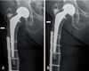
A 85-year-old woman who has history of taking oral bisphosphonate for more than 5 years underwent cemented bipolar endoprosthesis in 1998. Her initial diagnosis leading to operation was intertrochanteric fracture of right femur. She experienced Vancouver type C periprosthetic fracture at the ipsilateral femur in 2007. It was treated with open reduction and internal fixation. Since that time, the patient has been treated with 35 mg risedronate weekly for another 4 years. Forty one months after the union of periprosthetic fracture, she complained sharp pain on right hip during weight-bearing. She did not recall any history of trauma. The radiographs showed protruded lateral cortex with transverse, uni-cortical fracture line at subtrochanteric area around the radiographically loose cemented stem (Fig. 2A). Bone scan showed new increased focal uptake at the right subtrochanteric region of the femur. The patient was instructed to discontinue risedronate and to use two crutches with non-weight bearing. She was prescribed daily subcutaneous teriparatide injection for four months. Seven month after the initial treatment, her pain was markedly improved. Radiographs showed evidence of a healed fracture. One year after the onset of pain, her pain had resolved and the fracture was consolidated on the radiographs (Fig. 2B). She was allowed to bear weight as tolerated and had returned to her previous household ambulatory activity level.
DISCUSSION
There is a concern about the possibilities that periprosthetic insufficiency fractures can occur due to recently increased number of elderly patients with osteoporosis1). The occurrence of atypical femoral fractures in patients receiving long-term bisphosphonates therapy was well described in many reports5678). But regarding a periprosthetic fracture, there have been only a few reports that the fractures occur at distal part of a stable cementless stem. However, in contrast with those reports, our cases showed subtrochanteric fracture around the radiographically loose cemented stems with otherwise pain-free hip. In this regards, we believe that the clinical pattern of these fractures could not be easily classified as atypical or stress fractures. Because the fractures were found at the same area in elderly patients with almost identical clinical situation without any history of trauma and they were definitely different from commonly reported atypical fractures, we considered them as peri-prosthetic insufficiency fractures. Although a number of classification systems in periprosthetic fracture have been proposed in the past, the presence of these fractures and other reports with distal atypical fractures can challenge the current classification of periprosthetic femoral fractures.
Most atypical fractures were reported as having poor results with only conservative treatment. Withdrawing bisphosphonates therapy should be considered if investigations show early signs of such a fracture6789101112). Potent anabolic agent such as teriparatide may be an option in symptomatic patients before complete fracture1314). In our cases, both cases were treated nonsurgically with teriparatide injection, protected weightbearing, and discontinued bisphosphonates. The authors did not assure whether teriparatide critically accelerated healing of these fractures or not. However, both patients successfully escaped surgery with the same protocol, suggesting that the teriparatide might play a clinical role. After two years of follow up, fractures did not recur.
Because there are many elderly patient with osteoporosis who are undergoing hip replacement, surgeons should consider the possibility of periprosthetic insufficiency fractures when the osteoporotic patient with cemented stem showed sudden hip or thigh pain without a history of trauma. Although we are not convinced that our protocol is the only method to deal with this type of fracture, we believe that it can be treated non-operatively if it is not displaced.
 XML Download