

 PDF
PDF ePub
ePub Citation
 Print
Print


INTRODUCTION
The incidence of femoral neck fractures, which lead to severe complications such as avascular necrosis and non-union, is gradually increasing due to longer life expectancy and growing prevalence of osteoporosis. In particular, femoral neck fractures in elderly patients commonly occur after minor injuries, and undisplaced fractures graded as type I or II according to the Garden classification1) account for about 19% of all hip fractures2). The treatment principles of undisplaced femoral neck fractures are stabilized fracture by internal fixation and early exercises. Since displacement of bone fragments is common, multiple attempts have been made to achieve stable fixation using a variety of implant options3456789). Internal fixation with multiple cannulated screws is frequently used because of several advantages such as fracture fixation stability, lower complication rate, and the ease of operations using image amplifiers10). In dementia patients with femoral neck fractures, complications such as loss of fixation or reduction loss after surgical treatment are more likely to occur than in healthy elderly individuals because of their lack of understanding, communication problems with health care providers, and different responses to pain11). Thus, we considered proximal femoral nail antirotation (PFNA; Synthes, Solothurn, Switzerland) as an alternative plan for dementia patients who have difficulty with postoperative ambulation. We aimed to evaluate the clinical and radiological results, associated complications during the follow-up, and re-operation of internal fixation using PFNA in elderly dementia patients with undisplaced femur neck fractures.
MATERIALS AND METHODS
1. Subjects
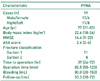
We retrospectively studied patients over 70 years of age who walked independently with a cane or crutches (household ambulation ability, according to the Koval classification12)) and who suffered from dementia and were diagnosed with undisplaced femoral neck fractures (Garden type I or II) from October 2006 to December 2012. Regardless of the presence or absence of dementia, we consulted patients with cognitive impairments as preoperative symptoms to the Department of Neuropsychiatry for preoperative and postoperative interdisciplinary cares (temporary control of aggressive behavior and lack of cooperation for rest). We determined the criteria for dementia patients by using interviews, two clinical psychology practitioners preoperatively evaluated subjects who were unable to cooperate in ambulation limitation or rehabilitation. We excluded patients with pathologic fractures or a history of surgery of the ipsilateral hip joint or femur. Of 22 patients, 2 patients who withdrew from outpatient follow-up and 1 patient who died within 1 year of operation were excluded. A total of 19 patients (13 men and 6 women) were enrolled in the study. Their mean age was 77 years (range, 71-82 years). According to the Garden classification, 11 patients had type I fractures and 8 had type II fractures. Most fractures occurred as a result of low-energy injuries including misstepping in 11 cases and falling from the height of a chair in 8 cases. Bone mineral density of the healthy side of the hip was measured using dual energy X-ray absorptiometry (DEXA), and T-scores were used as a reference. The mean T-score of the healthy side of the femoral neck was -2.75 (range, -1.5 to -4.7); it was below -2.5 in 2 cases and above -2.5 in 17 cases. The severity of dementia was evaluated using the Mini Mental State Examination (MMSE)13); the mean MMSE score was 14.4 (range, 9-20). The American Society of Anesthesiology (ASA) classification14) was used to assess each patient's preoperative risk, and the mean ASA score was 2.6 (range, 2-4). The mean follow-up was 53.3 months (range, 30-72 months). The mean body mass index was 22.6 kg/m2 (range, 18-26 kg/m2). Right side was affected in 10 cases and left in 9 cases (Table 1).
2. Surgical Methods
All operations were conducted under spinal anesthesia, and a fracture table and image amplifier were used. Surgery was performed within 24 hours except in patients with a high anesthesia risk due to underlying medical conditions; for such patients, surgery was delayed but not more than for 3 days after injury. The average time between injury and surgery was 39 hours (range, 24-72 hours). Prophylactic antibiotics and anticoagulants were administered to all patients preoperatively.
A patient was in a supine position while the upper body was supinated to make the intramedullary approach easier during operation. Before surgery, an image amplifier was used to assess displacement and then a 3-5-cm longitudinal incision was made 2-3 cm proximal to the greater trochanteric tip; the gluteus maximus was then split and the greater trochanteric tip was examined by palpation. A guide wire was placed on the insertion spot and lightly hit with a hammer for temporary fixation. A reamer was then used to expand the hole and a flexible intramedullary guide wire was inserted into the insertion hole. A reamer was then used again to expand the hole. Repeated expansion of the insertion hole allowed to prevent additional displacement of neck fractures.
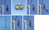
The size of PFNA was determined based on radiological images taken before surgery. When the PFNA was inserted into intertrochanteric fractures, a thick medullary nail was used to provide sufficient resistance for rotator power, but the optimal diameter of nails used for fixation of femoral neck fractures has not been firmly established. Hence, in this study, to prevent additional displacement, we used sufficiently small size of PFNA, which enables insertion of nails into the intramedullary space without resistance, and employed sufficient intramedullary reaming. In all cases, the caput-collum-diaphysis angle was 130° and the length of nail was 170 mm. To prevent any resistance and to carefully insert nails and spiral blades, it was carefully monitored using the anteroposterior view and lateral views if the guide wire insertion spot was not toward either anterior or superior area of the femoral head. Meanwhile, the guide wire was inserted from the lateral cortex of the femur to approximately 5 mm depth from the cortex of the femoral head. Once the desired position and insertion depth were confirmed, two additional wires were inserted into the femoral head. For this additional insertion, the aiming zig for antirotation wire, designed for convenient insertion using already placed guide wires, was used so that 1) further displacement of the femoral neck fracture was prevented when a spiral blade was inserted and 2) a spiral blade of a proper size was introduced. After the spiral blade was fixed, the compression instrument for the PFNA blade, which is included in the PFNA set, was kept ready for use in case of fracture line widening, but was not otherwise used (Fig. 1).
All surgical procedures were performed by one surgeon. For rehabilitation, patients were asked to start early ambulation using either a wheelchair or a walker, as much as possible. Active hip flexion-extension, femoral raising, and femoral quadriceps exercises were started once acute pain subsided. Depending upon patients' prognosis and the type of fracture, partial weight bearing in the standing position and walker-assisted ambulation were allowed 3-7 days after surgery. As we expected, despite strict instructions given to the patients and their caregivers, there were difficulties in ambulation control.
3. Assessment Methods
1) Clinical Assessment
Walking ability was assessed according to the Koval classification12) from the preoperative period to 24 months after surgery and was graded from independent community ambulatory (grade 1) to nonfunctional ambulatory (grade 7) status. Walking ability was evaluated every 4 weeks for the first 6 postoperative months and then every 3 months for the next 24 months.
2) Radiological Assessment
Preoperative, postoperative, and final follow-up radiographs of the hip were taken in the anteroposterior and lateral views. A picture archiving and communication system (PACS; Maro View; Marotech, Seoul, Korea) was used to minimize errors in radiographs. Patients were in the supine position to ensure that radiation was directed perpendicularly to the hip joint from a certain height (110 cm) after the femur had been internally rotated by approximately 15°. Radiological assessment was carried out by two independent observers (K1 and K2), and the mean values were calculated. To ensure the reliability of the measured values, the interobserver agreement was measured with Kappa coefficients (K1=0.88, K2=0.81). Radiological follow-up was performed regularly to confirm bony union. Bony union was defined as the absence of pain on full weight-bearing and the presence of three or more cortical callus bridges without any fracture line. The location of the blade within the head was recorded using the Cleveland method15) by dividing the anteroposterior view of the femoral head into the upper, middle, and lower zones, and each zone was then divided into anterior, middle, posterior zones. Tip-apex distance (TAD)16) was calculated as the sum of the distances from the tip of the lag screw to the apex of the femoral head on the anteroposterior and lateral radiographs. At the time point of the radiographic appearance of complete bony union, avascular necrosis of the femoral head of the proximal femur was analyzed on magnetic resonance imaging (MRI) scans. When bone single-photonemission computed tomography (bone SPECT) was conducted at the same time, the abnormally increased 99mTc-hydroxymethane diphosphonate (99mTc-HDP) uptake was visually determined based on a coronal image of the hip. Elevated uptake and cold spots indicating non-union at the fracture site were considered to indicate reduced femoral head viability. Bone SPECT images were interpreted by a nuclear medicine specialist without knowing clinical and test findings.
3) Assessment of Complications
Non-union was defined as healing failure without any clinical or radiographic signs of postoperative progression to union. Additional signs of non-union were pain, gradual osteolysis, change in fracture location of more than 10 mm, and excessive sliding (>20 mm) of internal fixatives17). Avascular necrosis of the femoral head was defined as the presence of pain, collapse of the femoral head, osteoporosis, increase in the bone shadow, subchondral sclerosis of the femoral head, or subsidence18). Early fixation failure was defined by the presence of varus deformity of more than 10 mm, posterior displacement of the femoral neck exceeding 20 mm, or screw penetration by comparing radiographs taken immediately after the operation and within the first 3 postoperative months17). In addition, we evaluated infection, implant fracture, and secondary fractures.
RESULTS
Duration of operation was defined as the time from the induction of anesthesia to the end of anesthesia. The mean operation time was 82.5 minutes (range, 50-120 minutes). The mean blood transfusion volume was 80 mL (range, 20-150 mL).
1. Clinical Results
Walking ability was evaluated using the Koval classification12) from the preoperative period to 24 postoperative months. The walking ability of patients was 2.6 points (range, 14 points) preoperatively and 2.8 points (range, 1-5 points) at the final follow-up, showing an average decrease of 0.2 points. If two patients with complications (one with non-union and one with avascular necrosis of the femoral head and non-union) were excluded, walking ability was 2.71 points (range, 14 points) preoperatively and 2.78 points (range, 15 points) at the final follow-up, showing a decrease of less than 0.1 points. Walking ability evaluated from before injury to 4 weeks after surgery showed an average decrease of less than 0.5 points, indicating the possibility of early ambulation (Table 2).
2. Radiologic Results
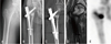
The blade was located within the femoral head on postoperative radiographs was recorded with the Cleveland classification15), in Cleveland zone 5 in 12 cases and in Cleveland zone 8 in 7 cases. There were no cases of inappropriate placement of the blade within the head. The mean TAD was 9.3 mm (range, 8-12 mm) (Table 2). The average time to bony union was 4.14 months (range, 2.5-7 months) in 17 out of 19 patients. In these 17 patients, MRI scans were performed when satisfactory radiological signs of bony union were seen, and no femoral head necrosis was observed. Bone SPECT revealed no case of increased uptake or a cold spot within the femoral head (Fig. 2).
3. Complications
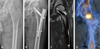
Of the two cases with non-union, avascular necrosis of the femoral head occurred in one case and was observed as a shadow or deformity in the femoral head on simple radiographs taken in the anteroposterior and lateral views 12 months after surgery. According to the Ficat and Arlet classification19), avascular necrosis was graded as stage 2. The patient with non-union only had the most severe symptoms of dementia with an MMSE13) score of 9. This patient is currently staying at a care facility and family members refuse to allow additional surgical treatment. This patient's inability to cooperate and the difficulty in rehabilitation were expected due to severe dementia, and there is a high risk of postoperative complications because of reduced lower limb muscle strength; the patient is dependent in wheelchair ambulation and currently under observation.
The other patient with non-union and avascular necrosis had the preoperative MMSE13) score of 15 and ambulation score of 2. Ambulation ability did not change until the 8th postoperative week in comparison with the preoperative status and slightly decreased afterwards to 3 points. Despite the diagnosis of these complications using radiological tests, the ambulation score was 3 at the final follow-up. Since the patient and family members have not complained of pain or discomfort severe enough to warrant additional surgery, the patient is under outpatient observation (Fig. 3). There were no other complications such as early failure of fixation within 3 postoperative months, or implant fracture, infection, thrombosis, or embolism at the final follow-up (Table 2).
DISCUSSION
The incidence of femoral neck fractures has been increasing along with the growing senior population, and fractures are common among the elderly because of severe osteoporosis at the femoral neck. Since chronic medical and mental diseases such as cerebrovascular disease and dementia in elderly patients are often associated with low treatment compliance11). In addition to systemic complications, complications due to the anatomical nature of the femoral neck frequently occur including avascular necrosis of the femoral head, non-union, traumatic coxarthritis, and reduction loss. Although the development of therapy methods and internal fixation devices has improved treatment outcomes, there are still problems to be solved2021), since some issues regarding appropriate management of femoral neck fractures are controversial.
When reduction holds the fracture within the acceptable range of alignment, internal fixation using multiple cannulated screws is the best method to treat femoral neck fractures because of stability, reduced complications, and easy surgical procedures using an image amplifier22). However, using screws only may result in limited weight bearing because of poor bone quality in the proximal femur in elderly patients23). When fixation with cannulated screws only is used for patients with dementia who lack the ability to understand instructions on performing postoperative physical therapy and exercises, the risk of reduction loss is high because of the failure of weight bearing. Although partial weight bearing using crutches for a certain period of time is required after cannulated screw fixation, there are difficulties in many aspects of rehabilitation of older patients because it is difficult for them to pay attention to partial weight bearing instructions11).
According to Heyse-Moore8), who fixed femoral neck fractures using dynamic hip screws (DHS) with a one-hole plate, bony union was obtained in all 37 patients with undisplaced fractures, and avascular necrosis of the femoral head occurred in 1 case. They recommended the use of fixation with DHS because they were cautious about spinning applied to the femoral head while inserting lag screws. The use of DHS increases fixation stability within the femoral head, ensures sustained compression of the fractured side and reduces the occurrence of screw penetration. However, compared with cannulated screw fixation, this technique has a high risk of avascular necrosis of the femoral head, may result in deformity of the head due to spinning during screw insertion and is associated with a large volume of blood loss. For these reasons, the use of DHS for basicervical fractures is limited.
The PFNA with a helical blade on the femoral head, provides rigid fixation through compression of cancellous bone, and has a greater resistance to varus or rotational deformity. Windolf et al.24) compared the stability of standard DHS and DHS-blade in a model of an unstable femoral neck fracture. They reported that a fixation device with a helical blade is mechanically more stable than a standard hip screw. However, Vidyadhara and Rao25) suggested that displacement can occur in initially undisplaced femoral neck fractures when an intramedullary nail is inserted into a wide proximal segment. To prevent femoral head rotation and additional displacement at the fracture site, caution is needed in reaming the medullary canal and inserting an intramedullary nail for PFNA insertion. Since the DHS blade we used was long and was placed over the fracture line, there were concerns about ensuring the initial compression force at the fractured side. However, in our study, femoral neck fractures were managed using sliding hip screw implants for fixation to allow better compression. Since successful outcomes were achieved by treating femoral neck fractures with length-stable fixation without compression caused by sliding2627), we conducted treatment and research focusing on length-stable fixation. To apply reduction to displaced femoral neck fractures and achieve mechanically stable fixation for patients with femoral neck fracture and poor bone quality, the development of implant which the length of the helical blade should not exceed the fracture line should be effiective. The insertion of a helical blade into the center of the femoral head is important for successful management of intertrochanteric fractures.28) However, the ideal position of the helical blade in a femoral neck fracture has not yet been established. We inserted the helical blade into the center of the femoral head, because we thought that the optimal position of the aiming zig for antirotation wire is the center of the femoral head. Fixation using an intramedullary nail with a helical blade can provide stable head fixation of undisplaced femoral neck fractures associated with osteoporosis in patients who have difficulties in postoperative ambulation because of dementia and impaired cognitive function. Moreover, the advantages of this approach are smaller impact of the rotation of the femoral head in comparison with lag screw fixation and better stability in comparison with cannulated screw fixation. Postoperative non-union or avascular necrosis of the femoral head can be managed with hip arthroplasty, but the risk of intra- or postoperative fractures has to be borne in mind if patients have a history of intramedullary nailing because of possible gluteus medius muscle injury, bone loss at the greater trochanter tip29), or stress on the distal screw30).
Since this study involved only a small number of patients and all of them had severe dementia, there was no control group(s) to compare the outcomes with those of cannulated screw fixation or DHS. Another limitation of this study was a relatively short follow-up period. For a more objective analysis, meta-analysis and further long-term studies are warranted. Since preoperative assessment revealed osteoporosis in two patients, the results of this study are not applicable to the general population with dementia and undisplaced femoral neck fractures.
CONCLUSION
We anticipate that mechanical insertion of a stable helical blade can reduce the risk of complications caused by patient noncooperation and by femoral head rotation due to standard DHS. The technique we used in this study may be particularly useful in elderly patients with undisplaced femoral neck fractures and poor bone quality in the femoral head, especially in those with dementia resulting in limitations of postoperative ambulation or lack of cooperation with rehabilitation.

 XML Download