

 PDF
PDF ePub
ePub Citation
 Print
Print


INTRODUCTION
The rapidly destructive coxarthrosis was first reported by Postel and Kerboull in 1970 and has been a rare disease ever since. Until the present day, the exact etiology has not been established123). Nevertheless, several studies have pointed out common features of rapidly destructive coxarthrosis.
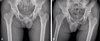
Initially, pain is the only symptom while the plain radiograph shows no abnormal findings. Narrowing of involved joint space proceeds more than 2 mm or more than 50% of the entire joint space within 6 to 12 months45). Eventually, total obliteration of joint space accompanied by necrosis and segmentation of femoral head and subchondral bone of acetabulum, and joint subluxation leads to joint destruction (Fig. 1, 2)3678910). Such radiologic findings are accompanied by severe pain in the hip joint and claudication28). However, the range of motion in the hip joint is relatively spared10).
Similar to the osteonecrosis of the femoral head, the total hip arthroplasty is often the treatment of choice for rapidly destructive coxarthrosis. Charrois et al.11) have mentioned that more amount of transfusion is expected in rapidly destructive coxarthrosis than that in the osteonecrosis of the femoral head. However, there is a shortage of concurring studies nor evidence based evaluation.
The purpose of this study is to compare the perioperative blood loss in primary non-cemented total hip arthroplasties for rapidly destructive coxarthrosis and typical osteonecrosis of the femoral head.
MATERIALS AND METHODS
The participants were selected among the patients who had received the primary non-cemented total hip arthroplasty from January of 2000 to December of 2013. The first group consisted of 24 patients who were diagnosed with the rapidly destructive coxarthrosis. Symptom duration of these patients was shorter than 12 months. The first group consisted of patients whose joint space got narrowed more than 2 mm or 50% within 6 to 12 months45). All of them were surgically and pathologically confirmed with femoral head necrosis and joint destruction. One hundred seventy-one patients, who had suffered the Ficat stage IV osteonecrosis of the femoral head, were included in the second group. The patients with preoperative usage of steroid or anticoagulant therapy and hemodynamic abnormality on the blood tests were excluded from the study (Table 1). Exclusion was based on the fact that those patients had tendency to bleed more, and might require transfusion due to difficulty of coagulation. After exclusion, total number of 19 patients was enrolled in the rapidly destructive coxarthrosis group (group 1) and 40 patients were enrolled in the osteonecrosis of the femoral head group (group 2). There was no patient with loss to follow-up in our study. For comorbidities, group 1 had 1 patient diagnosed with diabetes mellitus, 6 with hypertension, 1 with asthma, 1 with dyslipidemia and 2 patients had history of cerebrovascular accident. Group 2 had 5 patients diagnosed with diabetes mellitus and 8 with hypertension (Table 2). All operations were conducted under general endotracheal anesthesia and performed by a single surgeon with posterolateral approach. Three kinds of implants were used for two groups (Table 3). Patients were applied with elastic stocking and pneumatic compressor to avoid deep vein thrombosis. Physical exercises including tilting table, continuous passive motion, and parallel bar gaiting were prescribed three days after operation date.
The patients of group 1 (male:female, 13:6) had a mean age of 63.3 years (standard deviation [SD], 13.4), a mean weight of 59.3 kg (SD, 6.0), mean body mass index (BMI) of 21.8 kg/m2 (SD, 1.6), and mean surgery time of 188 minutes (SD, 30.0), total blood volume of 4,028 mL (SD, 493). The patients of group 2 (male:female, 27:13) had a mean age of 59.3 years (SD, 12.9), a mean weight of 62.5 kg (SD, 9.2), mean BMI of 23.1 kg/m2 (SD, 2.6), and mean surgery time of 162 minutes (SD, 31), total blood volume of 4,114 mL (SD, 618).
Measurement of the blood loss was done by the method proposed by Mercuriali and Inghilleri12), and Nadler et al13). It was calculated by comparing the hematocrits of 1 day before the operation and that of 5 days after the operation and adding the compensated blood loss and non-compensated blood loss. For the statistical analysis of the variables, chi-square test, unpaired t-test, and analysis of covariance (ANCOVA) test were used. All the statistical processing of data was done with the help of the Department of Statistics in College of Medicine, The Catholic University of Korea, and SAS version 9.1 statistical software (SAS Institute, Cary, NC, USA) was used. P-value below 0.05 was viewed statistically significant.
RESULTS
The non-compensated blood loss and compensated blood loss were 362 mL (SD, 187; range, 77-675) and 630 mL (SD, 180; range, 380-760) in group 1, respectively. In group 2, the non-compensated blood loss was 180 mL (SD, 145; range, 53-519) and the compensated blood loss was 503 mL (SD, 260; range, 190-1,505). The total blood loss was 992 mL (SD, 300; range, 457-1,434) in group 1 and 683 mL (SD, 360; range, 226-1,975) in group 2 showing a significant difference (P=0.002, Table 4).
Parameters other than BMI and operation time did not show significant difference with chi-square test and unpaired t-test (different implant used ratio, P=0.924; sex difference ratio, P=0.056; age, P=0.262; body weight, P=0.179; BMI, P=0.053; surgery time, P=0.003; estimated total blood volume, P=0.594). Thus, post-hoc analysis with ANCOVA test was conducted to evaluate whether BMI and operation time have any influence on different blood loss between two groups. After adjustment with ANCOVA test, influence of BMI and operation time on the fact that group 1 had larger amount of blood loss than group 2 did not have statistical significance (BMI, P=0.9466; operation time, P=0.721).
DISCUSSION
We had tried to calculate relatively accurate blood loss in this study. Compared to group 2, the blood loss occurring from the primary non-cemented total hip arthroplasty was significantly higher in group 1. We assert that results above can be explained with following 3 hypotheses.
First, the magnetic resonance imaging and other radiologic studies have revealed more extensive marrow edema in group 1, some extending to the peritrochanteric area (Fig. 2). Boutry et al.14) have mentioned that rapidly destructive coxarthrosis had more extensive bone marrow edema at peritrochanteric area, when compared with osteonecrosis of femoral head. These findings indicate the increased marrow pressure, and the blood loss may be a result of such fact15).
Next, the radiologic and operative findings showed more severe synovitis in group 1. This may have been caused by the superolateral displacement of femur head and the inferior displacement of hyaline body within the joint, creating stimulation to the synovium, and it can be speculated that neo-angiogenesis and activation of inflammatory cytokines had increased the hemorrhage. Komiya et al.6) had revealed that the activation of interleukin-1 and metalloproteinase-2 and 3 were significantly higher in the synovial fluid of the rapidly destructive coxarthrosis. Also, Matsumoto et al.16) have pointed out that decreased level of tissue inhibitor of matrix metalloproteinases in serum and joint fluid may indirectly proliferate pathogenesis of rapidly destructive coxarthrosis.
Lastly, the preoperative and postoperative transfusion may have contributed to the increased hemorrhage. In this study, the average packed red blood cell units received were 3.4 units in group 1, and 2.7 units in group 2. A previous study had reported that increased transfusion to patients with the primary total hip arthroplasty was related with more blood loss through the drain tube, and that finding corresponds with the relationship between the amount of transfusion and the blood loss in this study17). This means that while the transfusion keeps the homeostasis of the blood, it is related with increased blood loss after the surgery.
Aforementioned three hypotheses may have been the triggering factors for the increased blood loss in group 1 than in group 2, and it is our opinion that this study can contribute to estimating the blood loss of the two groups during and after the surgery and in developing the transfusion plan to the patients. Further studies should be conducted to evaluate relationship between these hypotheses and blood loss with statistical analysis.
Limitation of this study is that the number of enrolled patients with rapidly destructive coxarthrosis is quite few, due to its rarity. Moreover, comorbidities like diabetes and hypertension are not taken into consideration which might have influence on results.
CONCLUSION
Compared to the osteonecrosis of the femoral head group, the rapidly destructive coxarthrosis group had more blood loss when the primary non-cement total hip arthroplasty was performed. This may have been caused by marrow edema extended up to peritrochanteric area, severe synovitis, and increased blood transfusion during and after the surgery. Our results will make a great contribution to the estimation of blood loss during and after the surgery and the establishment of transfusion plan.


 XML Download