

 PDF
PDF ePub
ePub Citation
 Print
Print


The bisphosphonate class of drugs is among the most commonly utilized anti-resorptive agents for treating osteoporosis. Drugs in this class have demonstrated excellent clinical outcomes in osteoporotic fractures and their prevention1). However, it has been reported that long-term use of bisphosphonates may suppresses normal bone resorption and formation, thereby negatively impacting bone regeneration and remodeling. Although the degree of hardness may be improved, bone strength may worsen due to a reduction of durability and changes in micro structure23). Therefore, long-term use of bisphosphonates often results in atypical stress fractures due to repetitive stress. Affected regions include subtrochanteric femurs where stress is increased by concentrated bending force accompanied by rapid bone replacement. Additionally, insufficiency fractures are commonly observed in pelvic rings and sacra, yet rarely observed in the pelvic limb4). In the present case, we describe a female patient with insufficiency fractures on both sides of the femoral necks after long-term use of an anti-resorptive agent. Despite the performance of metal screw fixation, the fracture of the right femoral neck developed further and bone union failed. This case demonstrated that insufficiency stress fractures can be found on femoral necks, rather than subtrochanteric femurs and femoral shafts, in patients receiving long-term treatment with an anti-resorptive agent, and that the progress of bone union in such fractures was different from that observed in more common cases.
CASE REPORT
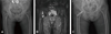
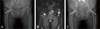
A 78-year-old female patient visited our hospital due to pain that developed in her left inguinal region without noticeable trauma five days before her visit. The pain was exacerbated by walking and limited the distance she could walk to less than 30 m. According to her medical history, she had been diagnosed as having osteoporosis per bone mineral density results showing a femoral neck T score of -2.8, and had received treatment with the bisphosphonate ibandronate (Bonviva®; Roche Pharm., Basel, Switzerland) through intravenous injection (3 mL/3 months) for the first year. This was followed by oral administration of ibandronate over the next three years (150 mg/month). In addition, she was treated with Cal-D-Vita (Cal-D-Vita tablet®; Bayer, Leverkusen, Germany) containing calcium carbonate (600 mg) and vitamin D (400 IU) once per day over a period of two years. Caltrate plus vitamin D (Caltrate D 400®; GSK Pharm., London, UK), which contains calcium citrate, was alternatively prescribed for an additional year following the discontinuation of Cal-D-Vita tablet®. She underwent total knee replacement surgery for both knees 20 years ago, followed by lumbar fusion surgeries for vertebrae 4-5 four years ago and vertebrae 3-4 two years ago. According to the biochemical examination results, no abnormal neurological findings were detected; however, a positive Patrick's test result on the right, as well as oppressive pain in the inguinal region, were observed. In radiographic examinations, an insufficiency fracture was suspected, as osteosclerosis and a slight perpendicular shade on the right femoral neck were observed and confirmed using computed tomography (CT) and magnetic resonance imaging (MRI). Her bone mineral density measurement as assessed using dualenergy X-ray absorptiometry (Lunar iDXA; GE Healthcare, Fairfield, CT, USA) revealed a T score of -2.9 for the femoral neck. Despite finding a reduction in bone hardness, as evidenced by her alkaline phosphatase level (16 µg/L), her osteocalcin level was found to be within the normal range (21.77 ng/mL). Furthermore, her level of cross-linked telopeptide of collagen type I was also found to be within the normal range (0.668 ng/mL). Two screws were percutaneously inserted and fixed as shown in Fig. 1, followed by treatment with Teriparatide (Forteo Inj®; Eli Lilly, Indianapolis, IN, USA) rather than a bisphosphonate. The patient was recommended to walk using crutches for three months and was discharged after two weeks, as nothing unusual was observed. Approximately seven months later, the patient visited the hospital with symptoms on her right side similar to those demonstrated in her left inguinal region. The same series of examinations were performed, and it was confirmed that she had a similar insufficiency fracture on the left femoral neck. The same surgical procedures used for her insufficiency fracture on the left femoral neck were performed (Fig. 2). Four weeks after discharge, the patient visited us again and complained of sudden and serious pain in her right inguinal region. The fracture shade on the right femoral neck became clearer based on the radiographic results, which showed that a gap between the bones had widened, as well as clear sclerotic changes of the fractured surfaces on the CT scan (Fig. 3). After concluding that the bone union was not successful, she underwent bipolar hemiarthroplasty. No callus was observed between the fracture fragments, and the gap was filled with fibrous tissues. Lastly, sclerosis of the fracture margin was found at the inferior portion. Microscopy findings revealed that the lamella of the cortical and cancellous bone adjacent to the sclerosis around the fractures were layered and thickened. Despite the presence of osteocytes, osteoclasts were barely detectable. At high magnifications, chondrocytes, cartilage, and osteoclasts were observed around the fractures, yet bone formation by osteoblasts was deemed insufficient (Fig. 4).
DISCUSSION
There are two types of stress fractures. Fatigue fractures are caused by unaccustomed, strenuous, repetitive and abnormal external force and stress on bones with normal tensile strength. Interestingly, it is often the result of heavy military training. Insufficiency fractures, on the other hand, are caused spontaneously by daily activities or even light exercise on bones that have weak tensile strength without specific trauma5).
General causes of insufficiency fractures include osteoporosis, osteomalacia, hyperparathyroidism, rheumatoid arthritis, fluoride treatment, diabetes, fibrous dysplasia, Paget's disease, osteogenesis imperfecta, radioactivity, and other mechanical factors. In general, these insufficiency fractures occur on the pelvic rings or sacra. Although there has been one previous report showing insufficiency fractures of the femoral neck and tibia4) the current study reports a case of insufficiency fractures on both sides of the femoral necks in a patient with usual osteoporosis (T score, -2.9) which is unprecedented considering that the fractures were caused by daily activity and occurred on both sides.
Insufficiency fractures of the femoral neck are further classified into two types, transverse and compressive. The transverse type is often found in elderly patients and is characterized by a slightly cement penetrated radiolucent line on the upper cortex of the femoral neck. The compressive type is common in young patients, is more stable, and is characterized by a radiopaque spot at the lower femoral neck. Although the patient in the current study had transverse type fractures, they were accompanied atypical radiological findings including sclerotic changes in the upper femoral necks on both sides, a low intensity signal band on the upper cortical bone of the femoral neck on a T1 MRI, and a high signal at the same location on the T2 image.
The bisphosphonate class of antiosteoporotic drugs lowers the incidence of fracture by maintaining bone strength through elevating bone density and inhibiting bone resorption as part of the overall bone metabolism of postmenopausal women. Due to its safety and effectiveness, it has been widely used to treat osteoporosis6). In particular, the ibandronate utilized in our hospital belongs to the highly potent nitrogen-containing bisphosphonate class, which effectively inhibits osteoclast actions by binding to the bone surface. Additionally, a single oral administration per month increases convenience. These agents inhibit bone resorption and thus elevate bone density. Because these bisphosphonates are chemically stable, they are barely metabolized and have more than a 10 year half life in the skeletal system7). Despite the preventive potential of bisphosphonates against fractures through the elevation of bone strength and suppression of bone resorption, the results from multiple previous studies have suggested that these agents may have a negative impact on certain sites where remodeling and osteogenesis are needed135).
In the current case, the patient had been prescribed an anti-resorptive agent for over four years, which may have inhibited bone remodeling for the right femoral neck where the insufficiency fracture was observed. Subsequently, a stress fracture resulted and bone union failed despite the use of conventional screw fixation and appropriate postoperative treatment, which were insufficient to overcome the retarded osteogenesis. When loading weights on the femoral neck, bending force is used repetitively, suggesting that the use of compressive hip screws may be warranted for better stability and strength. In 2005, Odvina et al.8) found a lack of bone formation and a delay of bone union in an iliac crest examination of nine cases for which patients were prescribed long-term treatment with alendronate and experienced abnormal stress fractures. In another study, Park et al.9) reported stress fractures of the subtrochanteric femurs on both sides after long-term exposure to alendronate. Furthermore, the subtrochanteric femurs were found to be most often fractured by relatively high energy damage. In 2012, Kim et al.10) also reported an insufficiency fracture of the ipsilateral femoral neck after six years of exposure to alendronate. Collectively, these findings suggest that more research on dose and administration periods for drugs in the bisphosphonate class may be timely and warranted in Korea.
Stress fractures on the femoral shafts or subtrochanteric femurs are characterized by several radiological properties including thicker cortical bone at fracture sites accompanied by thick sclerosis, a transverse fracture line of the cortical bone, and a spike of inner cortical bone. In contrast, insufficiency fractures of the femoral neck do not show such radiological findings. Therefore, insufficiency fractures can be diagnosed via physical examinations followed by MRI in cases involving osteoporosis patients complaining of hip pain while walking without noticeable trauma.
In our case, the patient complained of pain in her hip when walking, which led to suspicion of a femoral neck insufficient fracture. Although radiological examinations were performed, no clear evidence was demonstrated for fractures, and only slight sclerosis of the upper cortical bone of the femoral neck was confirmed. The immediate MRI examination was followed by additional imaging tests, which demonstrated a low signal on the femoral neck and a high signal band on T1 and T2 images, respectively, which supported the diagnosis of an insufficiency fracture. Such MRI findings indicated congestion, bleeding, and edema inside of the bones, which constituted sufficient evidence of fractures. In cases such as this, confirmatory bone scans are not considered to be mandatory11).
Regarding the association between the long-term effects of bisphosphonates and insufficiency fractures of both femoral necks, histological examinations utilizing a piece of bone obtained from the right femoral head prosthetic replacement surgery indicated that successful bone union should not have been expected for several reasons. These include the detection of sclerotic thick bone tissues on both cortical bones of the femoral neck fractures, and weakened mechanical tensile bone strength due to suppressed bone substitution (osteoclasts were barely present despite improved bone density). Furthermore, bone formation by osteoblasts was insufficient despite the observation of cartilage, chondrocytes, and osteoclasts on the fracture sites, all of which are often detected in general fractures. Lastly, no bone connection was present in the gap.
Although management of the insufficiency fracture of the femoral neck was attempted via both early and subsequent immediate screw fixation, fracture nonunion was not averted. The fractures observed in our patient, who had been exposed to long-term treatment with antiresorptive agents, displayed a different course of progress as compared with general insufficiency fractures, suggesting that further stable fixation may be required. Additionally, a weight-bearing delay should be considered in order to protect the fracture sites. As opposed to subtrochanteric and intertrochanteric femurs fractures, femoral necks are subjected to continuous and repetitive tensile stress, which makes it unclear as to whether these proactive actions resulted in better clinical outcomes (in this case, final bone union). Further investigations, case reports, and long-term follow ups may be warranted in order to clarify this point as well.


 XML Download