

 PDF
PDF ePub
ePub Citation
 Print
Print


INTRODUCTION
Acetabular fractures, typically attributable to high-energy trauma associated with upper or lower extremity fractures, as well as brain, chest, or abdominal injuries, are difficult to treat surgically and more prone to postoperative complications123456). Since Letournel7) proposed that surgical treatment was associated with a better prognosis than conservative management in patients with acetabular fractures and dislocations, open reduction with internal fixation and early ambulation have been recommended as basic management. Patient's age, delay to injury-related surgery (in days), the presence of hip dislocations, fracture types, the preoperative degree of displacement, the degree of postoperative reduction, and femoral head and associated injuries have been identified as factors that may influence the outcomes of surgical management for acetabular fractures7891011121314). However, a limited number of domestic studies have been conducted in order to explore factors affecting the treatment outcomes of acetabular fractures. Therefore, this study aimed to identify the factors that may influence postoperative clinical and radiological outcomes in acetabular fractures treated surgically.
MATERIALS AND METHODS
1. Patients
This study included 106 patients who underwent open reduction and internal fixation due to acetabular fracture by nine surgeons in Pusan National University Hospital (Busan, Korea) from January 2000 to December 2012. Factors examined included age, gender, injury mechanism, associated injuries, fracture type, the presence of dislocation, nerve injury, the preoperative degree of displacement, and delay to injury-related surgery (in days). Based on data from operation records, surgical approaches, surgical methods, femoral head injuries, and the degree of reduction were also examined. Clinical and radiological outcomes in acetabular fractures were evaluated at the end of the first postoperative year, and the outcomes were evaluated according to the Matta scoring system. After evaluating joint space, sclerosis severity, and the degree of osteophyte formation, radiological outcomes were classified into excellent, good, fair, and poor. Clinical outcomes were graded into excellent, good, fair and poor by evaluating pain, gait, and range of motion according to the modified Merle d'Aubigne and Postel clinical grading system11).
2. Predictors of Treatment Outcomes
Based on results from previous studies34571516), we determined that possible prognostic factors included the degree of postoperative dislocation, patient's age, associated injury (head, chest, abdomen, genitourinary system, spine, and extremities), femoral head injury, fracture type according to Letournel classification, the presence of hip dislocation, the preoperative degree of displacement, surgical approaches, and surgical methods. According to the Matta scoring system, the degree of postoperative displacement was classified into three categories by marking a maximum displacement on the anteroposterior and oblique radiographs in mm11). To evaluate the effect of the degree of initial displacement on postoperative radiological and clinical outcomes, patients were divided into two groups (>20 mm and ≤20 mm) according to the initial degree of displacement on radiographs of the articular surface. Fractures were classified according to the Letournel-Judet classification system17). Fracture types were divided into simple and associated fractures, which were then re-classified into five sub-categories.
3. Statistical Analysis
All statistical analyses were performed using IBM SPSS Statistics version 21.0 software (IBM Co., Armonk, NY, USA). P-values <0.05 were considered to be statistically significant. A univariable regression analysis was performed in order to determine factors associated with radiological and clinical outcomes, and a multivariable regression analysis was conducted in order to identify significantly associated factors. Additionally, a logistic regression analysis was conducted in order to analyze the incidence of osteoarthritis.
RESULTS
1. Baseline Characteristics
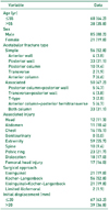
Patients included 85 men (80.2%) and 21 women (19.8%) with a mean age of 50.4 years (range, 17-78 years). The most common injury types were attributable to car accidents in 54 cases (50.9%), followed by falls in 29 cases (27.4%), other causes in 23 cases (21.7%). According to the Letournel fracture classification system, 56 cases were considered to be simple types (52.8%) and associated types in 50 cases (47.2%). There were 67 patients (63.2%) with an initial degree of displacement <20 mm and 39 patients (36.8%) with a degree of displacement >20 mm. Surgical procedures were carried out using a Kocher-Langenbeck approach in 56 cases (52.8%), an ilioinguinal approach in 21 cases (19.8%), and a combined ilioinguinal and Kocher-Langenbeck approach in 21 cases (19.8%; Table 1).
2. Clinical and Radiological Results
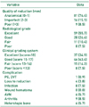
Clinical outcomes were evaluated according to the modified Merle d'Aubigne and Postel clinical grading system. Scores were excellent (18 points) in 37 cases (34.9%), good (15-17 points) in 46 cases (43.4%), fair (13-14 points) in 15 cases (14.2%), and poor (<13 points) in eight cases (7.5%). Radiological evaluation was conducted using the Matta scoring system. Scores were excellent in 59 cases (55.7%), good in 28 cases (26.4%), fair in 11 cases (10.4%), and poor in eight cases (7.5%; Table 2). According to the results of a Spearman correlation analysis, the clinical and radiological classification scores showed a statistically significant and strongly positive correlation (P<0.001).
In a univariate regression analysis, the degree of postoperative reduction (P<0.001), age (P=0.020), and the preoperative degree of displacement (P<0.001) showed significant associations with radiological results. Moreover, the degree of postoperative reduction (P<0.001), age (P=0.005), and the preoperative degree of displacement (P<0.001) were identified as factors significantly affecting clinical outcomes (P<0.001; Table 3). Through a multivariate regression analysis, the degree of postoperative reduction (P<0.001) and the preoperative degree of displacement (P=0.001) were found to be prognostic factors for postoperative radiological and clinical outcomes (Table 4).
Regarding patients belonging to the fair and poor groups according to the Matta scoring system, those complaining of hip pain and movement limitations were classified into the arthritis group. Osteoarthritis of the hip joint occurred in a total of nine patients (8.4%) with anatomical reductions in three cases, incomplete reduction in one case, and poor reductions in five cases. There were three patients with a preoperative degree of displacement <20 mm and six patients with a degree of dislocation >20 mm. By performing a logistic regression analysis, the postoperative degree of reduction was identified as a prognostic factor for the development of osteoarthritis (P=0.005).
DISCUSSION
Of the 106 patients with acetabular fractures treated surgically, 76.4% achieved anatomical reductions, 78.3% had excellent or good clinical outcomes, and 82.1% had excellent or good radiological outcomes. Statistical analyses revealed that the postoperative degree of reduction and the initial displacement were critical factors for predicting patient clinical and radiological outcomes, and the postoperative degree of reduction was found to be a predictive factor for the occurrence of arthritis.
Matta11) reported that preoperative displacements had a insignificant effect on prognosis in 262 acetabular fracture patients with a mean initial displacement of 20 mm. Conversely, Meena et al.18) demonstrated significantly poorer results in patients with an initial displacement of >20 mm. The results from the current study were comparable with those of the latter study, showing significantly (P=0.001) poorer radiological and clinical outcomes in the group with an initial displacement of >20 mm.
A large number of studies have addressed the importance of anatomical reductions in the management of acetabular fractures111219). Bhandari et al.20) evaluated the clinical outcomes of 109 patients with acetabular fractures according to the modified Merle d'Aubigne and Postel score. Of these, 84% had excellent or good outcomes, and the study authors suggested that the postoperative degree of reduction was the most important factor for prognosis. In the current study, near-anatomical reductions were associated with satisfactory clinical and radiological outcomes (P<0.001).
In order to achieve precise reductions, it is important to choose a surgical approach appropriate for each fracture type according to the AO fracture and dislocation classification21). In both-column acetabular fractures in particular, an extensile approach is considered as a better option for more precise reductions; however, the results of a previous study found satisfactory outcomes from indirect reductions of the posterior column using an ilioinguinal approach22). Isaacson et al.23) obtained satisfactory outcomes in anterior-column and both-column acetabular fractures using the Stoppa approach. However, that study was not designed to verify the effects of surgical approaches on treatment outcomes.
The results of a few studies have suggested that fracture types according to the Letournel classification showed significant effects on prognosis1624). Furthermore, the results from previous studies revealed an unfavorable prognosis for patients with posterior column, posterior column + posterior wall, and T-shaped fractures12131419). Since the Letournel system classifies fracture types according to fractures of the column or wall of the acetabulum, Meena et al.18) proposed that fracture types were helpful in guiding decisions regarding surgical approaches, but were not predictive of prognosis. Similarly, fracture type was not found to be a significant factor for predicting clinical and radiological outcomes or arthritis incidence in the current study.
In a study conducted by Moed et al.25), a poor prognosis was observed in 100 patients with posterior wall fractures and an age >55 years. In contrast, Matta.11) suggested that postoperative reductions were significant factors affecting treatment outcomes, as compared to age, which had an insignificant effect. Although age was associated with clinical and radiological outcomes in the current study, age was not found to be a prognostic factor for treatment outcomes or arthritis occurrence.
Osteoarthritis occurred in nine patients (8.4%). Of these, the outcomes of postoperative reductions were anatomical in three cases, incomplete in one case, and poor in five cases. In a study conducted by Chiu et al.1), osteoarthritis occurred in 13.2% of all satisfactory reductions and in 43.5% of unsatisfactory reductions. The likelihood of reduction achievement tended to reduce the risk of post-traumatic osteoarthritis. In the current study, postoperative reductions were found to be predictive of the risk of developing osteoarthritis after the surgical management of acetabular fractures, and the likelihood of reduction achievement significantly decreased the incidence of osteoarthritis.
The current study analyzed acetabular fractures by dividing them into simple and associated fractures due to the relatively small sample size. Therefore, further large-scale studies are warranted in order to analyze detailed fracture types according to the Letournel classification.
CONCLUSION
The results of the current study revealed that the initial degree of displacement and the quality of reductions were important factors affecting the prognosis of patients with acetabular fractures, and that the achievement of anatomical reductions was more likely to reduce the risk of osteoarthritis.


 XML Download