

 PDF
PDF ePub
ePub Citation
 Print
Print


Fractures of the acetabulum pose a difficult problem for the patient and the surgeon because of the commonly experienced late complications of posttraumatic osteoarthrosis, osteonecrosis (ON) of the femoral head, and heterotopic ossification. Acetabular fractures associated with hip dislocations have been well documented in literature. They are usually associated with osteonecrosis of the femoral head (ONFH) and have a high rate of morbidity and mortality. ON associated with isolated acetabulum fracture without dislocation of the hip is known to occur, but as per knowledge of the authors its occurrence has rarely been reported to present after using an anterior surgical approach. The purpose of this article is to bring to notice the occurrence of this rarely reported event. The exact cause of ON here eludes us, with possible mechanisms being hypo-volemia intra-operatively with a low mean arterial pressure, leading to decreased blood flow to the femoral head; nature and complexity of the injury and predisposition of the patient.
CASE REPORT
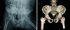
A 61-year male presented to the emergency department with a history of road traffic accident. He arrived hemodynamically stable with a blood pressure of 126/76 mmHg and a heart rate of 78 beats per minute. On plain radiograph (Fig. 1A) anteroposterior and two Judet 45° oblique view1) and computed tomography (CT) scan of pelvis (Fig. 1B), the findings revealed both column fracture of acetabulum without hip dislocation, but no presence of femoral head fracture or ONFH.
After patient's vitals were stabilized, we performed the surgical procedure. Buttress plating through ilioinguinal approach was performed using a reconstruction plate, which was supplemented by a compact hand plate. The surgical procedure lasted for 3 hours 44 minutes. The total blood loss intra-operatively was 3,000 mL. The preoperative hemoglobin (Hb) was 9.7 gm%. The patient was transfused 8 units of whole blood, 3 units of fresh frozen plasma and 8 units of packed red blood cells. Intra-operative Hb was 9.7 gm%; the average mean arterial pressure was 91.82 mmHg during the operative procedure. Post-operative blood loss via suction drain was 380 mL. Post-operatively the patient was transfused 2 units of whole blood and 1 unit of fresh frozen plasma. The Hb postoperatively was 10.3 gm%, the patient was shifted to intensive care unit for a day, later was transferred to the ward.
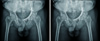
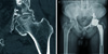
The post-operative X-ray (Fig. 2A) and CT scan revealed an acceptable reduction of the fracture fragments and a concentric hip. Sitting up was performed on the first postoperative day; the patient subsequently began formal physical therapy and active range of motion exercises. Partial toe touch weight bearing (20 to 30 lb; 9 to 13.6 kg) with a walker was maintained for 6-8 weeks. Progression to full weight bearing was started on the basis of the follow-up radiographs. Physical therapy was continued until muscle strength and range of motion were regained. The patient was then followed up on a regular basis, every monthly. His progress was monitored. On subsequent follow-up, the patient had complains of pain in the hip joint. The patient's 4-month postoperative X-ray revealed a radiolucent lesion in the superolateral part of femoral head, crescent sign, and sclerosis. The 10-month follow up X-ray (Fig. 2B) showed collapse and sclerosis, findings consistent with ONFH. A CT scan (Fig. 3A) confirmed this. ON was diagnosed only when the radiographic findings provided a clear differentiation from wear of the femoral head2). The joint pain increased due to the ONFH, we performed a total hip replacement (Fig. 3B) 12 months after the index surgery. Histopathology report of the femoral head confirmed ON.
DISCUSSION
Late complications of acetabulum fractures include heterotopic ossification and ONFH, which are present in less than 10% of the population3). The exact cause of these complications are not entirely understood. The incidence of ON described in literature varies from 3% to 53%3,4,5). The incidence of ONFH is known to be high in transverse and posterior wall fractures associated with posterior dislocation6). ON also occurs in conjunction with approximately 3% of anterior hip dislocations and in more than 13% of posterior hip dislocations. In a recent meta analysis of 3,670 surgically treated displaced acetabular fractures the incidence of ONFH showed an overall incidence of 5.6%3), suggesting that it is grossly overestimated and that most of the observed changes in the head of the femur are probably due to osteoarthritis5).
ONFH is caused by inadequate blood supply to the affected segment of the subchondral bone. When posterior surgical approaches have been used, ON rates as high as 42% within the first year after surgery have been reported7). The anterior surgical approach to the acetabulum theoretically leads to the least devascularization8). Many systemic conditions are associated with ON, but 25% of all cases are described as idiopathic and can contributes as a cause9). Trauma is one of the most common causes of ON, interruption of the blood supply to the affected segment of the bone being the cause of ischemia.
In this case the exact cause of ONFH eludes us, especially in the absence of any patient related predisposing risk factors, except presence of fracture without hip dislocation and subsequent intervention by an ilio-inguinal approach. A probable theory of etiology could be the intra-operative hypovolaemia, low mean arterial pressure, causing compromised flow to the femoral head being so as to act as the final blow. Alteration of the blood supply to vital organs during hypovolaemia is well established. With mean arterial pressure usually in the range of 50 to 60 mmHg, the flow to the femoral head is potentially compromised10) so as to act in an accumulative stress theory, as suggested by Kenzora and Glimcher9). It is questionable as to whether this alone would be enough to explain the development of ON. Patient also could have had other unknown risk factors for non-traumatic ON.
 XML Download