

 PDF
PDF ePub
ePub Citation
 Print
Print


INTRODUCTION
For several decades, the treatment of choice for unstable intertrochanteric fractures in elderly has been open reduction and internal fixation1). But, we frequently encounter failures even after successful fixation which subsequently require reoperation. So, nowadays, it is still debatable whether we should treat unstable intertrochanteric femur fracture associated with osteoporosis in elderlys with hip replacement or internal fixation. In its treatment, we should aim toward regaining preinjury walking ability and function as soon as possible. We'd like to report short term results of primary cementless hip arthroplasty in treatment of unstable intertrochanteric femur fracture.
MATERIALS AND METHODS
1. Patients
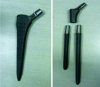
Between March 2009 and Feburary 2012, 47 arthroplasty cases were performed by single surgeon in one academic institute and followed up for more than one year. Twelve cases of them were lost by loss of contact (8 cases) or natural death (4 cases). So 35 cases were evaluated at one-year follow-up. They were 21 females and 14 males with mean age of 78 years (range, 71-92 years). The mean body mass index was 24.5 kg/m2 (range, 21-36 kg/m2). Selection criteria for arthroplasty was unstable pertrochanteric femoral fracture (AO type 31-A2.2, A2.3 and A3) in elderly more than 70 years having severe osteoporosis (Singh index <4). When treating unstable types with displaced posteromedial buttress or lateral wall fracture, we chose arthroplasty or osteosynthesis on alternate base, which was not strict. Arthroplasty was used for ones with severe comminution involving femoral neck and osteosynthesis was selected for those having anteromedial cortical fracture extending so distally that anteromedial cortical continuity was preoperatively determined to be able to be restored and fixated rigidly. The reduction tip for anteromedial cortical fracture is well described in one report about osteosynthesis of pertrochanteric fracture2). Preoperative evaluation of patient's status was performed by American Society of Anesthesia score. The femoral stem used was either tapered rectangular stem or modular fluted one of Wagner type. The tapered monoblock rectangular stem was C2 Hip (Lima-Lto®, Udine, Italy) (Fig. 1A). The modular long stem is cementless titanium alloy stem with a gritblasted surface that promotes long-term biological fixation through osseointegration (Lima-Lto®) (Fig. 1B). For total hip replacement, Delta-PF cup (Lima-Lto®) was used.
Retrospective evaluation was performed by operative time, transfusion amount, time to operation day, hospital stay and time to full weight bearing. Clinically, ambulatory ability was evaluated by Parker and Palmer (P&P) mobility score3) and functional ambulation was evaluated by four types of Hoffer et al.4). Hip function as a whole was appraised by Harris hip score (HHS). Complications related to hip arthroplasty were searched for its presence. Radiologically, bone healing of fractured trochanter and presence of subsidence, stress shielding or osteolysis were checked.
2. Operative Technique
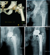
The patient was positioned in lateral decubitus position and the hip was approached posterolaterally. The piriformis was exposed by retracting tendon of gluteus medius inserting upon superoposterior tip of greater trochanter and cut, tagged and turned down to protect sciatic nerve. The other short external rotator muscles were cut with electocautery from its attachment to the intertrochanteric line and the joint capsule was opened along the femoral neck and tagged with absorbable suture. With hip moderately flexed, externally rotated and slightly abducted so as not to disengange the trochanteric fracture, the femoral neck was transsected with saw leaving fractured trochanteric area untouched and the femoral head was removed. Temporary cerclage wiring was performed through posteromedial and posterior fragment of trochanteric area. Remained portion of fractured posteromedial buttress was identified and marked with eletrocautery for indication of anteversion. For additional check, flexed leg with hip flexed, adducted and internally rotated was utilized for landmark for femoral anteversion in such a way that neck component was rotated clockwise in right hip and counterclockwise in left hip more than 90°starting from the point of flexed leg. In case with intact calcar, primary stability was checked after rasping and primary retangular stem could be inserted. In somes cases where primary stability can be achievable with this stem, reconstruction of femoral calcar was also performed with autogenous femoal head or neck fragment (Fig. 2A). If primary stability was not able to be obtained because of quality of bone or broken intramedullary calcar, modular Wagner type femoral stem was chosen. Medullary canal was broached manually starting from diameter of 12 mm and increasing sequentially (Fig. 2B). Femoral head autograft was occasionally used for reconstruction of calcar. Acetabular replacement was performed in case with preexisting concomittant hip joint arthritic change or when this is highly expected to occur because of high contact pressure between acetabular cartilage and prosthetic bipolar cup. Distal fixating femoral component of appropriate diameter was press-fit to the level of stem insertion preoperatively templated. This was able to be achieved by selecting a rasp so wide as to fit tight intramedullarily and rasping about 2 cm more distally than that level. If real distal component was to be inserted proud of premeasured level despite of impaction, the whole modular stem could not reduced easily after combined with the shortest trial modular neck component. In this case, the distal fixating femoral component was impacted carefully to be put downward or acetabular replacement was considered to accommodate femoral prosthetic head by producing a little high riding cup position with successive acetabular reaming. Patients were usually allowed of wheel chair ambulation before 1 week after surgery. After 1 week after operation, the partients were permitted to exercise standing gradually starting from titling table to tolerable weight bearing.
RESULTS
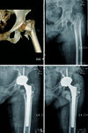
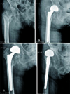
Follow-up period was at least 12 months for all patients (mean 15 months; range, 12-34 months). Fracture type was 11 cases of A2.2, 18 cases of A2.3 and 6 cases of A3.3. Femoral stem used was 8 case of rectangular tapered wedge type and 27 cases of fluted modular distal fixation type (Fig. 3, 4). Twenty-seven cases were treated by hemiarthroplasty and 8 cases by total hip replacement (Table 1). Average operative time was 92 minutes (range, 80-120 minutes), mean transfusion amount was 950 mL (range, 800-1,600 mL), mean time from admission to operative day was 3.8 days (range, 2-7 days) and mean duration of admission was 29 days (range, 17-38 days). Mean time to full weight bearing was 47 days (range, 24-79 days). P&P mobility score improved from mean preinjury score of 7.1 to mean postoperative last follow-up score of 6.5. Median HHS at last follow-up was 75 (range, 54-88; good/fair, 75%), which was not statistically significant compared with preinjury one (Table 2). According to functional ambulation, there were 13 community ambulators, 9 household ambulators, 10 nonfunctional ambulators and 3 wheel chair-bound patients at last follow-up. The proportion of community and household ambulators at last follow-up was comparable to preinjury one (Table 3). As immediate complications, there were one case of linear periprosthetic femoral fracture visible very faintly in postoperative simple radiogram and the other case of dislocation of prosthetic head from replaced acetabular cup which was manually reduced without any recurrency. There was one case of acetabular erosion developing to posterosuperior acetabular defect, 12 months postoperatively (Fig. 5). There were one case of thigh pain and two cases of inguinal pain at last follow-up, all of which were negligible. There wasn't any case of deep infection. Trochanteric bone fragments was fixed by cerclage wire in 18 cases, by absorbable suture in 9 cases and left untouched in 8 cases. There was solid bone union of fractured trochanteric fragment in every case without any case of subsidence of stem. Eight cases showed mild cortical bone erosion around cerclage wire and 9 cases showed wire breakage at last follow-up, but all of them were uneventful.
DISCUSSION
Primary prosthetic replacement has had limited use in the management of acute intertrochanteric fracture5). Generally accepted indication for primary hip replacement after intertrochanteric fracture is either symptomatic ipsilateral degenerative hip disease or attempted open reduction and internal fixation that cannot be performed because of extensive comminution and poor bone quality5,6). Arthroplasty was chosen in treatment of unstable pertrochanteric fracture because it allowed early full weight bearing and rehabilitation without any concern of healing complication such as malunion, nonunion and osteonecrosis. Aside from selection critera indicated, we chose it especially for one in which reoperation, which might be real in unstable fracture, was unconvincing to patient's guardains during informed consent. Bonnevialle et al.7) reported hip replacements are an excellent alternative to osteosynthesis in unstable trochanteric fracture in patients aged over 75 years. Kim et al.8) reported in unstable intertrochanteric femoral fracture, a long-stem cementless calcar-replacement arthroplasty showed comparable functional outcome to a proximal femoral nail.
It is reported that patients treated satisfactorily with internal fixation showed better functional results than those treated with hemiarthroplasty and unsatisfactory internal fixation9). Some authors even insisted that nearly all trochanteric area fracture can be and should be treated with fixation and preservation10). But, they considered only bone union that can be easily obtained in trochanteric area fracture and didn't take into account the hip function, walking ability and possibility of reoperation. Excessive sliding or medialization (loss of offset) has been one of major problems in sliding hip screw, while varus deformity and shortening in cephalomedullary nail11). In addition, conversion hip arthroplasty after failed osteosynthesis with intramedullary nail of pertrochanteric fracture would be difficult and burdensome to the patient12,13). Treatment with prosthetic replacement can make it possible to restore hip mechanics as normally as possible and is absolved of the risk of future malunion, nonunion and avascular necrosis14).
When choosing prosthetic replacement of trochanteric fracture, we should treat disturbed greater trochanter-abductor mechanism, which is discovered in even in type A2 fracture where posterior greater trochanteric area is usually fractured. We usually don't fix these area in performing either cephalomedullary nail or sliding hip screw, and these neglect often lead to proximal migration of fractured posterior greater trochanter and can be a cause of postoperative limping in treating trochanteric area fractures with osteosynthesis11). On the while, it is possible to make reconstruction of disturbed greater trochanter-abductor mechanism in treatment with prosthetic replacement. Posterior fragment of greater trochanter not involving trochanteric ridge could be just wired or sutured to remaining greater trochanter. In case of greater trochanteric fracture extending to vastus ridge, it was pierced with wire which was later anchored with tension to two cerclage wire around proximal femoral shaft.
It is also debatable whether to fix the posteromedial bone fragment or not. Chu et al.15) reported that in clinical treatment results of unstable intertrochanteric fracture treated by Wagner SL stem (Zimmer, Warsaw, IN, USA), all the trochanteric area fractures healed well regardless of fixation of trochanteric fracture fragment. Grimsrud et al.16) introduced a novel cerclage cable technique for unstable intertrochanteric hip fractures in cemented hip arthroplasty. Posteromedial fragment in lesser trochanteric area was considered to contribute to stability of bone implant complex in internal fixation of unstable pertrochanteric fracture, so we wired and tensioned this as anatomically as possible in early cases of our series. But, uneventable bone union was also achieved in some cases in which cerclage wiring was not performed. We attributed this phenomenon to the fact that load bearing occurred in hip implant itself and transmitted through endosteal surface of diaphysis. As long as load bearing occurs in this way alone, it may not be important to fix the posteromedial bone fragment. So importance of initial primary stability of femoral implant cannot be stressed too much.
If prosthetic replacement is chosen in peritrochanteric area fracture, cementless femoral stem is preferred to cemented one because of risk of cardiopulmonary complications in elderly patients17,18). Its disadvantages are intraoperative possibility of femoral fracture and less likeliness of smooth transition of load from prosthesis to bone. Because the femoral stem has the possibility of poor initial fitting caused by proximal calcar deficiency, we selected distally diaphyseal fixating fluted stem considering unstable intertrochanteric femoral fracture as one case of femoral revision with preserved femoral diaphysis (Paprosky type I-IIIa). Park et al.19) concluded that the clinical results and mechanincal stability obtained with this fluted and tapered modular distal fixation stem design are comparable with or even better than those obtained with other cementeless revision stem designs in femoral revision of proximal femoral deficiency of Paprosky type II, IIIA and IIIB. This type of femoral implant can be nicely fitting in femoral geometry of Dorr type III because it can have good scratch-fitting with even minimal endosteal surface15). Concomittant acetabular replacement may well have influence upon the clinical outcome, but its difference between total replacement and hemiarthroplasty was not extensively studied in this study. It was thought that total replacement should be selected in case with acetabular dysplasia or high activity and hemiarthroplasty should be reserved for less active case or one with high operative risk as salvage operation.
Concerning leg length discrepancy often encountered in hip replacement of intertrochanteric femur fracture, a few authors reported various method to obviate it. Lee et al.17) utilized a method comparing the tibial tuberosity of operated leg with that of contralateral one after reduction and insertion of trial implant in lateral decubitus position. Another Lee et al.20) either templated the distance between femoral head and lesser trochanter on contralateral side or made use of soft tissue tension after trial reduction. We templated appropriate implant size preoperatively and intraoperative roentgenogram also helped us to check leg length by comparing with the corresponding area of opposite proximal femur. In case of short leg, we chose either long neck head or longer (>60 mm) proximal neck segment. If the length is too long for trial component to be reduced snugly, the distal fixating femoral component was impacted carefully to be put downward or acetabular replacement was considered to accommodate femoral head by upward reaming.
The first limitation of this study is that this study is a retrospective one performed in a cohort of prospectively observed patient with short-term follow-up. However, long-term follow-up is hardly achievable considering old age of patients and decreased activity in postoperative period. The second is that it doesn't have control group of osteosynthesis or cemented prosthesis. But this study suggested one good treatment option for unresolvable unstable intertrochanteric fracture in elderly allowing early full weight bearing without concern of reoperation. Overall hip functions were nearly regained to preinjury level in cases showing good function before injury, which may explain superb average hip function even in all elderly cases with mean age of 78 years. On the while, reoperation rate was reported to be as high as 12% in osteosynthesis of these fractures21). Complications in our arthroplasty series were trivial. The linear fracture discovered immediately postoperatively healed without any intervention and the dislocation of femoral head reduced by closed method didn't recur anymore. This dislocation was thought to be related to both good preinjury range of hip motion and neglected fixation of anteromedial bone fragment followed by its displacement with subsequent loss of tension around hip prosthesis. One unfortunate case of acetabular erosion developing bony defect was attributable to acetabular dysplasia of mild degree, so we thought it might be the best policy to replace acetabulum as well in cases showing increased acetabular index or having degenerative change more than moderate degree (>Tönnis grade 1).
CONCLUSION
If carefully selected, cementless hip replacement arthroplasty could be a good option for unstable intertrochanteric femoral fracture in elderly in shortterm follow-up, but it is necessary to try to obviate rare complications such as postoperative dislocation or development of acetabular defect.


 XML Download