

 PDF
PDF ePub
ePub Citation
 Print
Print


Introduction
Alumina ceramic femoral heads were introduced for total hip arthroplasty (THA) in the early 1970s1) and due to the advantages of ceramic articulation such as low wear, scratch resistance, wettable surface and relatively low biological reactivity of the wear particle they gained popularity among joint reconstruction surgeons treating young patients who desire to maintain their active lifestyle. The fracture rate of the third generation ceramic is as low as 0 to 0.004%1,2). However, the incidence of ceramic fracture is higher than what it is thought to be and it is one of the most serious complications after ceramic THA. The recommended treatment for ceramic head fracture is urgent revision THA with complete synovectomy. Furthermore there are many concerns regarding the acceleration of third body wear after revision THA due to ceramic fracture. Contamination of the joint by ceramic particles is a recognized cause of revision THA after ceramic fracture3). These ceramic particles, in turn, lead to accelerated third body wear and osteolysis. However no consensus has been reached so far with respect to revision methodology in such cases, probably due to lack of numbers and long term follow up.
We report a case of extensive metallosis due to severe metal head wear after revision THA for THA ceramic liner fracture. The patient and his family members gave their consent for publication of the case details.
Case Report
A 63-year-old male was treated with THA because done for right femoral neck fracture at another hospital in 2002. At the time of surgery, a SPH-CONTACT® cup (Lima-Lto, San Daniele del Friuli (UD), Italy) acetabular component which comprises of sandwich type polyethylene backed ceramic liner and ceramic head, and a C2® (Lima-Lto, San Daniele del Friuli (UD), Italy) femoral stem were used. In November 2006, he underwent revision THA because of ceramic liner fracture at the same hospital (Fig. 1A). The surgeon exchanged the ceramic liner and head with a polyethylene liner and a 28 mm cobalt-chromium metal head, respectively. Four years after revision surgery, the patient visited the hospital complaining of right of knee and hip flexion and mass on right anterior thigh and pain on right dorsal foot. The hip pain started insidiously 3 years ago and the dorsal foot pain started 1 month ago. There was no history of trauma and fever.
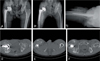
On physical examination, range of motion of the right hip was limited to 90° of flexion, 20° of abduction, 30° of adduction, 30° of external rotation, and 15° of internal rotation. The range of motion of the right knee was also limited to 100° of flexion. A large mass 8×15 cm was palpable in the right thigh but there was no local heatness or tenderness. Laboratory study showed normal blood cell counts, erythrocyte sedimentation rate (ESR), C-reactive protein (CRP), and coagulation profile. The blood Cromium (Cr) / Cobalt (Co) level was 4.93 ug/dL (normal range: 0-30 ug/dL)/68.2 ug/dL (normal range: 53 10 ug/dL), the serum BUN/Creatinine level was 10.5 mg/dL (normal range: 8-23 ug/dL)/0.8 mg/dL (normal range: 0.5-1.3 ug/dL). A radiograph of the hip showed the metal ball head was flattened and migrated. But, there was no evidence of osteolysis or component loosening. Bubble sign and peripheral calcification was seen around proximal femur (Fig. 1B, C). A pelvic CT scan revealed a mass around the posterior wall of acetabulum anterior to the rectus femoris (Fig. 1D, E) and ceramic debris of liner fracture (Fig. 1F). Based on these findings, diagnosis of metal femoral head wear after right revision THA with very severe metallosis was made.
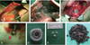
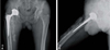
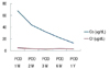
We did a second revision and exchanged the femoral head and the liner with Metasul® 32 mm head (Zimmer, Warsaw, USA) and Metasul® liner (Zimmer, Warsaw, USA) with cementing thereby doing a metal-on-metal type of of THA. Notable metallosis of the surrounding tissues was found during the operative procedure, and a black pseudotumor was present in the muscles; it extended anteriorly from the acetabulum to the proximal one third of the thigh (Fig. 2A, B). Intraoperatively we also found the sciatic nerve being compressed by the pseudotumor and hence debulking of compressing mass was done (Fig. 2C). Severe wear of metal femoral head and perforation on the top of metallic head was found (Fig. 2D, E). We could see multiple scratches inside the polyethylene liner, but the polyethylene wear was not that severe as that of the metal femoral head. We found four ceramic fragments during the surgery (Fig. 2F). We tried to remove the mass-like metallosis as much as possible (Fig. 2G). Postoperatively the pain on the right foot dorsum subsided. He was discharged 12 days postoperatively without any complications. He was followed up for 2 years. There was no pain and his knee range of motion had recovered 140° of flexion (Fig. 3A, B). The Cr and Co level checked at 1 year follow-up was within the normal range (Fig. 4).
Discussion
The presence of entrapped particles formed by ceramic fracture has been recognized as one of the main causes of severe wear of metallic bearing surfaces. And there were cases of ceramic head fracture after ceramic-on-ceramic THA and few cases of liner dissociation in total hip replacement with a sandiwich ceramic liner in Korea4,5). The stiffness, volume, and size of the debris are considered to have an effect on the degree of wear of the counter surface6). Ceramic particles from the recently manufactured ceramic bearing surfaces are considered to be of a small grain size and less concentrated7). However, in this case, large ceramic particles were embedded in the polyethylene liner, and these ceramic fragments caused a severe metal head wear. In rare cases of ceramic breakage, large ceramic debris can cause serious complications. Such a severe metallosis of a new steel head following THA was reported in 22 of 105 revision cases after ceramic femoral head breakage. Kempf et al8). reported severe metallosis with massive wear of the steel ball head after revision of a THA. Matziolis et al9). reported a case of massive wear of the metal head that occurred after total synovectomy as an additional surgical procedure during revision THA done for ceramic fracture. Abrasion of the hard bearing surfaces will occur if the hardness of the third body debris exceeds that of the metal or ceramic bearing surface6).
The metal-on-polyethylene articulation was changed to metal-on-metal articulation in this case. After changing to metal-on-metal articulation, although the ceramic particles were impinged onto the metal surface, the ceramic particles could not invade the metal surface. The change to ceramic on ceramic bearing is also associated with some risk of refracture of ceramics because, in cases of ceramic head fracture, the trunnion of the femoral stem is usually injured. Therefore, we thought that in revision THA due to ceramic fracture, after total synovectomy, metal-on-metal articulation will be better option than polyethylene bearings or ceramic-on-ceramic bearings. These are much more simple surgical procedures and we do not need to change the acetabular socket or the femoral stem. Recently, Biolox option® head (CeramTec AG, Plochingen, Germany) with a titanium neck sleeve is available. If we use this femoral head, we can manage these ceramic fractures with ceramic-on-ceramic bearing without stem revision.
In this case, we performed as meticulous a debridement as much as possible. If surgeons want to remove all the fragments of a fractured ceramic, the principles of tumor surgery-marginal excision should be applied. So, the resection borders are decided in healthy tissue at a sufficiently safe distance from the macroscopically visible focal margins. After the revision surgery for ceramic fracture, the patient needs to be followed up serially and the surgeon should look more carefully for any radiographic signs of early wear.
 XML Download