

 PDF
PDF ePub
ePub Citation
 Print
Print


INTRODUCTION
Inflammation, swelling, and recession in the gingiva sometimes occur around the tooth in conditions of occlusal hypofunction, for example, in the case of a high-displaced canine.123 The deteriorating gingival effect is caused by atrophic changes in collagen metabolism in the periodontal ligament and alveolar bone. Indeed, several studies have shown that alveolar bone resorption is accelerated, whereas bone formation is suppressed in conditions of occlusal hypofunction.45678
Recently, cone-beam computed tomography (CBCT) has been widely accepted as a useful tool for orthodontic treatment.91011 Moreover, using a three-dimensional (3D) structural analysis software enables gathering detailed quantitative information. However, to date, this analysis technique has been exclusively used in animal experimental models, including rodents.678
In the present report, we introduce 3D quantitative analyses of the alveolar bone before and after orthodontic treatment to evaluate the morphological condition of the human alveolar bone.
ETIOLOGY AND DIAGNOSIS
Etiology
The patient was a man aged 60 years and 1 month, whose chief complaint was anterior crowding because of arch length insufficiency.
Diagnosis
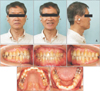
The patient had a straight profile, Class I molar relationship, and a normal mandibular plane angle (the ANB angle [ANB] = 1.6°; the Frankfort-mandibular plane angle [FMA] = 22.2°) (Figure 1A). Gingival recession around the canines was confirmed (Figure 1B). Intraoral examination revealed an overjet of +1.0 mm, and overbite of +1.0 mm (Figure 1B).
TREATMENT OBJECTIVES
The treatment objectives were to eliminate crowding. Careful treatment planning was essential to avoid the gingival recession observed on CBCT. CBCT images for evaluating the bone condition were acquired immediately before orthodontic treatment and at 1 year after active treatment, because some reports have suggested that relapse usually occurs within a few months to 1 year after active treatment.12131415 CBCT was performed using 3D Accuitomo FPD8 (Morita Corp., Kyoto, Japan) with an image intensifier. The field of view was 80 × 80 mm and the voxel size was 0.16 × 0.16 × 0.16 mm. We analyzed the adaptive threshold level to exclude the cortical/trabecular bone from the bone marrow according to the instructions provided by the manufacturer of the 3D image analysis software (TRI/3-D-BON; Ratoc System Engineering, Tokyo, Japan) and by using the discriminant analysis method.16 We then divided the enamel and bone by applying the same method using the 3D image analysis software.16 The study participants provided informed consent, and the study design was approved by the ethics review board of the Tokyo Medical and Dental University (permission numbers, 846 and 1254).
TREATMENT ALTERNATIVES
If we expanded the dental arch to eliminate crowding, the canines could protrude from the buccal alveolar bone base because CBCT revealed the alveolar bone surrounding the canine was thin in this patient. Thus, we tried not to expand the dental arch, but to make an interproximal reduction to eliminate crowding.
TREATMENT PROGRESS
The premolars and molars, but not the maxillary and mandibular incisors, were bonded using 0.018 × 0.025-inch (in) slot preadjusted edgewise brackets (Roth-type prescription). To aid leveling with a light continuous force, a 0.014-in improved superelastic nickel-titanium alloy wire (ISW) (SENTALLOY-Medium; Tomy, Tokyo, Japan) followed by a 0.016 × 0.022-in ISW (L&H Titan, Tomy) was placed. We performed orthodontic stripping from the initial stage on the interproximal surfaces between the canines (i.e., approximately 3.0 mm in total) in both the maxilla and the mandible.
After gaining space for the anterior discrepancy, both the maxillary and mandibular incisors were bonded using 0.018 × 0.025-in slot preadjusted edgewise brackets. The active treatment period was 2 years and 3 months.
For retention, the patient was instructed to wear removable retainers for 24 hours a day for the first year, and at night only for the next year. Occlusal rest for the right maxillary third molar was incorporated into the upper retainer to prevent overeruption.
RESULTS
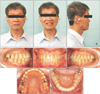
The anterior crowding was eliminated without gingival recession after active treatment (Figure 2). The change in maxillary and mandibular intercanine widths was from 36.0 mm to 36.5 mm and from 36.5 mm to 36.5 mm, respectively. This indicated that the treatment could successfully avoid gingival recession and alveolar bone resorption surrounding the maxillary and mandibular teeth.
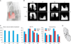
First, we examined the cortical bone before orthodontic treatment. The region of interest (ROI) was the buccal side of the canines, and the result of bone volume analysis before treatment showed that the cortical bone surrounding the right mandibular canine was lower than that in the other three quadrants (Figure 3A, 3B, 3C). Three-dimensional images of the cortical bone were constructed using a micro-CT analysis software (TRI/3-D-BON) according to methods described in the literature.678 We analyzed the adaptive threshold level to exclude the bone from the bone marrow according to the instructions provided by the manufacturer of the 3D image analysis software by using the discriminant analysis method; we also divided the enamel and bone by using the same method.16 We measured the percentage of bone volume per tissue volume (BV/TV, %) and the connectivity of the cortical bones (mm3), which indicated the mean volume of the bone as measured in all directions from a particular point inside the bone (Figure 3C).
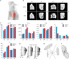
Furthermore, we examined the trabecular bone around the canine root. The ROI was 1.28 mm around the apex of the root, and we constructed the 3D images by using the micro-CT analysis software (Figure 4A and 4B). The percentage of BV/TV, trabecular thickness (Tb.Th, mm), trabecular number (Tb.N, per mm), marrow space star volume (Vm, mm3), and trabecular star volume (Vtr, mm3) were measured. The marrow space star volume (Vm) indicates bone rarefaction. The trabecular star volume (Vtr) indicates connectivity of the trabecular bone.171819 The trabecular bone parameters showed an improvement in the rarefaction of bone tendency after the orthodontic treatment (Figure 4C), which may be in line with previous findings regarding the changes caused by an occlusal hypofunctional condition.45678
Stability of retention was evaluated after 2 years, and the evaluations revealed no significant gingival recessions and relapse.
DISCUSSION
In animal experimental models, we have examined the alveolar bone microstructure by using micro-CT analysis.678 Using a 3D structural analysis software enables gathering detailed quantitative information; however, this software has been exclusively used in animal experimental models, including rodents. Therefore, in this report, we introduced 3D quantitative analyses of the alveolar bone before and after orthodontic treatment to evaluate the morphological condition of the alveolar bone in humans.
We selected this patient because inflammation, swelling, and recession in the gingiva are sometimes known to occur in patients with a high-displaced canine, especially in old age. The patient described in this report was 60 years and 1 month old before treatment. In this case, we analyzed the alveolar bone before and after orthodontic treatment by using a 3D structural analysis software for evaluating the morphological condition of the bone. Interestingly, the cortical bone did not show any sign of recovery unlike the trabecular bone (Figures 3C and 4C). This may be because the trabecular bone was affected easily within a short period, while the cortical bone was less affected, as shown in previous osteoporotic models.2021 Furthermore, a recent study suggested that the cortical bone and trabecular bone did not show the same response in osteopenia.22 If a reference phantom was simultaneously scanned when the data were acquired, bone mineral density analysis could be performed to obtain further information.2324
Studies have reported that the marrow space star volume is a bone structural parameter that can reveal trabecular connectivity.171819 Some reports have documented the marrow space star volume of the lumbar vertebra and iliac crest, and have compared the data, for example, before/after treatment. However, few studies have analyzed the alveolar bone. Therefore, future studies investigating and comparing the marrow space star volume in the alveolar bone of patients might yield interesting results.
When planning treatment using dental implants, CBCT data are now essential to obtain detailed information on the size of the alveolar bone and the location of the mandibular canal or maxillary sinus floor before implant surgery.2526 Likewise, in the orthodontic field, detailed information on the alveolar bone condition around the tooth prior to treatmentis helpful in preventing gingival recession and/or relapse.
 XML Download