

 PDF
PDF ePub
ePub Citation
 Print
Print


INTRODUCTION
Narrowing of the pharyngeal airway space (PAS) has been suggested as one of the causes of obstructive sleep apnea.12 PAS is associated with the tongue, the hyoid bone, and their adjacent muscles, and is affected by orthodontic treatment and orthognathic surgery. Previous studies have reported that with the inferoposterior movement of the tongue and the hyoid bone immediately after mandibular setback surgery, the PAS showed a corresponding decrease over longterm observation periods in patients with Class III malocclusion.34567
The effect of orthodontic treatment with incisor retraction on PAS dimensions arises from changes in intraoral volume. Contemporary orthodontic mechanics incorporating miniscrew-type temporary anchorage devices enable bodily retraction of the maxillary and mandibular incisors in cases of severe protrusion via the effective enforcement of the anchorage segment; however, narrowing of the PAS after treatment has been a concern.89 Previous studies have reported that, similar to mandibular setback surgery, orthodontic treatment with extraction decreases the PAS.101112
Most studies to date have measured the amount of incisor retraction achieved by incisal tipping movement rather than by bodily movement. The tipping movement of the incisors may have less effect on the tongue and PAS than does the bodily movement. To our knowledge, few studies have investigated the difference between PAS change caused solely by the posterior displacement of the mandibular incisors versus that caused by the simultaneous posterior displacement of both the mandibular body and incisors.
The aims of this study were to compare changes in the PAS caused by bodily retraction of the mandibular incisors and those caused by mandibular setback surgery without extraction.
MATERIALS AND METHODS
Subjects
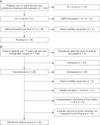
This retrospective study included 163 patients who had undergone orthodontic treatment with extraction at the Department of Orthodontics, Yonsei University Dental Hospital, Seoul, Korea, between 2006 and 2012. Among these patients, 33 (17 men and 16 women; mean age, 24.52 ± 6.15 years) who satisfied the following inclusion criteria were included in the incisor retraction (IR) group: over 17 years of age, history of four premolar extractions (a tooth per quadrant), more than 5 mm bodily retraction (reference point: estimated center of resistance located at 0.67 of the root length from the apex of each incisor as measured on a lateral cephalogram) of an incisor,1314 no severe dentofacial deformity such as a cleft lip or palate, no maxillary expansion using rapid maxillary expansion (RME), and less than 10° decrease in the incisor mandibular plane angle (IMPA) (Figure 1A).
All patients in the IR group were treated with preadjusted 0.018-inch edgewise brackets with the Roth prescription (Tomy, Tokyo, Japan). After leveling and alignment, tapered miniscrews with 1.8-mm diameter and 7.0-mm threaded length (Orlus No 18107; Ortholution, Seoul, Korea) were placed between the maxillary and mandibular second premolars and the first molar under infiltration anesthesia. Thereafter, 0.016 × 0.022-inch stainless steel rectangular archwires with additional labial crown torque (10°) on the incisor segment were placed in both arches, including the second molars. Short crimpable hooks (TP Orthodontics, LaPorte, IN, USA) were attached distally to the lateral incisor. A retraction force of 150 g was provided by using elastic chains (Ormco, Glendora, CA, USA), and the chains were replaced every 4 weeks. Space closure was performed independently in each arch.
We chose 36 patients who satisfied our inclusion criteria for the mandibular setback (MS) group from a pool of 143 patients who had undergone orthognathic surgery at our hospital between 2006 and 2012. These 36 patients met the following inclusion criteria: over 17 years of age, history of orthognathic surgery without extraction, no severe dentofacial deformity, no RME, no genioplasty, and no severe facial asymmetry over 4 mm of menton deviation from the facial midline. Patients who had over 1 mm anteroposterior movement of the maxilla were excluded, in order to remove the effect on the PAS.151617 Among the 36 patients chosen, six who had incisor retraction of over 9 mm were excluded in order to evenly match the amount of posterior displacement of the mandibular incisors between the two groups. The final MS group included in the study comprised 30 patients (15 men and 15 women; n = 3, one-jaw surgery; n = 27, two-jaw surgery; mean age, 22.78 ± 4.82 years) (Figure 1B). Patients in the MS group underwent bilateral one-piece Le Fort I osteotomy of the canine fossa and zygomatic buttress and bilateral intraoral vertical ramus osteotomy, as carried out for mandibular setback.
The study protocol conformed to the guidelines of the Declaration of Helsinki and was approved by the Institutional Review Board of Yonsei Dental Hospital, Seoul, Korea (2-2015-0033).
Methods
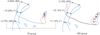
Lateral cephalograms were acquired using a Cranex 3+ (Soredex, Helsinki, Finland) in the natural head position without swallowing before (T1) and after (T2) treatment. The Frankfort horizontal (FH) plane at T1 was set as the horizontal reference plane (HRP). The vertical reference plane (VRP) was the plane that passed through the sella, perpendicular to the HRP. All cephalometric landmarks were digitized using the V-ceph program (Osstem Inc., Seoul, Korea). Landmarks and variables were set on the basis of the recommendations of previous studies111217 (Figure 2).
Reliability
All lateral cephalometric measurements were performed by the same investigator. Two weeks after the first digitization of the landmarks, all measurements were re-digitized by the same investigator. The intraclass correlation coefficient was greater than 0.94.
Statistical analysis
All statistical analyses were performed using IBM SPSS Statistics for Windows, version 21.0 (IBM Corp., Armonk, NY, USA). The Shapiro-Wilk test was used to verify the normality of data distribution. Sex distribution and mean age were not normally distributed; thus, non-parametric tests such as chi-square and Mann-Whitney U test were used. The paired t-test and independent t-tests were used to compare the changes in variables between T1 and T2 in each group and between the two groups, respectively. Pearson's correlation analysis was used to determine the relationship between skeletal, dental, and pharyngeal variables in each group.
RESULTS
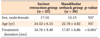
No difference was observed in sex distribution or mean age between the IR and MS groups. However, the mean treatment duration of the IR group (34.76 ± 9.48 months) was significantly longer than that of the MS group (17.87 ± 6.86 months; p < 0.001) (Table 1).
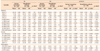
Significant intergroup differences were observed in the angle of the sella-nasion plane to point B (SNB); the angle of the lines connecting point A, the nasion, and point B; the angle of the FH plane to the mandibular plane; horizontal positions of point B (B-VRP), the hyoid bone (H-VRP), and the center of resistance of the lower incisor (L1-VRP); vertical position of the center of resistance of the upper incisor (U1-HRP); and the IMPA at T1 (Table 2). The inferior pharyngeal airway space (E-IPW) in the IR group (6. 65 ± 1.71 mm) was significantly smaller than that in the MS group (7.89 ± 2.45 mm; p < 0.05).
In the IR group, points A and B moved 0.28 ± 0.47 mm and 0.47 ± 0.95 mm posteriorly, respectively, and the hyoid bone moved inferiorly after treatment (p < 0.01). The middle pharyngeal airway space (U-MPW) decreased 1.15 ± 1.17 mm after treatment (p < 0.001) (Table 2).
In the MS group, point B moved superoposteriorly, and the hyoid bone moved posteriorly after surgery (p < 0.001). The upper and lower incisors moved posteriorly (p < 0.001), but the IMPA increased after surgery (p < 0.01). At T2, both the U-MPW and the E-IPW decreased 1.25 ±1.35 mm (p < 0.001) and 0.88 ± 1.67 mm (p < 0.01), respectively (Table 2).
After treatment, no significant intergroup differences were observed in the SNB, B-VRP, and H-VRP. Additionally, no significant intergroup difference was observed in the E-IPW at T2. The hyoid bone moved more posteriorly in the MS group than in the IR group (p < 0.001), but no significant intergroup difference was observed in the vertical movement of the hyoid bone. No significant intergroup difference was observed in the superior pharyngeal airway (PNS-SPW) and the U-MPW after treatment. However, a greater decrease in the E-IPW was observed after surgery in the MS group than in the IR group (p < 0.05) (Figure 3). Moreover, no significant correlation was observed between changes in the PAS and the skeletal and dental cephalometric variables in either group (data not shown).
DISCUSSION
In this study, superior impaction of the posterior maxilla (about 3.3 mm) was not considered a criterion for exclusion because previous reports have shown that superior repositioning of the posterior nasal spine (about 3.5–4.5 mm) does not have an effect on the dimensional change of the PAS.517
The E-IPW in the MS group was about 1.24 mm larger than that in the IR group before treatment. Some studies have reported that the skeletal classification has no effect on the PAS.1819 However, other studies have revealed that patients with skeletal Class III malocclusion have a larger PAS than do patients with Class I or Class II malocclusion.202122 In this study, neither the PNS-SPW, U-MPW, nor E-IPW showed significant differences between the two groups at T2 because the PAS in the MS group decreased because of mandibular setback surgery.
In the IR group, the U-MPW decreased by 10.39% during treatment (Figure 3). In this study, unlike in a previous study,12 the posterior movement of the mandibular incisor was measured at the estimated center of resistance rather than at the anterior tip to ensure that the measurement reflected bodily retraction of the mandibular incisors.1314 After treatment, the movement of the mandibular incisors measured to the mandibular plane showed that they had retracted by about 6.1°. Moreover, the posterior movement of the center of resistance of the mandibular incisor was 6.64 ± 0.89 mm, indicating a 1:0.96 ratio of incisal edge-to-apex displacement. This ratio was considered acceptable to represent the bodily retraction of the mandibular incisor based on the findings of previous studies.2324 Wang et al.12 reported that the U-MPW decreased by 7.88% in patients with skeletal Class I malocclusion who had undergone four-bicuspid extraction, and showed a significant correlation between the retraction of the lower incisor and the airway behind the soft palate, uvula, and tongue. Because the maxillary incisor is located above the mandibular incisor, the bodily retraction of the maxillary incisor is not expected to affect the PAS change compared with the bodily retraction of the mandibular incisor. Taken together, these results show that the PAS may decrease after mandibular incisor retraction, and it may occur because of a decrease in the intraoral volume and its compression by the posterior movement of the tongue and soft palate.
Many studies have described the decrease in the PAS after mandibular setback surgery, but the extent of decrease reported was slightly different in each study.345615 Consistent with the results of previous studies, the results of this study did not show any change in the PNS-SPW space in the MS group after surgery.6 However, the U-MPW of those patients decreased by 10.12% and their E-IPW decreased by 11.15% after surgery (Figure 3).
After treatment, the E-IPW decreased in the MS group, but no significant intergroup difference in the E-IPW was observed at T2. Additionally, no significant intergroup difference was observed in the anteroposterior position of point B at T2. This result suggests a correlation between the anteroposterior position of the mandible body and the E-IPW.2122 It is possible that the U-MPW decreased in both groups because of the effect of the posterior displacement of the mandibular incisors on the tongue and soft palate, but the E-IPW decreased only in the MS group because of the posterior displacement of the mandibular body.
Nevertheless, the extent of posterior movement of the incisors and/or the mandibular body does not have a direct correlation with the magnitude of decrease of the PAS. The U-MPW increased after treatment in 15.15% of the patients in the IR group (5 of 33) and 13.33% of the patients in the MS group (4 of 30). The E-IPW too increased in 23.33% of the patients in the MS group (7 of 30) after surgery. It is difficult to assume that, with increased posterior movement of the incisor and mandible, the PAS decreased correspondingly. This is because of the variations among individuals in the reaction of the tongue, pharyngeal airway, and adjacent muscles, as well as limitations in the precise evaluation of the PAS, as the transverse width of the pharyngeal airway could not be measured using lateral cephalometric radiograms.
In this study, the hyoid bone was observed to move more posteriorly, and inferiorly, in the MS group than in the IR group. However, no significant intergroup difference was observed in the extent of inferior movement of the hyoid bone. The impact of the posterior and inferior movement of the hyoid bone on the PAS remains controversial.25 Previous studies have reported the posteroinferior movement of the hyoid bone immediately after mandibular setback surgery, and subsequent partial reversion to its original position.5262728 The position of the hyoid bone is determined by the balance of the muscles attached to the cranial base and mandibular symphysis,12 and the inferior movement of the hyoid bone is an adaptation process to prevent the tongue from encroaching the PAS.3 Further long-term evaluation of the stability of the hyoid bone is required.
This study has several limitations, which should be taken into consideration when interpreting the data. The purpose of this study was to compare and evaluate the extent of PAS reduction by site when comparing the bodily retraction of the mandibular incisor to the posterior movement associated with the simultaneous posterior displacement of the mandibular incisor and body through mandibular setback surgery. Unfortunately, the two groups had significantly different anteroposterior skeletal relationships of the jaws before treatment. This is because most cases requiring bodily retraction of the maxillary and mandibular incisors are primarily those of skeletal Class I or II malocclusion with bimaxillary protrusion, whereas mandibular setback surgery is mainly performed in cases of skeletal Class III malocclusion with mandibular prognathism. An additional consideration is that the lateral cephalogram, used for evaluation here, has the limitation of allowing only a two-dimensional evaluation of the PAS. Although many previous studies45 have used two-dimensional lateral cephalograms to measure changes in tongue position, this study did not measure the change in tongue position, size, or volume before and after treatment in each group. This is because the position of the tongue may be affected by subtle changes in head position, even though all radiograms were acquired in a reproducible manner with unstrained position of the head.29 In fact, systematic reports of individual variability and reproducibility of the airway dimensions and tongue and hyoid position on lateral cephalometric radiograms at the same head position are scarce. However, initial tongue position and subsequent changes may predispose the effect of treatments on the upper airway. In the MS group, patients who underwent one-jaw and two-jaw surgeries were included. Although this study minimized the effect of maxillary surgery on the dimensional change of the PAS by limiting the anteroposterior movement of the maxilla, the MS group lacked homogeneity and this could constitute another limitation of this study. This study was designed retrospectively and evaluated only the PAS without subjective data such as patient questionnaires or objective data such as pulmonary function or polysomnography data. Further research is required to verify the conclusions drawn from this study. This should include a prospective design including the abovementioned subjective and objective data and should control for confounding factors or individual variations such as the reaction of the tongue, pharyngeal airway, and adjacent muscles, as well as the transverse width of the pharyngeal airway.
CONCLUSION
Within the limitations of this study, it still showed that the majority of patients studied experienced a decrease in the U-MPW because of the posterior displacement of the mandibular incisors and/or the mandibular body, thereby affecting the tongue and soft palate. The E-IPW decreased only in the MS group because of the posterior displacement of only the mandibular body. However, the amount of posterior movement of the incisors and/or mandibular body did not have a direct correlation with the amount of decrease of the PAS because the reactions of the tongue, pharyngeal airway, and adjacent muscles varied among subjects. Future prospective and well-controlled studies of individual variations are necessary to obtain more robust results.

 XML Download