This article has been
cited by other articles in ScienceCentral.
Abstract
Objective
This randomized controlled trial aimed to compare the stability of mandibular arch orthodontic treatment outcomes between passive self-ligating and conventional systems during 6 months of retention.
Methods
Fortyseven orthodontic patients with mild to moderate crowding malocclusions not requiring extraction were recruited based on inclusion criteria. Patients (mean age 21.58 ± 2.94 years) were randomized into two groups to receive either passive self-ligating (Damon® 3MX, n = 23) or conventional system (Gemini MBT, n = 24) orthodontic treatment. Direct measurements of the final sample comprising 20 study models per group were performed using a digital caliper at the debonding stage, and 1 month, 3 months, and 6 months after debonding. Paired t-test, independent t-test, and non-parametric test were used for statistical analysis.
Results
A significant increase (p < 0.01) in incisor irregularity was observed in both self-ligating and conventional system groups. A significant reduction (p < 0.01) in second interpremolar width was observed in both groups. Mandibular arch length decreased significantly (p = 0.001) in the conventional system group but not in the self-ligating system group. A similar pattern of stability was observed for intercanine width, first interpremolar width, intermolar width, and arch depth throughout the 6-month retention period after debonding. Comparison of incisor irregularity and arch dimension changes between self-ligating system and conventional system groups during the 6 months were non-significant.
Conclusions
The stability of treatment outcomes for mild to moderate crowding malocclusions was similar between the self-ligating system and conventional system during the first 6 months of retention.
Keywords: Self-ligating system, Conventional system, Stability, Malocclusions
INTRODUCTION
Treatment stability is one of the most important objectives in the field of orthodontics; however, despite decades of research, the stability of aligned teeth is variable and largely unpredictable.
1
Post-treatment Changes In Dentition Can Be Affected By Numerous Factors Such As Alteration Of The Original Arch Form, Periodontal And Gingival Tissues, Mandibular Incisor Dimensions, Environmental Factors, Neuromusculature, Growth, Post-treatment Tooth Positioning, Establishment Of Functional Occlusion, Third Molar Development, And The Original Malocclusion Element.
2 Additional Factors That May Influence The Stability Of Orthodontic Treatment Include The Type, Duration, And Timing Of The Retention Appliance.
3 The Various Elements Leading To The Relapse Of Treated Malocclusions Are Not Fully Understood, Resulting In Wide Variation In Retention Protocols Among Clinicians.
456
Self-ligating brackets have been gaining increasing popularity; every major orthodontic manufacturer has introduced a self-ligating bracket into the market.
7 Some researchers have claimed that the lower forces produced by self-ligating bracket systems might result in greater physiological tooth movement and produce more stable treatment results.
89101112 A previous study reported that physiological expansion was possible with the use of self-ligating systems, enabling a greater number of non-extraction treatment cases and reducing use of expansion auxiliarie.
13 This raises concern regarding the stability of treatment outcomes compared with conventional systems. This also applies to the use of the same broad expanded arch form for both maxillary and mandibular arches. Studies on stability after treatment with self-ligating brackets are lacking at present. Based on a systematic review of self-ligating brackets
14 and a recent literature review, no studies have compared the stability of treatment outcomes between self-ligating and conventional brackets. An improved understanding of the clinical evidence behind the impact of different types of appliance systems, either self-ligating or conventional, on orthodontic treatment stability could assist in evidence-based selection of appliance systems. This understanding is required in combination with the consideration of several other influencing factors in the decision making process such as cost, oral hygiene, chairside time, comfort, treatment interval, patient demand, accessibility.
In light of the increasing trend in self-ligating system use, this study aimed to compare stability outcomes after mandibular arch orthodontic treatment for mild to moderate crowding malocclusions between a passive self-ligating system (0.022-in slot; Damon® 3MX, Ormco, Orange, CA, USA) and a conventional system (0.022-in slot; Gemini MBT, 3M Unitek, Monrovia, CA, USA), using Hawley and vacuum-formed retainers. The specific objectives were to investigate the pattern of change and to compare changes in incisor irregularity, intercanine width, interpremolar width, intermolar width, arch length, and arch depth during a 6-month retention period between both systems.
MATERIALS AND METHODS
This randomized clinical trial study was monitored by the Universiti of Malaya's institutional review boards and ethical approval was obtained from the Medical Ethics Committee Research (DF OT 1005/0033[P]). Patients were recruited based on the following selection criteria: treated only with a fixed orthodontic appliance using either the passive self-ligating or conventional system, comprehensive orthodontic treatment involving both arches, presenting with mild to moderate crowding malocclusions without the need for extraction, all permanent teeth erupted except for third molars, and no previous orthodontic treatment. Patients were excluded for the following reasons: prescribed single arch or sectional fixed appliance treatment, still growing (< 18 years old), presenting with congenitally missing teeth, poor periodontal status, craniofacial anomalies (e.g., cleft lip or palate patient), severe skeletal discrepancies requiring orthognathic surgery, severe crowding requiring extraction(s), and spaced dentitions.
To determine the sample size and power of the study, PS Sample Size Calculation Software (United States;
http://biostat.mc.vanderbilt.edu/wiki/Main/PowerSampleSize) was used. At the significance level of 0.05 and power of study at 85%, to detect a clinically meaningful difference (incisor irregularity) of 2.0 mm
51516 as clinically significant, the power analysis calculated that 19 patients in each group were sufficient with a ratio of 1:1. Therefore, a total sample size of 38 patients was required. To compensate for potential sample attrition during the follow-up studies, an additional 20% of patients were included.
Written informed consent was obtained and a written information sheet explaining the research was given to each selected volunteer participant. This randomized clinical trial comprised a two-arm parallel study with 1:1 allocation ratio. The selected 47 orthodontic patients (mean ± standard deviation [SD] age = 21.58 ± 2.94 years) were randomized into two groups to receive either a passive self-ligating (Damon® 3MX 0.022-in slot, n = 23) or a conventional system (Gemini MBT 0.022-in slot, n = 24).
A standardized treatment protocol template, that had been calibrated via discussion among three clinicians regarding the active orthodontic treatment and retention phase was produced and strictly adhered to during the clinical trial. For the passive self-ligating system, Damon® archwires were used in the following sequence; 0.014-in copper-nickel-titanium (CuNiTi), 0.016 × 0.025-in CuNiTi, and 0.019 × 0.025-in stainless steel (SS). For the conventional system, Ortho Form™ II (3M Unitek, Monrovia, CA, USA) archwires were applied during treatment. The sequence of archwires used for the conventional system was followed; 0.014-in nickel-titaium (NiTi), 0.018-in NiTi, 0.017 × 0.025-in NiTi, and 0.019 × 0.025-in SS. Following completion of the treatment, patients from both groups were randomly assigned (stratified randomization) to receive either Hawley or vacuum formed retainers; the most commonly used retainers.
17 Since there are wide variations in retention protocols among clinicians,
46 the duration of the retainer wear was standardized for each retainer type based on the studies by Destang and Kerr,
4 and Thickett and Power,
16 because these protocols were the most similar to those practiced in Malaysia. Both types of retainers were issued on the same day as the debonding.
The retention protocol for each type of retainer was clearly explained to the participant during retainer delivery and reinforcement of instructions was performed at each review appointment during the retention period. To ensure compliance during the retention review period, a reminder telephone call was made the day before the appointment. Orthodontic study models were collected at the debonding stage (T1), 1 month (T2), 3 months (T3), and 6 months (T4) after debonding. Each study model was labeled by an assistant and measured randomly by one researcher who was blinded to the appliance system, ensuring that none of the patients' staged models were measured consecutively. It was agreed that the poor quality casts would be excluded from the analysis, for example, if there were voids or blebs on the plaster model obscuring the teeth or if the teeth on the models were fractured or missing.
The stability of orthodontic treatment outcomes between the self-ligating and conventional system was compared during the 6-month retention period. Data was collected from direct measurements of the final sample comprising 20 study models per appliance system group, using a calibrated digital caliper. The mandibular arch variables assessed were as follows:
i) Little's index of irregularity (IR): the sum of the distances between the anatomic contact points from the mesial of the left canine to the mesial of the right canine.
ii) Intercanine width (ICW): the distance between cusp tips of the right and left canine. In cases of cusp attrition, the wear facet center was used.
iii) First and second interpremolar width (1st and 2nd IPW): the distance between the cusp tips of the right and left first and second premolars. In cases of cusp attrition, the wear facet center was used.
iv) Intermolar width (IMW): the distance between the mesiobuccal cusp tips of the right and left first molars. In cases of cusp attrition, the wear facet center was used.
v) Arch length (AL): the sum of the right and left distance between the midpoint of mesioincisal edges of central incisors to the mesiobuccal cusp tips of the right and left first molars.
vi) Arch depth (AD): the distance measured from the midway point between the mesioincisal edges of the central incisors and the point bisecting the line connecting the mesiobuccal edges of the cusp tips of the right and left first molars.
To test the reliability of the method, one examiner (Norma Ab Rahman) measured all variables on eight randomly selected models and repeated those measurements 2 weeks later. Measurements were also repeated by a second examiner (D.Z) on the same eight orthodontic casts that had been measured by the first examiner. The variable measurements of the study models were repeated 2 weeks after the first measurement by the same examiner or researcher. Intra and inter-observer reliability coefficients were then calculated.
Histograms were visually inspected for assessment of data normality. Skewness and kurtosis testing were further utilized in order to evaluate the amount and direction of histograms relative to a standard bell curve. The distribution of the data was found to be skewed for incisor irregularity but was normally distributed for the arch dimensions. The median and the interquartile range were therefore calculated for the skewed variables, while mean and SD were determined for variables that were normally distributed. Paired and independent t-tests were used to investigate the pattern of change and to compare the changes, respectively. For variables that violated the normality assumption, the nonparametric Mann-Whitney U-test and Wilcoxon signed rank test were employed to achieve the same objectives. Bonferroni adjustment for family wise error was undertaken due to multiple comparisons. The level of statistical significance was pre-specified for the pattern of change at p < 0.01 and for the comparison of the change at p < 0.008 after Bonferroni adjustment.
RESULTS
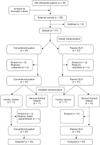
A total of 47 subjects were selected, with 23 randomized to the passive self-ligating system group and 24 to the conventional system group. There was a 12.8% (n = 6) drop-out rate in this study; 8.5% (n = 4) during treatment and 4.3% (n = 2) during the 6-month retention period, resulting in a final sample size of 20 subjects in each group (
Figure 1). The statistical test showed reasonably good agreement between repeated measurements for both intra-examiner and inter-examiner calibrations. The intra-class correlation coefficient (ICC) for intra-examiner calibration was in the range of 0.908–0.995, whereas for inter-examiner calibration, the ICC ranged between 0.770–0.911, demonstrating that the method had good reliability and repeatability. Independent
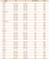
t-tests showed that there were no significant differences in age, sex, crowding, types of retainers, IR, ICW, IPW, IMW, AL, and AD between the two groups, reflecting the adequate randomization of subjects to each group, and therefore ensuring comparability (
Table 1).
Incisor irregularity (
Table 2 and
Figure 2) for both the conventional and self-ligating systems increased significantly (
p < 0.01) during the 6-month retention period. The total amount of mean ± SD relapse from dedondding to 6 months of retention was 0.69 ± 0.76 mm and 0.43 ± 0.63 mm in the conventional and self-ligating system, respectively. For both systems (
Tables 3 and
4), the overall changes in ICW, 1st IPW, IMW, and AD from T1 to T4 were not statistically significant (
p > 0.01). However, the 2nd IPW significantly decreased in both systems over the 6-month retention period (
Figure 3). The total amount of 2nd IPW relapse over the 6-month retention period was 0.41 ± 0.12 mm (
p = 0.008), and 0.59 ± 0.25 mm (
p < 0.01) with the conventional and self-ligating system, respectively. The AL (
Figure 4) significantly decreased (
p < 0.01) in the conventional system by 0.89 ± 0.42 mm but not in the passive self-ligating system.
No statistically significant differences in incisor irregularity or arch dimensions were recorded during the 6-month retention period between the self-ligating and conventional systems (
p > 0.008,
Table 5). The changes in ICW, 1st IPW, 2nd IPW, IMW, AL, and AD during the 6 months in both systems were less than 1 mm, with AL showing the largest difference between both groups (0.80 mm at T2 to T4) and mandibular IMW exhibiting the smallest difference (0.001 mm at T1 to T3).
DISCUSSION
This study investigated the effects of two orthodontic bracket systems, a passive self-ligating system and a conventional system, on the stability of mandibular arch orthodontic treatment outcomes after a 6-month retention period. A similar proportion of Hawley and vacuum-formed retainers were used during the retention period for both groups because they are the most commonly used retainers in Malaysia.
15 Since there is no firm evidence regarding the best retainer for use after active orthodontic treatment, in this study, the use of the most popular retainers in this country allows for a better representation of an average clinical setting and removes the influence of different types of retainers on study findings.
Treatment modality was restricted to non-extraction therapy of mild to moderate crowding malocclusion, to ensure that the changes in arch dimensions were not affected by dental extractions that have the potential to distort results. In the self-ligating system group, Ortho Form™ II is a square-shaped archwire from 3M Unitek™. This type of archwire was used in the conventional group because its shape is most similar to the Damon® archwire. The type of archwire used corresponded to the appliance system in order to reflect the true effect of the advocated passive self-ligating system, as recommended in Damon® guidelines.
18 Therefore eliminated the possible confounding factor from wire type. Since there is no definitive conclusion regarding this issue, it is best to use the type of archwire that corresponds to the specific appliance systems.
Due to paucity of published research investigating the impact of the bracket system (conventional and self-ligating systems) on stability during the retention period, it is difficult to compare our findings with previous studies. The relapse in both systems was confined to incisor irregularity and 2nd IPW, whereas the instability in AL was observed only for conventional system; although this may not be clinically significant. There was equal maintenance of the ICW, 1st IPW, IMW, and AD between the two systems. There was not recorded difference in stability at T4 between the two systems.
The incisors in the conventional system group were unstable and relapse occurred between T3 to T4. However, for the self-ligating system, relapse occurred throughout the 6-month retention period. The median differences for both systems were unlikely to be clinically significant (conventional system, 0.69 ± 0.76 mm; self-ligating system, 0.43 ± 0.63 mm). However, the systems were only distributed throughout the labial segment; the differences may have been clinically significant if applied to a single tooth displacement.
Furthermore, the 2nd IPW with both the self-ligating and conventional systems also showed instability. The relapse of the 2nd IPW with the conventional system occurred between T3 and T4, while with the self-ligating system, relapse occurred throughout the 6-month retention period. One probable cause for this pattern of relapse may be due to the duration of the archwires used during active treatment. In the conventional system group, four sequences of archwire changes were utilized compared with only three in the self-ligating system group; this enabled a longer duration of use for each archwire during active treatment. However, there was insufficient data to support this hypothesis.
The relapse of AL with the conventional system was first observed at T1 and occurred throughout rest of the 6-month retention period. In total, AL relapsed by 0.89 ± 0.42 mm with the conventional system but not with the passive self-ligating system. Despite the significant increase in incisor irregularity with the self-ligating system, this was not reflected in the AL changes, probably due to a combination of differences in labial segment changes and posterior transverse changes. However, statistical analysis showed no significant differences between the two systems. The findings of this study demonstrate that the treatment stability of the self-ligating system in terms of occlusal changes, incisor irregularity and arch dimensions, during a 6-month retention period was comparable to that of a conventional system. Therefore, the reports of lower forces associated with the use of self-ligating bracket systems, resulting in greater physiological tooth movement and more stable treatment outcomes,
1112131415 require more evidence and cannot be upheld within the limitations of this study. In the present study, patients suitable for non-extraction therapy were carefully selected (with mild to moderate crowding and stability), but were only investigated within the first 6 months of retention. Four patients dropped out during treatment; two subjects from both systems were excluded because extractions were performed to address patients' perception of deteriorated facial profile. Though equal in number, elimination of drop-outs due to extraction could possibly have introduced bias in terms of relapse from incisor proclination. Another limitation of this study was patient failure to attend the review appointments and retainer wear compliance. During the retention phase, two patients were discontinued due to loss of retainer and follow-up loss during the 6-month retention period. A further study to lengthen the retention period would enhance understanding of the effect of bracket systems on stability. A comparison of arch dimensions before the start of active orthodontic treatment and at the debonding stage should also be performed to explore the effect of bracket systems on stability.
CONCLUSION
The stability of treatment outcomes after using passive self-ligating and conventional systems were found to be similar for mild to moderate crowding malocclusions during the first 6 months of retention. Within the limitation of this study, the similar finding during retention produced by both systems suggested treatment stability may not be a major influence factor in the choice of fixed appliance systems. Alternatively other factors such as cost, oral hygiene, chairside time, treatment interval and other related factors may play a primary role in decision making.
Figures and Tables
Figure 1
CONSORT flowchart of the study.
UM, University of Malaya; SLS, self-ligating system.

Figure 2
Pattern of change in incisor irregularity after treatment with passive self-ligating (SLS) and conventional systems (CS) during the first 6 months of retention.
T1, At debond; T2, 1 month after debond; T3, 3 months after debond; T4, 6 months after debond.
*p < 0.01.

Figure 3
Pattern of change in second interpremolar width after treatment with passive self-ligating (SLS) and conventional systems (CS) during the first 6 months of retention.
T1, At debond; T2, 1 month after debond; T3, 3 months after debond; T4, 6 months after debond.
*p < 0.01.

Figure 4
Pattern of change in arch length after treatment with passive self-ligating (SLS) and conventional systems (CS) during the first 6 months of retention.
T1, At debond; T2, 1 month after debond; T3, 3 months after debond; T4, 6 months after debond.
*p < 0.01.

Table 1
Demographic and clinical characteristics of subjects

Table 2
Pattern of change for incisor irregularity in conventional system and self-ligating system

Table 3
Pattern of change for arch dimensions in conventional system

Table 4
Pattern of change for arch dimensions in passive self-ligating system

Table 5
Comparison of changes in mandibular arch dimensions between self-ligating (SLS, n = 20) and conventional systems (CS, n = 20)

ACKNOWLEDGEMENTS
We thank the participants, clinicians, examiner (D.Z), and all supporting staff involved in this study.
References
1. Freitas KM, de Freitas MR, Henriques JF, Pinzan A, Janson G. Postretention relapse of mandibular anterior crowding in patients treated without mandibular premolar extraction. Am J Orthod Dentofacial Orthop. 2004; 125:480–487.

2. Melrose C, Millett DT. Toward a perspective on orthodontic retention? Am J Orthod Dentofacial Orthop. 1998; 113:507–514.

3. Nanda RS, Nanda SK. Considerations of dentofacial growth in long-term retention and stability: is active retention needed? Am J Orthod Dentofacial Orthop. 1992; 101:297–302.

4. Destang DL, Kerr WJ. Maxillary retention: is longer better? Eur J Orthod. 2003; 25:65–69.

5. Shawesh M, Bhatti B, Usmani T, Mandall N. Hawley retainers full- or part-time? A randomized clinical trial. Eur J Orthod. 2010; 32:165–170.

6. Sheridan JJ, LeDoux W, McMinn R. Essix retainers: fabrication and supervision for permanent retention. J Clin Orthod. 1993; 27:37–45.
7. Eliades T, Pandis N, Johnston LE, White LW. Selfligation in orthodontics. 1st ed. Wiley-Blackwell;2009.
8. Damon DH. The rationale, evolution and clinical application of the self-ligating bracket. Clin Orthod Res. 1998; 1:52–61.

9. Khambay B, Millett D, McHugh S. Evaluation of methods of archwire ligation on frictional resistance. Eur J Orthod. 2004; 26:327–332.

10. Griffiths HS, Sherriff M, Ireland AJ. Resistance to sliding with 3 types of elastomeric modules. Am J Orthod Dentofacial Orthop. 2005; 127:670–675.

11. Henao SP, Kusy RP. Frictional evaluations of dental typodont models using four self-ligating designs and a conventional design. Angle Orthod. 2005; 75:75–85.
12. Kim TK, Kim KD, Baek SH. Comparison of frictional forces during the initial leveling stage in various combinations of self-ligating brackets and archwires with a custom-designed typodont system. Am J Orthod Dentofacial Orthop. 2008; 133:187.e15–187.e24.

13. Franchi L, Baccetti T, Camporesi M, Lupoli M. Maxillary arch changes during leveling and aligning with fixed appliances and low-friction ligatures. Am J Orthod Dentofacial Orthop. 2006; 130:88–91.

14. Chen SS, Greenlee GM, Kim JE, Smith CL, Huang GJ. Systematic review of self ligating brackets. Am J Orthod Dentofacial Orthop. 2010; 137:726.e1–726.e18.
15. Rowland H, Hichens L, Williams A, Hills D, Killingback N, Ewings P, et al. The effectiveness of Hawley and vacuum-formed retainers: a single-center randomized controlled trial. Am J Orthod Dentofacial Orthop. 2007; 132:730–737.

16. Thickett E, Power S. A randomized clinical trial of thermoplastic retainer wear. Eur J Orthod. 2010; 32:1–5.

17. Ab Rahman N, Low TF, Idris NS. A survey on retention practice among orthodontists in Malaysia. Korean J Orthod. 2016; 46:36–41.

18. Damon D, Bagden MA. Damon system: The workbook. Glendora, CA: Ormco Corporation;2004.


 PDF
PDF ePub
ePub Citation
 Print
Print


 XML Download