

 PDF
PDF ePub
ePub Citation
 Print
Print


INTRODUCTION
Maxillary molar distalization is one of the most frequently used orthodontic treatment modalities for the correction of a Class II molar relationship and/or space gaining. Appliances including headgear, pendulums, Jones jigs, and distal jets have conventionally been used for maxillary molar distalization.1234 However, distalization is often associated with adverse effects such as anchor loss and uncontrolled tipping. Currently, skeletal anchorage, including orthodontic miniscrew-assisted upper molar distalization is the standard treatment modality. It is not limited by patient compliance, and it can prevent dental anchor loss.
There are numerous clinical skeletal anchorage options available for upper molar distalization. One method utilizes miniplates inserted into the buccal maxillary, zygomatic, and palatal bones.567 The utilization of miniscrews inserted into the buccal interdental alveolar bone, maxillary tuberosity, and palatal area for the distalization of upper molars has also been reported.8910 Furthermore, combinations of conventional maxillary molar distalizing appliances and skeletal anchorages such as miniscrew-assisted pendulum appliances have been proposed.111213 All of these treatment modalities produce similar but different dental effects, with potentially varying levels of convenience for both clinicians and patients. Clinicians are required to acquire a full understanding of the effects of each appliance prior to treatment selection; therefore, the effects of each distalizing appliance require analysis.
A treatment system using midpalatal miniscrews has previously been reported.814 Among the proposed biomechanical options, maxillary molar distalization can be obtained using two distinct appliance designs, lingual arches and pendulum arms. The aim of this current study was to compare the patterns of tooth movement associated with these two different designs, using midpalatal miniscrews with the simultaneous use of buccal fixed orthodontic appliances.
Maxillary molar distalization with two midpalatal miniscrews
Midpalatal miniscrew placement
A multi-purpose miniscrew-supported biomechanical system to control maxillary dentition three-dimensionally has previously been described.8 This system utilizes two miniscrews placed in the midpalatal area, each with a 0.0215 × 0.0250-inch rectangular slot on its head. Miniscrews are inserted 2–3 mm lateral to the midpalatal suture on both the left and right sides, and in the sagittal position around the line connecting the right and left first upper molar. Placement is performed using a conventional miniscrew insertion technique. Under local anesthesia, miniscrews are inserted with a speed-reduction contra-angle hand-piece under saline irrigation. Through the insertion of various foms of wire into the miniscrews' rectangular slots, different types of maxillary tooth movement, including distalization, can be achieved.
Lingual arch type appliance for upper molar distalization
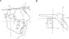
The lingual arch type of biomechanical system utilizes a lingual arch (0.8–0.9 mm) connecting the right and left first molars with hooks soldered onto the mesial part (Figure 1). A rectangular wire with hooked ends is inserted into the miniscrews' rectangular slots to connect two midpalatal miniscrews (Figure 1). Elastomeric chain or coil springs are engaged between the lingual arch hook and midpalatal miniscrew connecting wires to deliver a distalizing force to the molars. By adjusting the vertical length of the midpalatal miniscrew connecting wires, the maxillary molar distalizing pattern can be controlled: shorter lengths (hooks placed near the palatal roof) can provide greater root distalizing movement, while longer lengths (hooks placed near the tooth crown) can provide greater crown distalizing movement. In all of the cases included in the present study, the midpalatal miniscrew connecting wires and the lingual arches were adjusted to direct the distalizing force vector through the furcation of the maxillary first molar and parallel to the occlusal plane.
Pendulum type appliance for maxillary molar distalization
The pendulum type of midpalatal miniscrew-supported maxillary molar distalizer adopts the wire design of the conventional Hilgers pendulum appliance.15 A stainless steel 0.0215 × 0.0250 inch midpalatal-miniscrew connecting wire with a helix and a horizontal loop is utilized, with its end inserted into the lingual sheath of the first molar (Figure 1). The activation mode is the same as that for the conventional pendulum appliance: the helix is activated to distalize the whole pendulum arm and then inserted into the lingual sheath of the first molar.
MATERIALS AND METHODS
To investigate the molar distalization patterns associated with lingual arch type and pendulum type appliances, 14 patients who each received one of the two appliances were enrolled into the study. All patients exhibited a bilateral or unilateral Class II molar relationship at the start of treatment. Seven patients (14 molars, age 19.2±4.4 years) were treated with the lingual arch type appliance, and seven (12 molars, age 20.9±7.6 years) were treated with the pendulum type appliance. All patients received full fixed orthodontic appliances.
With brief regard to treatment mechanics, alignment and leveling was performed after placing full fixed appliances until 0.016 × 0.022-inch stainless steel wire could be engaged passively. Molar distalization was performed until a super Class I relationship was achieved. Molar distalizing force was subsequently decreased to hold the position while retracting premolars, canines, and incisors.
Initial (pre-orthodontic treatment) and final (post-orthodontic treatment) lateral cephalograms were obtained and analyzed. Maxillary molar movement was analyzed using measurements described by Cha and Ngan.16 The X-Y coordinate was constructed using a horizontal rotating sella-nasion line in a downward direction at 6° (X-axis) and a vertical line perpendicular to the horizontal line passing through the sella point (Y-axis). Using the X-Y coordinate, a 0–0 point was set at point A and a new X'-Y' coordinate was constructed (Figure 2A). Measurements recorded included the Y' line to maxillary first molar mesial cusp tip distance (mm), X' line to maxillary first molar mesial cusp tip distance (mm), X' line to maxillary first molar distal cusp tip distance (mm), and angle (°) between the X' line and a line starting from the mesial point of the maxillary first molar crown tangent to the root (Figure 2B).
RESULTS
Pre-treatment measurements did not differ statistically significantly between the two groups (Table 1). Post-treatment, first molar angulation differed significantly between the groups (Table 1). Post-treatment, the pendulum appliance group showed greater distal angulation than the lingual arch appliance group.
In the lingual arch appliance group, pre- and post-treatment comparisons yielded a mean distalization of 2.4 mm (Table 2). The vertical position exhibited slight intrusion (0.3 mm); however, this was not statistically significant. A statistically significant reduction in angulation to 0.8 mm indicated the occurrence of crown mesial tipping or root distal tipping.
For the pendulum type appliance, pre- and post-treatment comparisons yielded a mean distalization of 1.8 mm, and intrusion of the first molar differed statistically significantly (Table 2). The amount of intrusion differed between the mesial cusp and the distal cusp: distal cusp intrusion (X'-distal cusp distance −1.1 ± 0.4 mm) was greater than mesial cusp intrusion (X'-mesial cusp distance −0.8 ± 0.5 mm). Additionally, angulation changed significantly to +1.5° ± 1.3°, indicating the occurrence of distal tipping during distalization.
DISCUSSION
The utilization of midpalatal miniscrews as an absolute anchorage mechanism offers several advantages, and can aid in the achievement of optimal treatment outcomes. The most essential advantage is the low failure rate. The midpalatal area lacks critical anatomical structures such as large sized vessels and nerves, and dental roots which are reportedly responsible for increasing the risk of miniscrew failure when they are implanted in close proximity.1718 In contrast, the midpalatal area has an abundance of keratinized gingiva with an excellent quality of cortical bones, favoring the stability of miniscrews.19 However, the utilization of midpalatal miniscrews is not as popular as interdental miniscrews because the midpalatal area is far from maxillary dentition. Several articles describing the biomechanics of midpalatal miniscrew utilization for the control of maxillary dentition have been published.81420 The current report analyzes maxillary molar distalizing patterns as a follow-up study.
Distalizing force vector was adjusted to pass through furcation of the upper first molar for the lingual arch type appliance group. As expected, the results showed almost bodily distal movement: mean 0.8° mesial crown tipping or root distal tipping occurred, while a mean distal movement of 2.4 mm was achieved. This equates to approximately 0.3° of mesial crown tipping per 1 mm of distal movement, and is clinically negligible. The clinical advantage of the lingual arch appliance is the control of molar tipping; however, it is disadvantaged by its complex design (two wires in the palate) which can increase patient discomfort. Furthermore, it can only be applied in bilateral distalizing cases.
The pendulum type appliance produced significant distal tipping of the maxillary molars during distalization. This was anticipated based on previous studies investigating the effects of the conventional pendulum appliance, which included distal tipping of the maxillary molars during distalization.3421 Mean distal crown tipping of 1.5° occurred during a mean distalization of 1.8 mm, which equates to approximately 0.8° distal tipping per 1 mm of distalization. The extent of distal tipping was lower than that reported in previous studies investigating the pendulum appliance; however, direct comparison is not possible as total distalization was much smaller in the present study. In addition, the movement pattern of the upper first molars measured in the present study was actually the result of a combination of the effects of the midpalatal miniscrew-supported distalizer and the labial-fixed orthodontic appliance with continuous wire. Engagement of the continuous wire on the labial side may have reduced distal tipping. Subsequent distalization of premolars, canines, and incisors may also have affected the position of distalized maxillary molars. Such tooth movement may have influenced the first molar position with regard to mesio-distal angulation and transverse rotation (particularly, mesial-in rotation could have occurred). This effect seems to have been minor however, because the resultant molar movement pattern was as expected. The reduced distal tipping of the upper molar associated with the pendulum type appliance may be a result of this effect.
There was significant intrusion of the molar during distalization using the pendulum type appliance. Several previous studies have reported intrusion of the first molar after distalization with the conventional pendulum appliance; however, statistical and clinical significance was lacking.3422 In contrast, other studies have reported extrusion of the molar, but also without statistical or clinical significance.1213 One study by Byloff and Darendeliler23 reported significant maxillary molar intrusion during the application of the conventional pendulum appliance (1.68 ± 1.33 mm intrusion occurred in conjunction with 3.39 ± 1.25 mm distalization). They suggested that intrusion was attributable to the effect of the tongue and/or prevention of vertical growth by the rigid bonded appliance, and also noted the potential influence of the design and activation trajectory of the titanium molybdenum alloy loop. The pendulum type appliance in the present study showed significant intrusion both statistically and clinically; 1.1 mm intrusion (at the distal cusp) during 1.8 mm distalization. Unlike the study by Byloff and Darendeliler,23 the patients in the present study were adults, in whom vertical growth of the dentoalveolar unit cannot be expected. Furthermore, in contrast to the conventional pendulum appliance, the pendulum type appliance investigated in the present study lacked a resin button via which the tongue could exert intrusion pressure on the molars. We presume that the reason for the significant intrusion is related to the absolute anchorage source provided in the midpalatal area, which is directly connected to the first molar through a wire. Further studies are required to elucidate the relationship between skeletal anchorage position and upper molar distalization pattern.
Several studies have investigated the effects of bone anchorage-supported pendulum appliances.121324 These studies used miniscrews implanted in the anterior paramedian region of the midpalate area, embedded in the resin plate of a conventional pendulum appliance, and lacked a premolar anchorage component. Successful distalization of the maxillary molars with significant distal tipping and without significant vertical changes was a consistent finding in all of these studies. The difference in vertical movement between previously reported studies and the present study is presumably due to variations in the position of the miniscrews.
In summary, the lingual arch type appliance is capable of bilateral bodily molar distalization, while the pendulum type appliance can distalize molars with distal tipping and intrusion. The midpalatal miniscrew-supported pendulum type appliance reported in this study can be applied for both bilateral and unilateral molar distalization. Clinicians should be aware of the differences in molar distalization patterns between appliances to facilitate effective treatment decisions. Nevertheless the buccal fixed orthodontic appliance was used simultaneously with the molar distalizers, the expected movement pattern occurred but with reduced distal tipping of the molar,when pendulum type appliance was used. Therefore, when used properly the clinician can control distal tipping of the molar resulting from the pendulum type appliance by adjusting the buccal fixed orthodontic appliance. Additional well designed studies with larger sample sizes are required to elucidate the patterns of maxillary molar distalization associated with different appliance types.


 XML Download