Abstract
The aim of this case report is to describe the treatment of a patient with skeletal Class III malocclusion with maxillary retrognathia using skeletal anchorage devices and intermaxillary elastics. Miniplates were inserted between the mandibular lateral incisor and canine teeth on both sides in a male patient aged 14 years 5 months. Self-drilling mini-implants (1.6 mm diameter, 10 mm length) were installed between the maxillary second premolar and molar teeth, and Class III elastics were used between the miniplates and miniscrews. On treatment completion, an increase in the projection of the maxilla relative to the cranial base (2.7 mm) and significant improvement of the facial profile were observed. Slight maxillary counterclockwise (1°) and mandibular clockwise (3.3°) rotations were also observed. Maxillary protraction with skeletal anchorage and intermaxillary elastics was effective in correcting a case of Skeletal Class III malocclusion without dentoalveolar side effects.
In recent years, the use of skeletal anchorage for the orthopedic treatment of maxillary retrognathia has increased in order to avoid the dentoalveolar and skeletal side effects of tooth-borne devices and also to enhance maxillary protraction.1,2,3,4,5,6,7 De Clerck et al.5 brought a new perspective to the orthopedic treatment of Class III malocclusions and achieved maxillary protraction by using intermaxillary elastics between miniplates in the maxillary zygomatic crests and in the anterior mandibular region. With this approach, the extraoral face mask is no longer needed and intermaxillary traction can be applied 24 hours a day.8 Thereafter, this intraoral approach became very popular among the available orthopedic treatment alternatives. However, to our knowledge, skeletal anchorage using miniplates and miniscrews for orthopedic treatment has not yet been reported. This case report illustrates the use of 2 surgical miniplates and 2 miniscrews as anchorage for maxillary protraction in the pre-peak period of growth.
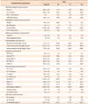
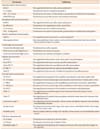
A male patient aged 14 years 5 months with an anterior crossbite was referred to our clinic with the complaint of a protruding lower jaw. It was confirmed from the patient's medical history that his general health was good and that he had no systemic or congenital diseases that could affect the orthodontic treatment. Cephalometric parameters used in this case report are defined in Table 1.
On clinical examination, it was observed that the patient had skeletal Class III malocclusion with a concave profile and a collapsed midface area (Figure 1). Cephalometric analysis (Nemoceph NX; Nemotec, Madrid, Spain) confirmed that he had a Skeletal Class III relationship (ANB: ??.7°) with maxillary retrusion (SNA: 77.5°; Table 2). The mandibular plane angle was low (GoGn/SN: 24.2°; FMA: 18.2°) and the maxillary incisors were in normal position while the mandibular incisors were lingually inclined (L1-NB: 2.7 mm). An anterior crossbite with a 1-mm negative overjet and a 1-mm overbite was observed. A 3-mm midline deviation with mandibular shifting to the left was also determined.
It was recorded that he had Angle Class I molar relationships on both right and left sides with a ??0-mm arch length discrepancy in the upper arch, which probably showed mesialization of the posterior teeth. When skeletal maturation was analyzed, it was found that the patient was in the pre-peak pubertal stage (sesamoid), according to hand-wrist radiography.
Three treatment alternatives were considered for maxillary advancement. The first option was to delay treatment and perform orthognathic surgery. The second was to apply a face mask. The third was to use skeletal anchorage with intermaxillary elastics for maxillary protraction. The patient and his parents preferred to try orthopedic traction from skeletal anchorage.
Miniplates (Titanium miniplate; Trimed, Ankara, Turkey) were inserted between the mandibular lateral incisor and canine teeth on both sides. Both miniplates were fixed to the bone with 2 titanium miniscrews (2.3 mm diameter, 5 mm length) (Figure 1C). The surgical procedures were carried out under local anesthesia (Ultracain D-S Forte; Sanofi-Aventis, Istanbul, Turkey). The sutures were removed after 1 week, and in the same session, self-drilling mini-implants (1.6 mm diameter, 10 mm length; Absoanchor; Dentos Inc., Daegu, Korea) were installed between the maxillary second premolar and molar teeth under the mucogingival line. One week later, Class III elastics delivering 75 g of force were applied on both sides. After 3 weeks, the force was increased to 200 g on both sides. Removable appliances were used to eliminate occlusal interferences and facilitate bite jumping (Figure 2C). The patient used the appliances for at least 18??0 hours each day, and treatment was continued until a positive overjet was obtained. Within 18 months of treatment, end-to-end molar relationships (sagittally) were achieved (Figure 2). The patient continued to use intermaxillary elastics for retention for 6 months, until growth and development had reached peak levels and his permanent dentition was complete. Thereafter, the patient was treated using fixed appliances in the permanent dentition. Prior to starting the fixed appliance therapy, the miniplates were removed to prevent the bone from remodeling over them, but the miniscrews were retained. After initial leveling and alignment, 0.018-inch (in) stainless steel archwires were used to correct protrusion of the upper incisors. Thereafter, rectangular 0.016 × 0.016 in and 0.016 × 0.022 in nickel-titanium archwires were used with Class III and box elastics. The fixed orthodontic treatment duration was 24 months, and proper overjet and overbite were achieved.
Positive overjet was achieved for the patient after 18 months. His soft tissue profiles improved, with the anterior displacement of the whole midface reducing the paranasal concavity (Figure 2A). Cephalometric analysis showed 2.7-mm forward movement of point A and 0.9-mm backward movement of point Pg. The ANB angle was increased by 5.2°, and the Wits value dramatically changed by 6.9 mm. The palatal plane showed 1.3° of counterclockwise rotation, and the vertical plane angle increased by 3.3°. The upper incisors exhibited approximately 1 mm of protrusion, and the lower incisors exhibited 0.8 mm of protrusion (Table 2 and Figure 2C).
The changes in the ANB angle and Wits value in the first phase were maintained after fixed orthodontic treatment. Class I canine and molar relationships were achieved, and the vertical plane angle (GoGn-SN) returned to its original value (Table 2 and Figure 3C). The maxillary and mandibular incisors were slightly more protruded (1 mm and 0.6 mm, respectively) at this stage. The malocclusion was partly camouflaged by the orthodontic treatment. Soft tissue balance was maintained, and the patient was satisfied with his facial appearance (Figure 3).
In the case under discussion, the treatment outcomes were a combination of skeletal and dental effects. Skeletal responses of the maxilla were obtained by means of modified skeletal anchorage. After obtaining positive overjet, intermaxillary elastics were continued for 6 months for the purpose of retention until the patient had his permanent dentition and had reached the peak of his growth and developmental stage. This period also facilitated the fixed orthodontic treatment phase, because the dental movements were performed to solve the arch length discrepancy rather than to provide camouflage. In most of the earlier studies, dental compensations using face mask therapy constituted half of the total correctionsafter using face mask therapy, and even these dental effects continued to increase depending on the patient's age.9
In the present case, approximately 1° counterclockwise rotation of the palatal plane and approximately 3° increase in the mandibular plane angle were observed, which corresponds to findings of previous studies.5,10,11,12,13,14 Although the vertical plane angle increased, it returned to its original value after the fixed orthodontic treatment. Therefore, this method can be considered promising for use in Class III patients with increased vertical plane angle or vertical facial height.
A disadvantage of tooth-borne devices is the loss of anchorage due to the inability to apply the orthopedic force directly to the maxilla, especially when preservation of arch length is necessary.10 In the present case, arch length discrepancies were maintained after the orthopedic treatment, in contrast to results of face mask treatment. Incisor movements were performed in order to solve this arch length discrepancy.
Lower incisor protrusion was observed in both treatment periods, as was the case in similar skeletal anchorage studies.5,6,13 This can be explained by the increasing tongue pressure on the lower incisors after eliminating the anterior crossbite.5 However, the absence of the chin-cup effect can also be considered a contributing factor.
The side effects in the anchorage regions caused by traditional face masks were avoided with this skeletal anchorage method. As for the use of zygomatic anchorage, the inability to place plates in the zygomatic region owing to limited space, particularly in patients in the pre-pubertal growth period, and the 8-step local surgery involved (placement and removal of miniplates) are considered to be the clinical disadvantages of this method.1 It can be considered an alternative treatment approach in skeletal Class III anomalies. However, upper second premolar and lower canine teeth are required to be erupted, or at least to have started erupting, for inserting the mini-implants and miniplates.
The modified skeletal anchorage treatment with 2 miniplates and 2 miniscrews was effective for maxillary protraction in this patient, who had maxillary retrognathia. Side effects that would be encountered with tooth-borne appliances were minimal. This treatment protocol was very comfortable and minimally invasive for the patient. The achieved protraction was maintained throughout the fixed orthodontic treatment.
Figures and Tables
Figure 1
Pretreatment extraoral (A), and intraoral (B) photographs of the patient. C and D, Miniplate inserted between the lower lateral and canine teeth. E, Pretreatment lateral cephalometric film. F, Pretreatment panoramic film.

Figure 2
Post-orthopedic extraoral (A), and intraoral (B) photographs of the patient with Class III elastics applied between the miniplates and miniscrews. C, Postorthopedic lateral cephalometric film. D, Postorthopedic panoramic film.

Figure 3
Posttreatment extraoral (A), and intraoral (B) photographs of the patient. C, Posttreatment lateral cephalometric film. D, Posttreatment panoramic film.

Table 2
Cephalometric analysis of the patient in pretreatment (T0), postorthopedic (T1), and posttreatment (T2) periods

Refer to Table 1 for the definition of each cephalometric parameter.
References
1. Klempner LS. Early orthopedic Class III treatment with a modified tandem appliance. J Clin Orthod. 2003; 37:218–223.
2. Chun YS, Jeong SG, Row J, Yang SJ. A new appliance for orthopedic correction of Class III malocclusion. J Clin Orthod. 1999; 33:705–711.
3. Enacar A, Giray B, Pehlivanoglu M, Iplikcioglu H. Facemask therapy with rigid anchorage in a patient with maxillary hypoplasia and severe oligodontia. Am J Orthod Dentofacial Orthop. 2003; 123:571–577.

4. Kircelli BH, Pektas ZO. Midfacial protraction with skeletally anchored face mask therapy: a novel approach and preliminary results. Am J Orthod Dentofacial Orthop. 2008; 133:440–449.

5. De Clerck HJ, Cornelis MA, Cevidanes LH, Heymann GC, Tulloch CJ. Orthopedic traction of the maxilla with miniplates: a new perspective for treatment of midface deficiency. J Oral Maxillofac Surg. 2009; 67:2123–2129.

6. Cevidanes L, Baccetti T, Franchi L, McNamara JA Jr, De Clerck H. Comparison of two protocols for maxillary protraction: bone anchors versus face mask with rapid maxillary expansion. Angle Orthod. 2010; 80:799–806.

7. Ge YS, Liu J, Chen L, Han JL, Guo X. Dentofacial effects of two facemask therapies for maxillary protraction. Angle Orthod. 2012; 82:1083–1091.

8. De Clerck EE, Swennen GR. Success rate of miniplate anchorage for bone anchored maxillary protraction. Angle Orthod. 2011; 81:1010–1013.

9. Koh SD, Chung DH. Comparison of skeletal anchored facemask and tooth-borne facemask according to vertical skeletal pattern and growth stage. Angle Orthod. 2014; 84:628–633.

10. Cha BK, Choi DS, Ngan P, Jost-Brinkmann PG, Kim SM, Jang IS. Maxillary protraction with miniplates providing skeletal anchorage in a growing Class III patient. Am J Orthod Dentofacial Orthop. 2011; 139:99–112.

11. Ahn HW, Kim KW, Yang IH, Choi JY, Baek SH. Comparison of the effects of maxillary protraction using facemask and miniplate anchorage between unilateral and bilateral cleft lip and palate patients. Angle Orthod. 2012; 82:935–941.

12. Kaya D, Kocadereli I, Kan B, Tasar F. Effects of facemask treatment anchored with miniplates after alternate rapid maxillary expansions and constrictions; a pilot study. Angle Orthod. 2011; 81:639–646.


 PDF
PDF ePub
ePub Citation
 Print
Print


 XML Download