

 PDF
PDF ePub
ePub Citation
 Print
Print


INTRODUCTION
In orthodontic practice, comprehensive diagnosis and treatment planning are essential for a successful treatment outcome.1 Stone casts are one of the tools that serve this purpose, and have long been the gold standard for measuring mesiodistal tooth dimensions, calculating indices such as the Bolton index, and determining the efficacy of orthodontic treatment.2,3,4 However, these study models have the disadvantages of being prone to degradation, breakage, and loss.5
With the introduction of digitized laser-scanned dental impressions that produce a three-dimensional (3D) image of the teeth and dental arches, the disadvantages of the study model have been surmounted. Moreover, the inconvenience of having to pour and trim plaster casts and the need to store and retrieve the models each time a patient is seen have been obviated.6 Now it is possible to view the dentition on a computer screen by rotating virtual models to provide a 3D view, as with hand-held models.6
In parallel with the use of 3D cephalometrics and 3D digital photography, the popularity of digital models has increased, and the paperless office has represented a great advance in practice efficiency.6 Currently, there are a number of companies that digitize dental models and offer software programs for the analysis of both linear and angular parameters, such as OrthoCad (Carlstadt, NJ, USA), OrthoProof (Albuquerque, NM, USA), O3DM (O3DM, Aarhus, Denmark), and Orthomodel (Orthomodel Inc., Istanbul, Turkey).6
Previously, the reproducibility of Bolton analysis, tooth width, and dental arch width measurements were evaluated.1,2,3,4,6,7,8,9 These studies revealed that measurements made with calipers on plaster casts demonstrated less inconsistency than measurements based on software programs using digital models.1,2,3,4,6,7,8,9 Zilberman et al.7 compared the precision of tooth size and arch width measurements made on plaster models with measurements made on OrthoCAD® virtual models and stated that both methods were clinically satisfactory. In 2004, Quimby et al.2 verified the accuracy and reproducibility of measurements performed on digital models for mesiodistal tooth widths, arch length, arch width, overjet, and overbite. They were found to be as precise and reliable as traditional plaster model measurements.
To correct a molar relationship or crowding, molar distalization is frequently performed with various appliances.10,11 To assess the efficacy of these appliances, superimpositions of serial cephalometric radiographs, study model photographs, and photocopies have been used in previous studies.10,12 To our knowledge, there is no study that compares the superimposition of 3D digital models with cephalometric radiography and model photocopy methods in cases of molar distalization.
The aim of this study was to evaluate the reliability of measurements performed for the evaluation of upper molar distalization on 3D digital models by comparing them with measurements obtained from lateral cephalometric radiographs and photocopies of study models.
MATERIALS AND METHODS
The samples were collected from the archive of the Department of Orthodontics, Faculty of Dentistry, Cumhuriyet University, Sivas, Turkey. The pre-treatment and post-treatment maxillary study casts and lateral cephalometric radiographs of 20 Class II patients (10 male and 10 female subjects; mean age, 16 years) whose maxillary first molars were distalized with an intraoral distalizer were used in the study. The study protocol was reviewed and approved by the Medical Scientific Ethics Committee of the university. The posterior movements of the maxillary first molars were evaluated on cephalometric radiographs (group CP), photocopies of plaster models (group PH), and 3D digitized models (group TD). To assess the methodological error of both techniques, 10 sets of randomly selected measurements were repeated 1 month after the first measurements. For this purpose, all reference points were removed, marked again, and re-measured by the same orthodontist a second time.
Cephalometric analysis
All lateral cephalometric radiographs were taken using the Proline PM2002 CC model (Planmeca Oy, Helsinki, Finland). The focal median plane distance was 152 cm and standardized at 73 kV and 15 mA for 0.64 s of exposure; the radiographic film used was 18 × 24 cm, Kodak MXG (Eastman Kodak Company, Rochester, NY, USA).
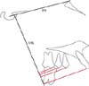
A sheet of transparent acetate was placed over the lateral cephalometric radiographs and the anatomical structures were outlined. To form a vertical reference plane, a line was drawn perpendicular to the sella-nasion (SN) plane from the intersection of the anterior wall of the sella turcica and the anterior clinoid process because these structures do not move with growth changes.13 To determine the distalization amounts, lines were drawn from the central incisors, premolars, and molars perpendicular to this plane (Figure 1). The difference between the pre-treatment measurement and post-treatment measurement revealed the amount of distalization for each tooth. All cephalometric measurements were corrected because of a magnification error of 10% for a more accurate comparison with the photocopy and 3D digital model measurements.
Measurements from photocopies of study models
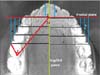
The occlusal surfaces of study models were photocopied on a photocopier (Toshiba eStudio 200; Toshiba, Singapore) with the contrast set at the darkest setting, as described by Champagne.14 The same operator (RN) performed the photocopy measurements using a Vernier caliper (0.1 mm precision). A frontal line perpendicular to the midsagittal plane and passing through the most anterior point of the incisive papilla was constructed on the photocopies to determine the distalization amounts of the central incisors, canines, premolars, and molars (Figure 2). As seen in the figure, lines were drawn from each tooth perpendicular to the frontal line to measure the amount of distalization for each tooth. To determine the amount of expansion of the canines, premolars, and molars, a sagittal line passing through the most anterior and posterior points of the incisive papilla was constructed on the model photocopies, and lines were drawn from the teeth perpendicular to this plane. The calculation of molar rotation is also demonstrated in Figure 2.
3D digital model analysis
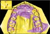
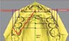
For the 3D digital model analysis, dental casts obtained before and after molar distalization were sent to the O3DM laboratory (Ortolab Sp., Czestochowa, Poland) for 3D surface laser scanning. The 3D scanning equipment used was a 3 Shape R700 model (3Shape A/S, Copenhagen, Denmark). The pre- and post-treatment scans of the dental models were superimposed on three points in the incisive papilla area (the most anterior point, the most prominent point, and the most posterior point of the incisive papilla) by using O3DM version 2 software (O3DM) (Figure 3). It should be noted that these points are not in the same line, so the program can make a reliable and reproducible superimposition. With this program, a frontal line perpendicular to the midsagittal plane and passing through the most prominent point of the incisive papilla was constructed on the superimposed 3D models to determine the distalization amounts of the central incisors, canines, premolars, and molars. As seen in Figure 4, lines were drawn from the teeth perpendicular to this plane, and the differences between the measurements taken before and after molar distalization were calculated. To determine the amount of expansion of the canines, premolars, and molars, a sagittal plane passing through the most anterior and posterior points of the incisive papilla was constructed on the superimposed 3D model. As seen in Figure 4, lines were drawn from the teeth perpendicular to this plane, and the distances were measured with the aid of software. The calculation of molar rotation is also demonstrated in Figure 4.
Statistical analysis
Because only the amount of distalization could be evaluated on cephalometric radiographs, all three groups were compared only in terms of this parameter. Group PH and group TD were compared in terms of the amount of distalization; expansion of the central incisors, canines, premolars, and first molars; and the degrees of molar rotation. The data were analyzed using SPSS for Windows, version 13.0 (SPSS Inc., Chicago, IL, USA). A Friedman test was used to compare the three groups, and a Wilcoxon test was used for the comparison of group PH and group TD. The accuracy and repeatability (intra-observer reliability) of the measurements were evaluated with the aid of Cronbach's alpha.
RESULTS
For all measurements, Cronbach's alpha value was very close to the ideal value of 1: 0.934-0.980 for group CP, 0.939-0.979 for group PH, and 0.941-0.982 for group TD. This indicates that intra-observer reliability was adequate for all measurements.
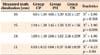
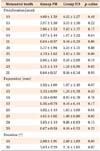
According to the 3D digital model measurements, the mean amounts of upper incisor and molar distalization were 0.56 mm and 4.52 mm, respectively (Table 1). Moreover, the upper molars demonstrated 1.87 mm of expansion and 2.88° of rotation resulting from distalization forces (Table 2).
No significant difference was observed in terms of the amount of distalization of the upper molars, premolars, or incisors among the three groups (Table 1). Measurements of the amount of distalization, expansion of the teeth, and degrees of molar rotation did not reveal any significant difference between group PH and group TD (Table 2).
DISCUSSION
In the literature, tooth movement is usually assessed via measurements made on lateral cephalometric radiographs or photocopies of study models.14 Recently, the use of 3D digital models has increased, and also can be used for this purpose.
Cephalometric radiographic measurement is a standard method used by orthodontists to assess skeletal, dental, and soft tissue relationships, as well as the results of orthodontic treatment. This approach, however, is based on two-dimensional (2D) views of 3D structures, which causes information loss and identification errors due to blurring, overlapping of anatomic structures, and the superimposition of bilateral structures.15 Magnification errors, subjecting patients to radiation exposure, and the inability to evaluate tooth movement in the transverse direction are other disadvantages of cephalometric assessments. In addition, errors associated with measuring small linear distances on cephalometric tracings further compromise a quantitative assessment of the orthodontic movements of each tooth.16
The photocopy method developed by Champagne14 is another method that requires a plaster model, but provides only a 2D projection of a 3D structure. It has many clinical drawbacks, such as difficulties in establishing reference points, a complicated measurement process, and a 2D measurement of the 3D curvature of the palatal vault.17 Despite the limitations of the cephalometric and photocopy methods, both are used in the assessment of molar distalization because there has been no other method that permitted superimposition.
In recent years, 3D digital models have gained increasing acceptance as an alternative to traditional plaster models in orthodontics. Unlike plaster models, 3D digital models are not subject to loss, fracture, or degradation. Digital storage eliminates the need for storage space, which is required for traditional models.18 In addition, tooth position can be measured accurately in three dimensions; the measurement of inclination, which is unreliable with plaster models, is especially accurate when this method is used. Furthermore, 3D mapping of tooth movement is possible by superimposing dental changes on stable reference structures with the use of digital sectioning techniques. The validity and efficiency of linear and angular measurements created with 3D digital models have been investigated, and it was confirmed that digital models offer a valid alternative to plaster models.5,19
Laser scanners are one of the devices capable of constructing 3D shapes of the dentition and occlusion20 with adequate accuracy and reliability.21 The disadvantages of the laser scanning method are the relatively long times required for the 3D scanning and analysis of dental casts, as well as the purchase costs of the scanner and software. Additionally, analyzing the casts requires special training in order to establish accuracy.19 Studies comparing direct measurements made from dental casts with those made from 3D digitized models produced by surface laser scanners have shown that the latter method is highly accurate for dental cast analysis.22 In this study, the laser scanning technique was preferred to a mechanical digitizer.
In this study, comparisons of measurements obtained from lateral cephalograms, photocopies of study models, and 3D digital models did not reveal any significant difference. These findings are in accordance with the results of Thiruvenkatachari et al.19 and Mavropoulos et al.22 Although no significant difference was found among the groups in the distalization amounts of the first molars and premolars, the fact that measurements from the photocopies and 3D models were closer to each other than measurements from cephalometry was remarkable. As a limitation of the methodology of our study, this difference might depend on the fact that the reference plane used for cephalometric measurements was vertical, while the reference planes used for the other two types of measurements were horizontal. Furthermore, because rotation and expansion could not be assessed on cephalometric radiographs, these parameters were compared between group PH and group TD.
The reliability of linear measurements obtained from plaster and 3D digital models was investigated by Bell et al.23 and Keating et al.24 The mean differences reported in these studies were 0.14 mm and 0.27 mm, respectively. We observed similar mean differences ranging from 0.02 mm to 0.24 mm, except for measurements of the second premolars, which showed differences of 0.5 mm and 0.62 mm for the left and right sides, respectively. This may be due to the fact that the second premolars presented more distal rotation, which might have affected the point location.
The agreement between transverse dimensional readings obtained using digital and plaster models has been assessed previously,23 and the mean differences between the approaches ranged from 0.04 mm to 0.4 mm.2 In this study, intercanine, interpremolar, and intermolar distances were measured, and the mean differences ranged from 0.01 mm to 0.33 mm, which was compatible with those in the literature. These small differences had no clinical significance.
The evaluation of orthodontic tooth movement requires the superimposition of certain reference points or lines on either cephalometric radiographs or plaster models. The cranial base, maxilla, or mandible is used as the reference point for the superimposition of serial cephalometric radiographs,25 while the superimposition of plaster models has limitations due to a lack of anatomic reference points or areas. With the development of 3D measuring devices, some investigators have performed 3D superimpositions of dental models to analyze tooth movement.12,17,26 The use of palatal rugae as reference points for measuring tooth movement on both serial dental models27,28 and 3D digital models 12,15 has been investigated and reported to be a suitable reference structure when studying serial models. On the other hand, Simmons et al.29 performed a longitudinal study (from primary dentition to young adulthood) of the anteroposterior stability of the medial rugae region and concluded that rugae landmarks did not seem to be stable reference points for the investigation of tooth migration. Moreover, Choi et al.30 suggested that future research should evaluate the 3D positional stability of the palatal rugae by using another stable reference plane. Consequently, we preferred to superimpose the models on three points that were not in a straight line and formed a plane located at the incisive papilla area rather than the palatal rugae, and our results were consistent with those obtained using conventional methods. Indeed, O3DM (O3DM) and some similar programs have the ability to perform a true superimposition with integration of digital dental models and cone-beam computed tomography (CBCT) images. However, to produce this model, we would need CBCT images of the patient in addition to the standard materials. This is a limitation of our study; however, the reason we did not use this method was the high radiation dose from CBCT compared with cephalometric radiography. In the near future, with a decrease in the radiation dose from CBCT, a 3D digital model analysis method presumably will be the gold standard for the superimposition of records.
CONCLUSION
The measurement differences among the 3D digitized model, cephalometric radiography, and plaster model photocopy methods were insignificant. The use of 3D digital modeling to assess the results of upper molar distalization is a reliable and valid alternative to conventional measurement methods.
 XML Download