

 PDF
PDF ePub
ePub Citation
 Print
Print


INTRODUCTION
Recently, several reports on orthodontic surgery without pre-operative treatment have been published.1,2,3,4,5 This mandibular setback surgery-first approach offers advantages over conventional orthodontic-surgical treatments, including enhanced patient cooperation, efficient and effective decompensation, and reduced total treatment time.3 Liou et al.4 maintained that the phenomenon of accelerated postoperative orthodontic tooth movement achieved by the surgery-first approach shortened the treatment period, and partially resolved the issue of dental decompensation, which minimized the number of postoperative orthodontic treatments.
However, disadvantages of the surgery-first approach have been reported. Baek et al.5 noted that considerable clinical experience is required for accurate assessment of skeletal discrepancies and the prediction of overall postoperative orthodontic treatment results. Further, postoperative occlusal instability can cause skeletal instability and associated postoperative orthodontic treatment difficulties.
According to previous surgery-first studies, surgery-first orthodontic treatment postoperative occlusal instability results primarily from premature contact of the extruded upper second molar. Premature cusp-to-cusp premolar contact is often associated with postoperative surgical occlusion. Additionally, premature contact induces postoperative occlusal instability, increased vertical dimension, and postoperative forward mandibular movement.5,6 Hwang et al.7 suggested that in addition to occlusal factors, proximal segments can affect postoperative skeletal stability. Therefore, a practical planning prerequisite for surgery-first orthodontic treatment is the analysis of both skeletal (including proximal segment) and dental (including vertical dimension) changes. For accurate analysis, the cone-beam computed tomography (CBCT) superimposition method and CBCT-generated cephalograms with ray-cast and maximum intensity projection (MIP) images could be used for simultaneous acquisition of skeletal and dental information.8,9
The aim of the present study was to investigate postsurgery-first-treatment skeletal and dental changes in skeletal Class III malocclusion, by superimposition of CBCT volumes representing the pre-treatment (T0), immediate-postoperative (T1), and post-treatment (T2) stages. Likewise, an additive correlation analysis of skeletal and dental changes was performed. The operative hypothesis was the existence of a correlation between vertical dimension changes and skeletal changes after surgery-first orthodontic treatment.
MATERIALS AND METHODS
Subjects
To test the proposed hypothesis, a retrospective study was conducted using data from 34 patients (23 men, 11 women; mean age: 26.2 ± 6.6 years) that presented to the Department of Orthodontics, Pusan National University Dental Hospital (Yangsan, Korea)between January 2010 and July 2012. The exclusion criteria were extraction history, severe facial asymmetry, cleft lip and/or palate, and temporomandibular joint disorder. The inclusion criteria for mandibular setback surgery-first orthodontic treatment were skeletal Class III malocclusion with no need for extractions. Accordingly, the patients selected suffered from skeletal Class III deformities that were corrected using a surgery-first without orthodontic treatment approach. The surgery-first orthodontic treatment protocol was as follows: First, a surgical occlusal setup was performed, taking into consideration the final position of the upper and lower anterior teeth. Second, a single surgeon performed a mandibular setback sagittal split ramus osteotomy (SSRO) with rigid internal fixation achieved by miniplates on all of the patients. Next, inter-maxillary fixation was applied for one week, followed by physiotherapy involving muscle and mouth-opening exercises using vertical elastics. One month after surgery, active orthodontic treatment was resumed. Last, the surgical stent was adjusted during the postoperative orthodontic treatment, and temporary anchorage devices were used to improve premature occlusion. The Institutional Review Board of Pusan National University Hospital reviewed and approved this study (E-2011069).
Data acquisition
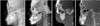
To evaluate statistically significant differences, all of the measurements were analyzed at three time points on CBCT-generated Half-Cephalograms (DCT pro; Vatech Co., Hwaseong, Korea). The Half-Cephalograms were obtained by reformatting the CBCT raw data to three-dimensional (3D) images using 3D imaging software (OnDemand 3D; Cybermed Co., Seoul, Korea). After superimposing the CBCT data on the anterior cranial base according to the maximization of mutual information theory, ray-cast images and MIP images were acquired from right side of the CBCT data (Figure 1).10 The measurement points and reference planes are defined in Tables 1 and 2. To evaluate the correlation between vertical dimension changes and skeletal changes, vertical dimension was defined as the distance from the furcation of the upper first molar to that of lower first molar. All of the CBCT-generated Half-Cephalograms were digitized using cephalometric analysis software (VCeph version 6.0; Osstem Implant Co., Seoul, Korea) at three time points including pre-treatment (T0), immediate-postoperative (T1),and post-treatment (T2).
Statistical analysis
All statistical analyses were performed using a statistical software package program (SPSS ver. 12.0; SPSS, Chicago, IL, USA). The same investigator repeated all of the measurements two weeks after initial data collection, and the mean of the two values was used in the statistical analysis. The systematic intra-examiner error between the two measurements was determined by a paired t-test, and the magnitude of the corresponding error was assessed using the intraclass correlation coefficient (ICC). Repeated-measures analysis of variance was used to determine the skeletal and dental changes at T0, T1, and T2, and multiple comparisons were performed with the Bonferroni's test. Spearman correlation coefficients were calculated to assess the relationship between the vertical dimension and skeletal changes after the surgery-first orthodontic treatment.
RESULTS
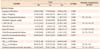
The means and standard deviations of the CBCT-generated Half-Cephalometric measurements taken at T0, T1, and T2 for all of the subjects are summarized in Table 3. Analysis of the systematic intra-examiner error revealed no statistically significant differences, and the ICC indicated robust reproducibility (ICC range: 0.945-0.989).
Changes from the pre-treatment to postoperative stage (T0 to T1)
The postoperative measurement changes from T0 to T1 are listed in Tables 3 and 4. The skeletal change at the Pog point manifested as -9.24 ± 3.97 mm horizontal and -0.33 ± 3.00 mm vertical mandibular movement. Further, the horizontal skeletal changes at the Pog point were statistically significant (p < 0.05). No statistically significant differences were detected in the proximal segment changes (Cd and Cp to FH plane/Poperpendicular plane; p > 0.05). Similarly, no significant postoperative dental changes were detected in the vertical dimension (p > 0.05).
Changes from the postoperative to post-treatment stage (T1 to T2)
No statistically significant differences were detected in the skeletal changes from T1 to T2 (p > 0.05). However, the mandible tended to move forward 1.22 ± 2.02 mm. Regarding the angular change of the proximal segment, the condylar position (Cd to Po-perpendicular plane) moved backward, and the coronoid process (Cp to FH plane) moved vertically. The dental changes in the vertical dimension were statistically significant (p < 0.05), and included the upward movement of the lower 1st molar, and a 1.62 ± 1.35 mm reduction from L6 to FH (Table 3 and 4).
DISCUSSION
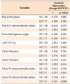
Our aim in the present study was to search for correlations between dental changes (vertical dimension) and skeletal changes (proximal segment) after the application of a surgery-first orthodontic approach to treat skeletal Class III malocclusion. We initially hypothesized that a correlation existed between vertical dimension and skeletal changes. Consequently, the study results revealed statistically significant correlations between T1 to T2 vertical dimension changes and T1 to T2 Cd to Po-perpendicular plane changes, T1 to T2 Cp to FH plane changes, and T0 to T1 Cp to Po-perpendicular plane changes.
There have been several reports of the surgery-first approach in orthognathic surgery cases,1,2,3,4,5,6,11 but the proper setup for the surgical model remains unclear. Liou et al.6 suggested that the maxilla and mandible be arranged in a proper molar relationship and with a positive overbite, which has been applied to non-extraction cases of Class I malocclusion.11 Baek et al.5 insisted that in order to obtain a normal post-surgical overbite and overjet, it was better to increase the extent of mandibular setback than to perfectly incline the lower incisor. For accurate prediction and simulation, the establishment of the appropriate setup and surgical models is critical.8,12 Nonetheless, in their surgical model setup, orthodontists empirically increase the vertical dimension and the extent of mandibular setback movement due to the occlusal interference.
Unfortunately, there are no specific guidelines for any case. Likewise, it is unclear how to resolve the increased vertical dimension resulting from premature contact in premolar or molar areas during the post-treatment stage. In the present study, the vertical dimension tended to increase at the immediate-postoperative (T1) stage, and decrease at the post-treatment (T2) stage. At T2, the mandible tended to move forward. Similarly, Baek et al.5 reported significant backward mandibular repositioning due to surgery, but significant forward relapse during postoperative orthodontic treatment. Additionally, Hwang et al.13 stressed that the increased extent of the vertical dimension changes affected T1 to T2 Pog point movement. For effective treatment planning, the extent of the increased vertical dimension and mandibular setback movement should be considered in surgical treatment objective.
However, consideration should also be given to other factors that influence increased vertical dimension change that emerged in the present Spearman correlation analysis, including T1 to T2 Cd to Po-perpendicular plane changes, T1 to T2 Cp to FH plane changes, and T0 to T1 changes in Cp to Po-perpendicular plane. During the period from the T1 to T2 stage, the condylion (Cd) and Cp points were significantly altered, even though the Pog point was not. Specifically, the Cd and Cp points, but not the Pog point, were significantly correlated to vertical dimension changes. Hence, the results indicated that the position of the proximal segment should be controlled.
Prior reports have emphasized that surgery-first postoperative instability stems from occlusal instability of the premature occlusal contacts and from proximal segment changes. However, a surgical stent can be used to control occlusal instability after surgery-first orthodontic treatment. In fact, several authors have recommended this strategy as a means of preventing occlusal interference, and have asserted that skeletal relapse caused by proximal and distal segment positional changes can be overcome by rigid fixation.1,2,3,4,5,6,8,11 However, Kim et al.14 reported proximal segment displacement during the healing-period, despite the application of rigid fixation systems. Therefore, even when rigid fixation is applied to mandibular setback SSRO, the proximal segment should be securely managed for skeletal stability, as well as in surgery-first orthodontic treatment cases. After careful management of skeletal stability and proximal segment position in the T1 stage, a surgical stent could be utilized as an effective tool for resolving premature occlusal contacts in the premolar or molar region, and for maintaining the T2 stage position of the Pog point. To this end, Hwang et al.7 maintained that the intended manual condylar position could improve skeletal stability in the short term.
However, the study had several design limitations. First, there was no comparison to a control group. Second, most of the cases involving an average of 2 mm crowding that were not extraction cases were investigated. Consequently, additional multi-centered studies that include extraction cases and control groups is recommended. Likewise, the changed vertical dimension or "transitional" occlusion was established postoperatively in the surgery-first approach, and orthodontics were an adjunctive postoperative treatment used to transfigure transitional occlusion to solid final occlusion.6 However, in surgery-first orthodontic treatment, in order to improve postoperative instability and achieve skeletal stability, not only surgical occlusion but also proximal segment management should be considered.
CONCLUSION
A correlation existed between vertical dimension changes and skeletal changes after mandibular setback surgery-first orthodontic treatment. Larger post-surgical vertical dimension changes were related to a greater number of skeletal changes during the post-treatment stage. Examining the mandibular position in relation to the post-surgical vertical dimension served to emphasize the integral importance of vertical dimension control and proximal segment management to the success of surgery-first orthodontic treatment.

 XML Download