This article has been
cited by other articles in ScienceCentral.
Abstract
Objective
This prospective clinical study aims to determine the differences between two treatment modalities for anterior open bite in growing patients. The treatment modalities involved the use of magnetic bite-blocks (MBBs) or rapid molar intruders (RMIs) applied with posterior bite-blocks.
Methods
Fifteen consecutive patients with a mean age of 11.2 (standard deviation [SD] = 1.6) years and a mean open bite of -3.9 mm were treated with MBBs. Another 15 consecutive patients with a mean age of 10.9 (SD = 1.8) years and a mean open bite of -3.8 mm were treated with RMIs applied on bite-blocks. Cephalometric radiographs were obtained before (T1) and immediately after appliance removal (T2). The treatments lasted four months, during which the appliances were cemented to the teeth. The morphological changes were measured in each group and compared using logistic regression analysis.
Results
The MBB group exhibited significantly greater decreases in SNA angle, ANB angle, overjet, and maxillary incisor angle (p < 0.05). The MBBs induced greater effects on the maxilla and maxillary dentition. The MBBs restrained maxillary forward growth and retracted the maxillary incisors more effectively than did the RMIs. Consequently, changes in the intermaxillary relationships and overjets were more distinct in the MBB group.
Conclusions
The anteroposterior differences between the appliances suggest that MBBs should be preferred for the treatment of patients with Class II open bites and maxillary incisor protrusions.
Keywords: Early treatment, Anterior open bite, Rapid molar intruder, Magnets
INTRODUCTION
The orthodontic treatment of skeletal open bite remains a challenge because it requires not only stable closure of the open bite but also an improvement in facial balance. The accepted approach to achieving these goals is to control or even reduce the posterior dimensions by intruding the posterior teeth.
1 The early treatment of open bite is favorable, because such early treatment affords the opportunity to control the direction of growth and might enable the patient to avoid a more aggressive treatment in the future.
2 Moreover, treatment at an early age improves the child's self-confidence by improving his appearance.
3 However, treatment becomes more challenging during the growth period owing to issues of compliance. Posterior bite-blocks have been found to control posterior dentoalveolar heights in the maxilla by intruding the maxillary molars or at least ceasing their eruption.
4 The posterior bite-block is a passive appliance that depends only on the biting force of the patient. The adjustments that have been made to this treatment modality include the addition of springs or magnets to apply additional force to the posterior teeth.
5,
6,
7
Magnets were used in the treatment of open bite for the first time by Dellinger
8 in 1986, who called the appliance the active vertical corrector. Dellinger considered anterior open bite to be a symptom of excessive eruption of the posterior teeth. He introduced the appliance as a reason-oriented treatment and assumed that the intrusion of these teeth would not only correct the open bite but also achieve good facial balance by allowing the mandible to rotate superoanteriorly and decrease anterior facial height. Previous animal models of magnetic bite-blocks (MBBs) have reported distinct morphological changes in the gonial angle of the mandible.
9,
10 Clinical studies of this appliance have revealed obvious corrections of anterior open bites and improved facial proportions. The authors of these studies have attributed the closure of open bites to dental and skeletal changes and attributed the changes in anterior facial height to counter-clockwise rotation of the mandible.
11,
12,
13,
14,
15
Another treatment for open bite was introduced a decade ago by Carano et al.,
16 who called his device the rapid molar intruder (RMI). This device consists of elastic modules with coil springs inside. The device is applied to the first molars via bands that are fixed to the molars. RMIs have been shown to perform well in the correction of open bites with mixed dentition and early permanent dentition. RMIs also improve patient appearance through counter-clockwise rotation of the mandible and advancement of the chin.
17
Both appliances (i.e., the MBBs and the RMIs) are noncompliant treatment modalities and have been found to be effective in the treatment of anterior open bite with mixed and early permanent dentition. The purpose of this study was to compare the growth and treatment changes elicited by these two appliances. We tested the null hypothesis that the extent of morphological changes following treatment would not be related to the treatment modality.
MATERIALS AND METHODS
This study was carried out at and funded by the Damascus University, Syria. The trial was approved by the Scientific Research Committee of the Dental Faculty at Damascus University. The sample consisted of 30 patients who had anterior open bites and were over the age of eight years and under the age of 14 years. The patient inclusion criteria were as follows: free of all systematic and developmental disorders; clinical anterior open bite; and cephalometric radiograph showing a mandibular plane angle (SN-MP) exceeding 36° or an S-Go/N-Me ratio below 57%. Fifteen consecutive patients (six boys and nine girls; age between 8.5-13.5 [mean 11.2, standard deviation (SD) 1.6 years]) were treated with MBBs. Another 15 consecutive patients (four boys and 11 girls; age between 8.1-13.5 [mean 10.9, SD 1.8 years]) were treated with RMIs attached to posterior bite-blocks. The nature of the treatment and the study were explained to the parents, and signed consent was obtained.
Magnetic bite-blocks
Samarium-cobalt (SmCo) magnets (U.S. Magnetix, Plymouth, MN, USA) with dimensions of 0.069×0.434 inches were coated with Ni-Cu-Ni and then embedded in posterior bite-blocks. Buccal wings were added to the lower bite blocks to prevent the development of a lateral crossbite (
Figure 1). The bite registered for making the appliance was taken such that the inter-occlusal distance was 4.5 mm in the area of the first permanent molar. Lingual and transpalatal arches were added to the bite-blocks to control the buccal inclination. Both arches were at least 2 mm away from the soft tissue. The magnets generated a 600-g repelling force when the gap between them was zero. We measured the force between the upper and lower cast models while they were mounted on an articulator. This force ranged from 350 to 450 g.
Rapid molar intruder
The RMIs (American Orthodontics, Sheboygan, WI, USA) were applied with a new technique that is illustrated in
Figure 2. Posterior bite-blocks were made in a manner similar to that used for the MBBs, then connected by lingual arch in the mandible and transpalatal arch in the maxilla. However, rather than magnets, 0.060-inch tubes were added to the buccal side of the bite-blocks, and the RMI was attached to these tubes. The application of the RMI to the bite blocks precluded bite opening from playing a role in the differences between the two appliances examined in this study. Moreover, the bite-blocks distributed the force of the RMI across all of the posterior teeth rather than only the first molars and thus avoided the need to extract or grind the deciduous teeth, which is required when the RMI is applied only to the first permanent molars. Therefore, all of the posterior teeth were simultaneously subjected to the intrusive force. No acrylic buccal wings were added to the bite blocks in this treatment.
In both groups, the bite-blocks were cemented to the teeth for four months. The patients were examined every three weeks to examine the stability of the appliance and to assure that the lingual and transpalatal arches remained sufficiently far away from the soft tissues. Many patients required adjustments of the lingual arch at some point during the treatment period. The patients were asked to wear vertical bands at night to prevent their mouths from opening. The patients were also encouraged to maintain a seal between their lips and trained to do so. After removal of the appliances, the patients were provided with removable passive bite-blocks as retainers for four months. The changes examined in this study include only the changes that were observed immediately after removal of the first appliance (i.e., the MBBs or the RMI).
Cephalometric measurements
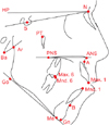
Cephalometric radiographs were taken for each patient before (T1) and immediately after removal of the MBBs or the RMI (T2). The radiographs were traced by a single author, and the parameters obtained are presented in
Figure 3.
Statistical analysis
Depending on the overbite value, the power of the study reached 75% with an effect size of 1 mm and a significance level of 0.05. The data were normally distributed according to Shapiro-Wilk tests, so parametric tests were used. To investigate any significant differences between the groups prior to the treatments, we used independent sample t-tests. The null hypothesis of the present study was that the changes observed after treatment would not be related to treatment modality, and this hypothesis was tested using logistic regression analysis. We used JMP Pro 9.0.2 (SAS Institute Inc., Cary, NC, USA) for all statistical analyses.
Method error
To check the reliability of the tracing process, 20 radiographs were selected randomly and re-traced again by the same author. The method error (δ) was evaluated according to Dahlberg's formula (δ=

), where d is the difference between two measurements, and n is the number of re-traced radiographs.
18 The method error ranged from 0.12 to 0.37 for linear measurements and from 0.13 to 0.56 for angular measurements.
RESULTS
All participants completed the intended period of treatment (four months), during which time the appliances were fixed to the posterior teeth. All of the patients exhibited increases in overbite (confidence interval [CI]: 2.6-4.0 mm in the MBB group and 2.4-3.9 mm in the RMI group). None of the cases treated with either of the appliances presented with posterior crossbites when the treatment was completed.
Independent sample
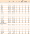
t-tests revealed no significant intergroup differences in any of the cephalometric parameters before treatment. The results of these tests and descriptive statistics for both groups are presented in
Table 1.
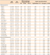
The skeletal and dental changes following treatment for each group are presented in
Table 2. Logistic regression analysis detected relationships between five parameters and the treatment modality type (
Table 2). The MBBs elicited significantly greater decreases in the SNA and ANB angles. Regarding the dento-alveolar parameters, the MBBs elicited greater decreases in the maxillary incisor angle and overjet. The occlusal plane angle decreased to a greater extent in the RMI group.
DISCUSSION
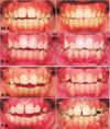
The appliances investigated in this study were effective in closing or improving open-bites. Although only five patients in the RMI group and four patients in the MBB group achieved positive overbite at the time of appliance removal, all of the patients exhibited increases in overbite (
Figure 4). Our study results revealed no significant differences between appliances with regard to the magnitude of the increase in overbite. At the time of appliance removal, we noticed that, in some cases, the patients exhibited premature contact particularly at the canines, which might indicate that overbite improvement may have been underestimated. Both appliances hindered oral hygiene, necessitating special care during the treatment duration. This problem is common to all types of cemented splints and bite blocks, including the McNamara-type rapid expander.
19 The major significant differences between appliances were related to changes in the maxilla as well as the positions of maxillary anterior teeth.
The increases in overbite can be attributed to the effects of minor but important changes in skeletal and dental structures. Increased posterior dento-alveolar height is a common problem among hyperdivergent subjects.
20 The intrusion or suppression of excessive vertical growth in dento-alveolar segments (-0.4 and 0.1 mm in the MBB and RMI groups, respectively) induced anterior rotation of the mandible (SN-MP changed -1.4 and -1.1 degrees in the MBB and RMI groups, respectively). Such rotation has a dramatic effect on the overbite. Moreover, continuous increases in anterior dento-alveolar height allow the incisors to elongate and exacerbate the overbite. We noticed growths of approximately 1 mm in the mandibular and maxillary incisors of both groups. Our study did not detect differences between treatment modalities with regard to these vertical changes.
Previous studies of animals have reported skeletal changes achieved using posterior bite-blocks with magnets. These changes include the remodeling of sutures around the maxilla and changes in the direction of maxillary growth.
9,
10 In the present study, we found that the effects on maxillary bone differed significantly between the group that received the MBBs and the group that received the RMIs with bite-blocks. The SNA angle decreased in the MBB group to a greater extent than in the RMI group (-1.1 and -0.2 degrees, respectively). As the magnitude of these changes differed between groups, these changes resulted in significantly different changes in the intermaxillary relationships. The ANB decreased by -1.7 degrees in the MBB group and by -1.1 degrees in the RMI group. These differences can be attributed to the variation in consistency of the applied force between groups. We observed that in the RMI group, the elastic modules were apparently deformed at the time of appliance removal, thereby indicating that the applied force was substantially reduced over the course of the treatment.
21 On the other hand, the magnitude of the force is consistent in the case of the magnets. Thus the maxillae were more susceptible to the intrusive forces applied by the MBBs than those applied by the RMIs. This difference also caused the patients to apply more muscular tension to achieve a lip seal, and the maxillae of these patients showed more effects attributable to labial pressure (
Figure 5). These differences in force seemed to restrain the forward growth of the maxilla to a greater extent with the MBBs than with the RMIs.
Both appliances were effective in retracting the maxillary incisors. However, the appliances differed significantly in the extent of this effect. The maxillary incisors in the MBBs group were retracted by more than twice the amount observed in the RMI group. Other studies of MBBs and RMIs have reported similar effects on the maxillary incisors.
11,
12,
13,
16,
17 Similar to the changes in the anteroposterior positions of the maxillae, the inclinations of the maxillary incisors can be attributed to the increased lip pressures generated by the children in the attempt to keep their lips sealed. The differences between the MBBs and RMIs in terms of restriction of forward growth of the maxilla and retraction of the maxillary incisors manifested as changes in overjet. The decrease in overjet in the MBB group reached -2 mm; this decrease was -1 mm in the RMI group. Such an effect is usually favorable in open-bite cases, which are frequently accompanied by a Class II relationship and an increased overjet due to backward rotation of the mandible. The occlusal plane was determined based on the intercuspation of the first molars and the mid-distance between the maxiallry and mandibular incisor edges. The significant difference between groups in occlusal plane angle might be explained by differences in the changes in dento-alveolar height. Such differences might have been too small to detect with our sample size. These differences could involve more intruded maxillary molars or more elongated maxillary incisors in the MBB subjects.
These effects of the MBBs on the maxillae indicate that this type of appliance may be preferred over RMIs for cases of open bite with a Class II component or a tendency toward a Class II component.
CONCLUSION
Significant differences were found between the fixed MBBs and the RMI with fixed bite-blocks. These differences included the following changes, which were observed after a four-month period of treatment.
1. The MBBs restricted the forward growth of maxillae to a greater extent.
2. The MBBs retracted the maxillary incisors to a greater extent.
3. The decreases in overjet and the intermaxillary relationship were more noticeable with the MBBs.
4. The RMIs with bite-blocks elicited greater upward rotation of the occlusal plane.
These differences suggest that the MBBs might be preferred for open-bite cases that are accompanied by Class II intermaxillary relationships and protrusion of the maxiallry incisors.
 ), where d is the difference between two measurements, and n is the number of re-traced radiographs.18 The method error ranged from 0.12 to 0.37 for linear measurements and from 0.13 to 0.56 for angular measurements.


 PDF
PDF ePub
ePub Citation
 Print
Print


 XML Download