Journal List > Korean J Orthod > v.45(6) > 1081154

Alessandri-Bonetti, Ippolito, Bartolucci, D'Antò, and Incerti-Parenti: Cephalometric predictors of treatment outcome with mandibular advancement devices in adult patients with obstructive sleep apnea: a systematic review

Abstract

Objective

The efficacy of mandibular advancement devices (MADs) in the treatment of obstructive sleep apnea (OSA) ranges between 42% and 65%. However, it is still unclear which predictive factors can be used to select suitable patients for MAD treatment. This study aimed to systematically review the literature on the predictive value of cephalometric analysis for MAD treatment outcomes in adult OSA patients.

Methods

The MEDLINE, Google Scholar, Scopus, and Cochrane Library databases were searched through December 2014. Reference lists from the retrieved publications were also examined. English language studies published in international peer-reviewed journals concerning the predictive value of cephalometric analysis for MAD treatment outcome were considered for inclusion. Two review authors independently assessed eligibility, extracted data, and ascertained the quality of the studies.

Results

Fifteen eligible studies were identified. Most of the skeletal, dental, and soft tissue cephalometric measurements examined were widely recognized as not prognostic for MAD treatment outcome; however, controversial and limited data were found on the predictive role of certain cephalometric measurements including cranial base angle, mandibular plane angle, hyoid to mandibular plane distance, posterior nasal spine to soft-palate tip distance, anterior nasal spine to epiglottis base distance, and tongue/oral cross sectional area ratio thus justifying additional studies on these parameters.

Conclusions

Currently available evidence is inadequate for identification of cephalometric parameters capable of reliably discriminating between poor and good responders to MAD treatment. To guide further research, methodological weaknesses of the currently available studies were highlighted and possible reasons for their discordant results were analyzed.

INTRODUCTION

Obstructive sleep apnea (OSA) is characterized by repetitive episodes of complete or partial closure of the upper airway during sleep that lead to sleep fragmentation and oxygen desaturation.1 This sleep-related breathing disorder is associated with daytime sleepiness, impaired quality of life, poor work performance, neurocognitive decline, increased risk of motor vehicle accidents and, in the long term, an increased risk of cardio-vascular disease and mortality.2 Nasal continuous positive airway pressure (nCPAP) maintains a positive pressure in the upper airway through a nose mask worn during sleep and is currently the most effective treatment option for OSA patients.3 Nevertheless, adherence to this therapy is low, with rates between 60% and 80%. The availability of alternative treatment options is therefore of the utmost importance.4 Mandibular advancement devices (MADs), which hold the mandible forward with the aim of preventing collapse of the upper airway during sleep provide a less invasive, more comfortable, and less costly treatment alternative for patients with mild to moderate OSA who do not tolerate, do not respond to, or are not appropriate candidates for treatment with nCPAP, or those who fail behavioral measures such as weight loss or sleep position change. MADs can also be used in patients with severe OSA who fail treatment attempts with nCPAP or who are not appropriate candidates for upper airway surgery.56
Patients undergo a repeat sleep study with MAD in situ to determine its effectiveness, which usually ranges between 42% and 65%.7 Clinical prediction of MAD treatment outcome would allow advanced selection of suitable candidates for this treatment before manufacturing the device, thus avoiding inappropriate delays in therapy and waste of resources. Accordingly, this topic has been defined by the American Academy of Sleep Medicine as an important area for future research.56
Previous studies have suggested that lateral cephalometry can identify craniofacial characteristics that could have an impact on treatment response.58910 Cephalometry is a low-cost, simple, and widely available radiographic technique, and it is therefore suitable as a screening procedure. Nevertheless, the clinical utility of cephalometric measurements in the prediction of MAD treatment outcomes in OSA remains controversial. A recently published review by Saffer et al.11 found no clear predictors of MAD treatment success. However, cephalometric and anatomical factors were not investigated because no randomized controlled trials addressing this issue were included in the review. The aim of this study was to fill this gap by conducting a systematic review of published studies examining the ability of cephalometric parameters to predict MAD treatment response in adult patients with OSA.

MATERIALS AND METHODS
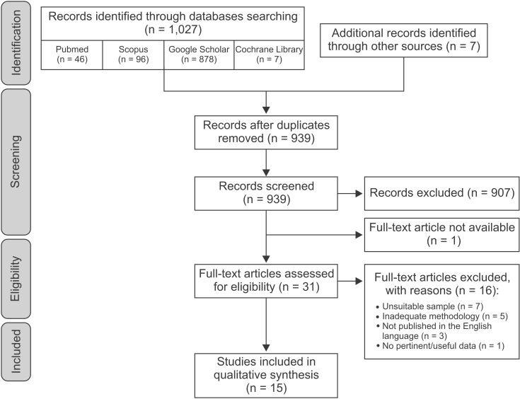
Search strategy

An electronic literature search was carried out on the following databases: MEDLINE, Google Scholar, Scopus, and the Cochrane Library. To identify the relevant studies the following search terms were used: "obstructive sleep" AND (apnea OR apnoea) AND (predict* OR outcome OR effect OR efficacy) AND ("oral appliance" OR "mandibular advancement device" OR "mandibular repositioning appliance") AND (craniofacial OR skeletal OR cephalometr*). The reference lists of all relevant publications were checked for additional studies. Searches were updated to December 2014.

Screening and study selection

In the first phase of selection, duplicates were removed and irrelevant articles were excluded by reviewing the titles and abstracts from the search results. In the next phase, the full texts of potentially relevant papers were evaluated to determine if they met the eligibility criteria. The inclusion criteria were:
  • Type of study: Randomized or non-randomized controlled trials, cohort or case-control studies (with a minimum sample size of 10 patients in each group) addressing the research question of the predictive value of cephalometric analysis in oral appliance treatment outcomes in adult OSA patients. Studies had to be published in English in an international peer-reviewed literature

  • Population: Male or female adult patients (≥ 18 years old) with a polysomnographic diagnosis of OSA (i.e., 5 or more respiratory events [apneas or hypopneas] per hour of sleep)

  • Intervention: Treatment with any MAD for OSA The exclusion criteria were:

  • Lack of a clear description of inclusion/exclusion criteria

  • Previous and/or current surgical or pharmacological interventions

  • Limitation to severe OSA patients

Two reviewers (DRI, SIP) independently screened paper titles and abstracts, with access to full texts where necessary to select studies into the review. Disagreements were resolved by discussion. Where resolution was not possible, a third reviewer (GAB) was consulted. The selected studies underwent data extraction and quality assessment.

Data extraction and quality assessment of selected studies

Two review authors (DRI, SIP) independently performed the data extraction. Extracted data included: first author, year of publication, study design, sample size, patient demographic and clinical characteristics, MAD type, degree of protrusion and vertical opening, time interval between polysomnographic evaluations, outcome (i.e., cephalometric parameters that differed significantly between good and poor responders). Selected studies were assigned to a class of evidence according to the classification of study designs by Jovell and Navarro-Rubio (Table 1).12

Data synthesis

A narrative synthesis was carried out. Data were sorted according to MAD type (1-piece or 2-piece appliances). Given the lack of homogeneity in the study settings, a quantitative synthesis seemed inappropriate. Therefore no meta-analysis was performed.

RESULTS

The electronic database search and the review of the relevant publication reference lists yielded 939 potentially relevant titles and abstracts after duplicates were removed from a total of 1,034 records. Following the first phase of evaluation, 907 publications were rejected based on the title and the abstract. One further study was excluded because full text was not obtained by searching paper and digital format sources, nor after attempting to contact the authors by e-mail correspondence.13 In the second phase, analysis of the full text of the remaining 31 studies led to the exclusion of 16 additional publications. Fifteen studies were therefore selected for the systematic review.589101415161718192021222324 The relevant data from each study are reported in Table 2. The PRISMA flow diagram (Figure 1) shows the number of articles reviewed in each phase of this systematic review.25
Cephalometric variables that were analyzed in the selected studies are listed in Table 3. The landmarks and reference lines necessary to define these parameters are shown in Figure 2.

Cephalometric skeletal measurements

Cranial base

  • 1-piece MAD: Svanholt et al.24 found that the distance between the sella turcica and the deepest point in the posterior cranial fossa was lower in the group that responded positively to MAD therapy.

  • 2-piece MAD: Both cranial base and anterior cranial base lengths were reported to be non-predictive of MAD treatment outcome.5822 Two studies addressed the predictive value of cranial base angle with contrasting results; one found an increased cranial base angulation to be predictive of MAD treatment success22 and the other did not.5

Sagittal jaw relationship

  • 1-piece MAD: Data concerning sagittal jaw relationship were controversial. SNA and ANB were recognized either as predictive of treatment success when decreased1024 or as non-predictive101418 of treatment outcome. SNB was found to be non-predictive1418 or predictive of treatment success when increased10 or decreased.24

  • 2-piece MAD: The majority of papers indicated that sagittal jaw relationship cephalometric parameters were not suitable for predicting treatment outcome (SNA,58919202122 SNB,519202122 ANB,517202122 Wits appraisal20). However, a decreased SNB value89 or an increased ANB value8 were occasionally reported as predictive of treatment success.

Vertical craniofacial dimensions

  • 1-piece MAD: Vertical craniofacial dimension parameters were usually recognized as non-predictive of treatment outcome,1424 with the exception of lower anterior face height,18 lower posterior face height,14 and mandibular plane angle,18 which were occasionally identified as predictive of treatment success when decreased.

  • 2-piece MAD: Saddle angle, articular angle, gonial angle, palatal plane angle, posterior face height, and the ratio between upper anterior face height and lower anterior face height were not useful for predicting MAD treatment outcomes.920212223 The majority of the papers ascribed a non-predictive role to anterior face height, upper anterior face height, lower anterior face height, and the ratio between posterior face height and anterior face height, but a lower anterior face height value9 as well as higher values of upper anterior face height8 and of the ratio of posterior face height to anterior face height23 were also occasionally found in good responders. Data on the predictive role of mandibular plane angle were conflicting: 3 out of 7 studies found it to be non-predictive of treatment outcomes,8921 2 studies indicated an increased value as a predictor of treatment success,522 and 2 studies reported a decreased value as a predictor of treatment success.2023

Maxillary and mandibular lengths

  • 1-piece MAD: Endo et al.10 found that maxillary and mandibular lengths were not predictive of MAD treatment outcome.

  • 2-piece MAD: There was general agreement that maxillary and mandibular lengths, ramus height, and corpus length were not suitable for prediction of MAD treatment outcome.517202122

Hard palate

  • 1-piece MAD: Hard palate length did not appear to be predictive of MAD treatment outcome.1016

  • 2-piece MAD: Ng et al.22 claimed that hard palate length was not predictive of MAD treatment outcome.

Hyoid bone

  • 1-piece MAD: Endo et al.10 found decreased values of MP-H, H-Me, and ANS-H to be predictive of MAD treatment success, whereas Kim et al.14 supported a non-predictive role of H-Me and ANS-H.

  • 2-piece MAD: There was general agreement that H-RGN, C3ia-H, H-Go, Go-H-Me were not suitable for predicting MAD treatment outcomes.9172021 Rose et al.23 found that decreases in H-Me and ANS-H were predictive of treatment success, while Hoekema et al.8 did not. Data on MP-H were conflicting. Five out of 7 studies found it to be non-predictive,5891921 while the two others reported that decreased MP-H20 and increased MP-H23 were predictive of treatment success.

Cervical vertebrae

  • 1-piece MAD: Svanholt et al.24 found that the prevalence of and severity of morphological deviations of the upper spine were greater in the group of patients that failed MAD.

  • 2-piece MAD: Neither the linear distance between RGN and C3ia21 nor the craniocervical angle2122 seemed to be predictive of MAD treatment outcome.

Cephalometric dental measurements

  • 1-piece MAD: Studies investigating dental parameters agreed on the lack of predictive value of these measurements for treatment outcome.1424

  • 2-piece MAD: Overjet and overbite were widely recognized as non-predictive of treatment success.917202223 However, Hoekema et al.8 found that increased overjet and overbite were prognostically favorable. Increased maxillary molar height also seemed to be associated with a better chance of successful treatment.17 None of the other cephalometric dental measurements exhibited predictive value.2123

Cephalometric soft tissue measurements

Soft palate

  • 1-piece MAD: Soft palate depth and thickness and palatal angle were identified as non-predictive of treatment outcomes,1014 whereas data concerning soft palate length, which, when decreased,16 was recognized both as non-predictive of treatment outcome1415 and predictive of treatment success, were controversial.

  • 2-piece MAD: Shen recognized soft palate thickness as non-predictive of treatment outcome.9 The role of soft palate length was controversial. Two out of 6 studies found that decreased soft palate length was predictive of treatment success922 while 4 did not.8192021

Tongue

  • 1-piece MAD: There was general agreement that the cephalometric variables of tongue length, height, and cross-sectional area were not useful for predicting MAD treatment outcomes.101415

  • 2-piece MAD: Tongue-related cephalometric variables were widely recognized as non-predictive of MAD treatment outcome9172223 with the sole exception of tongue/oral enclosure cross-sectional ratio, which Mostafiz et al.21 found was increased in complete responders.

Epiglottis

  • 1-piece MAD: Only 1 study examined the predictive role of the distance between ANS and Eb in treatment outcomes, finding that the greater this distance, the less effective the treatment.10

  • 2-piece MAD: No study investigated the predictive role of epiglottis parameters.

Upper airway

  • 1-piece MAD: Upper airway parameters were unanimously recognized as non-predictive of MAD treatment outcome.101416

  • 2-piece MAD: Neither vertical length nor cross-sectional area were found to be predictive of outcome. 91720 Concerning the upper airway widths, only 1 study (Shen et al.9) found significantly decreased retroglossal width in good responders. The majority showed a non-predictive role for retropalatal width, although Mehta et al.5 and Liu et al.17 found an increased retropalatal space in good and poor responders, respectively.

Facial contours

  • 1-piece MAD: Kim et al.14 found no differences in profile measurements between good and poor responders.

  • 2-piece MAD: No profile measurements were found to be predictors for treatment outcome with 2-piece MAD.20

DISCUSSION

MADs are increasingly used for treatment of mild to moderate OSA because they provide a less invasive, more comfortable, and less costly alternative to nCPAP. Nevertheless, MADs are not as efficacious as nCPAP, and the treatment success rates range from 42% to 65%.7 As certain craniofacial characteristics, including reduced posterior airway space, abnormally long soft palate, and low position of the hyoid, are commonly found in OSA patients it seems reasonable to assume that the efficacy of MAD may relate to morphological factors.26
Several modalities for assessing upper airway morphology have been recommended, including magnetic resonance, nasopharyngoscopy (in an awake state or during drug-induced "sleep"), computed tomography, and lateral cephalometry.89272829 Cephalometery is low-cost, simple, and widely available, and these advantages may offset the disadvantages of the cephalogram being a 2-dimensional projection of a 3-dimensional structure that is performed in an awake state and in an upright position whereas the pathology of OSA arises with the patient lying down during sleep. Therefore, we aimed to provide a systematic review of cephalometric parameters predictive of MAD treatment outcome.

Summary of the evidence

Within the limitations of the selected studies, some consideration can be made with regard to cephalometric parameters predictive of MAD treatment response. Data on the skeletal cephalometric measurements were conflicting. Among the cranial base cephalometric values, cranial base angle and the distance between the sella turcica for 2-piece MADs and the deepest point in the posterior cranial fossa for 1-piece MADs were suggested as possible prognostic factors. However, the data are still too limited to draw any conclusion.
The lack of predictive value of sagittal jaw relationships (SNA, SNB, SN-Pg, ANB, Wits appraisal) as well as linear parameters related to vertical jaw dimensions (anterior face height, lower anterior face height, lower posterior face height, upper anterior face height, ratio between posterior face height and anterior face height) was almost unanimously confirmed. On the other hand, the mandibular plane angle was associated with widely conflicting results, with some studies reporting a non-predictive role and other studies reporting both increased and decreased predictive values for treatment success. It is therefore impossible to draw any definite conclusion on this parameter based on the available data. Hyoid bone position also showed widely conflicting results, with some studies reporting a non-predictive role for MP-H and others reporting both increased and decreased predictive value for treatment success. Available data for H-Me and ANS-H were rather limited. Therefore, the prognostic value of hyoid bone parameters needs further investigation. A possible predictive role of upper spine morphological deviations for 1-piece MAD was suggested. Further study will be required to evaluate these findings.
With regard to dental cephalometric parameters, overjet and overbite, although being reported as predictive of MAD treatment outcome by Hoekema et al.8 alone, were mostly considered unlikely to carry any prognostic significance. Furthermore Liu et al.17 found that in 2-piece MADs, efficacy was decreased with greater eruption of the maxillary molars. However, being as this study was the only one examining this parameter, the recognition of a predictive role for the distance between the maxillary first molar and Frankfort plane seems premature. Therefore, further research on this parameter is also warranted.
Cephalometric soft tissue measurements did not seem to be very useful in predicting MAD treatment response. Soft palate length was recognized both as predictive of treatment success when decreased and as not significantly different between good and poor responders. Therefore, prediction of treatment outcome based on this measurement is not yet unjustified and will require further investigation. Greater ratio between tongue and oral cross sectional area21 and shorter distance between Eb and ANS10 have been shown to be associated with treatment success in 2- and 1-piece MAD, respectively; however additional studies will be needed to confirm these results. Although some studies have suggested a predictive role for retropalatal and retroglossal widths in treatment with 2-piece MAD, the results were controversial. The inconsistency of predictive values of upper airway cephalometric parameters was foreseeable because cephalometry is not the preferred imaging technique for evaluation of these anatomic structures. Indeed, there appears to be a strong linear relationship between 2-dimensional cephalometric and 3-dimensional computed tomography reconstructions for tongue, soft palate, and nasopharynx, while the configuration of oropharynx and hypopharynx appears to be less consistent.30

Limitations

Study level

The question of the predictive role of cephalometric parameters has not been addressed by randomized clinical trials because the variables of interest are not under the control of the investigators. The inclusion of observational studies therefore appeared necessary. The study design adopted in all of the studies selected for this review was case-control (either prospective and retrospective) rather than cohort: 2 or 3 groups differing in MAD treatment outcome (good and poor responders or nonresponders, partial and complete responders) were prospectively or retrospectively identified and compared on the basis of cephalometric parameters. As a result, the findings score low in terms of strength of evidence. A selection bias may have occurred because the sample was not randomly selected. Moreover in some of the prospective studies patients were included after they had been allowed to choose whether or not they wanted to participate or after they had decided entirely for themselves not to accept treatment with nCPAP (self-selection bias).1819 Finally, a few studies reported that some patients dropped out (attrition bias).5819
MAD treatment success has been associated with female sex, a younger age, a lower body mass index (BMI), and a lower baseline apnea-hypopnea index (AHI) but, due to the design of the studies included in this review, these baseline characteristics could not be properly addressed and may actually show systematic differences between good responders and poor responders (confounding bias).1722 In the 13 studies that reported male-female ratio, males had considerably more representation, ranging from 67.9% to 100% of the sample.591415161718192021222324 With the exception of the study by Mehta et al.,5 none of the others investigated the differences in sex distribution between good responders and poor responders. Six studies found no age difference between good and poor responders,51014151924 while 4 studies found age to be significantly lower in good responders. 17202122 Eight studies failed to find a predictive role for BMI,5,10141519202223 while 3 studies reported lower BMI in good responders.82124 Only 1 paper addressed the issue of weight gain or loss between the initial and the final evaluations.19 Six out of 14 studies did not account for potential differences in baseline AHI,81618192023 whereas comparison between good and poor responders in terms of AHI or apnea index led to significant differences in 2 studies, with lower values associated with treatment success,1421 and no significant differences in 6 studies.5910151722
With regard to observer bias, only 3 studies reported that the operator who performed the cephalometric analysis or the polysomnography was blinded to the treatment outcome.51422 None of the selected studies included a power analysis or a sample size calculation. The presence of potentially underpowered studies could lead to inconsistent or misleading results, because they may find no significant difference between study samples even in the presence of a real difference in the general population.

Review level

The findings of the present systematic review were also limited by the heterogeneity in the methodology of the included studies. Respiration was analyzed in terms of AHI in the majority of the investigations, but some of the earlier studies evaluated the respiratory disturbance index (RDI).19 The definition of sleep apnea was based either on AHI > 5, AHI ≥ or > 10, AHI ≥ or > 15, or RDI ≥ 10. Success criteria included RDI reduction of ≥ 50% and RDI ≤ 20, AHI < 5, AHI < 15, or AHI reduction of > 50% alone or in association with AHI < 20, < 10, or < 5. A certain degree of variability was present with respect to the type of MAD (monobloc or two pieces), as well as to the amount of protrusion (from 50 or 60% of maximum protrusion to the most advanced position without causing any discomfort) or vertical opening (ranging from "minimum" to 15 mm). The time interval between diagnostic and with-appliance polysomnography ranged from few weeks to several months. The cephalometric parameters also varied substantially between different studies. Because of the lack of homogeneity among the studies, any attempt to summarize the study results seemed unjustified. This, a narrative synthesis was carried out without performing a meta-analysis.
No "grey literature" or articles in non-English languages were included in the present review. Therefore the review is also subject to publication bias and language bias.

CONCLUSION

This review found controversial and limited data on the predictive role of certain cephalometric parameters for MAD treatment outcome. Therefore selection criteria for suitable candidates for MAD treatment by cephalometric analysis are currently inadequate. Although no definitive clinical recommendations can be made, this systematic review highlights the methodological weaknesses of the currently available studies, analyzes the possible reasons for their discordant results, and encourages and guides further research in this field. Prospective cohort studies in large samples with cephalometric prediction made prior to the MAD construction are required to clarify the clinical utility of lateral cephalometry for adult OSA patients who are considering oral appliances as a therapeutic option.

Notes

The authors report no commercial, proprietary, or financial interest in the products or companies described in this article.

References

1. Epstein LJ, Kristo D, Strollo PJ Jr, Friedman N, Malhotra A, Patil SP, et al. Clinical guideline for the evaluation, management and long-term care of obstructive sleep apnea in adults. J Clin Sleep Med. 2009; 5:263–276. PMID: 19960649.
2. Skomro RP, Kryger MH. Clinical presentations of obstructive sleep apnea syndrome. Prog Cardiovasc Dis. 1999; 41:331–340. PMID: 10406327.
crossref
3. Sullivan CE, Issa FG, Berthon-Jones M, Eves L. Reversal of obstructive sleep apnoea by continuous positive airway pressure applied through the nares. Lancet. 1981; 1:862–865. PMID: 6112294.
crossref
4. Waldhorn RE, Herrick TW, Nguyen MC, O'Donnell AE, Sodero J, Potolicchio SJ. Long-term compliance with nasal continuous positive airway pressure therapy of obstructive sleep apnea. Chest. 1990; 97:33–38. PMID: 2403899.
crossref
5. Mehta A, Qian J, Petocz P, Darendeliler MA, Cistulli PA. A randomized, controlled study of a mandibular advancement splint for obstructive sleep apnea. Am J Respir Crit Care Med. 2001; 163:1457–1461. PMID: 11371418.
crossref
6. Kushida CA, Morgenthaler TI, Littner MR, Alessi CA, Bailey D, Coleman J Jr, et al. Practice parameters for the treatment of snoring and Obstructive Sleep Apnea with oral appliances: an update for 2005. Sleep. 2006; 29:240–243. PMID: 16494092.
crossref
7. Ferguson KA, Cartwright R, Rogers R, Schmidt-Nowara W. Oral appliances for snoring and obstructive sleep apnea: a review. Sleep. 2006; 29:244–262. PMID: 16494093.
crossref
8. Hoekema A, Doff MH, de Bont LG, van der Hoeven JH, Wijkstra PJ, Pasma HR, et al. Predictors of obstructive sleep apnea-hypopnea treatment outcome. J Dent Res. 2007; 86:1181–1186. PMID: 18037652.
crossref
9. Shen HL, Wen YW, Chen NH, Liao YF. Craniofacial morphologic predictors of oral appliance outcomes in patients with obstructive sleep apnea. J Am Dent Assoc. 2012; 143:1209–1217. PMID: 23115150.
crossref
10. Endo S, Mataki S, Kurosaki N. Cephalometric evaluation of craniofacial and upper airway structures in Japanese patients with obstructive sleep apnea. J Med Dent Sci. 2003; 50:109–120. PMID: 12715927.
11. Saffer F, Lubianca JF, Rösing C, Dias C, Closs L. Predictors of success in the treatment of obstructive sleep apnea syndrome with mandibular repositioning appliance: a systematic review. Int Arch Otorhinolaryngol. 2015; 19:80–85. PMID: 25992156.
crossref
12. Jovell AJ, Navarro-Rubio MD. Evaluation of scientific evidence. Med Clin (Barc). 1995; 105:740–743. PMID: 8523956.
13. Liu Y, Lowe AA. Factors related to the efficacy of an adjustable oral appliance for the treatment of obstructive sleep apnea. Chin J Dent Res. 2000; 3:15–23. PMID: 11314530.
14. Kim YK, Kim JW, Yoon IY, Rhee CS, Lee CH, Yun PY. Influencing factors on the effect of mandibular advancement device in obstructive sleep apnea patients: analysis on cephalometric and polysomnographic parameters. Sleep Breath. 2014; 18:305–311. PMID: 23928957.
crossref
15. Lee CH, Mo JH, Choi IJ, Lee HJ, Seo BS, Kim DY, et al. The mandibular advancement device and patient selection in the treatment of obstructive sleep apnea. Arch Otolaryngol Head Neck Surg. 2009; 135:439–444. PMID: 19451462.
crossref
16. Lee CH, Kim JW, Lee HJ, Seo BS, Yun PY, Kim DY, et al. Determinants of treatment outcome after use of the mandibular advancement device in patients with obstructive sleep apnea. Arch Otolaryngol Head Neck Surg. 2010; 136:677–681. PMID: 20644062.
crossref
17. Liu Y, Lowe AA, Fleetham JA, Park YC. Cephalometric and physiologic predictors of the efficacy of an adjustable oral appliance for treating obstructive sleep apnea. Am J Orthod Dentofacial Orthop. 2001; 120:639–647. PMID: 11742309.
crossref
18. Marklund M, Franklin KA, Stenlund H, Persson M. Mandibular morphology and the efficacy of a mandibular advancement device in patients with sleep apnoea. Eur J Oral Sci. 1998; 106:914–921. PMID: 9786320.
crossref
19. Menn SJ, Loube DI, Morgan TD, Mitler MM, Berger JS, Erman MK. The mandibular repositioning device: role in the treatment of obstructive sleep apnea. Sleep. 1996; 19:794–800. PMID: 9085488.
crossref
20. Milano F, Billi MC, Marra F, Sorrenti G, Gracco A, Bonetti GA. Factors associated with the efficacy of mandibular advancing device treatment in adult OSA patients. Int Orthod. 2013; 11:278–289. PMID: 23867336.
crossref
21. Mostafiz W, Dalci O, Sutherland K, Malhotra A, Srinivasan V, Darendeliler MA, et al. Influence of oral and craniofacial dimensions on mandibular advancement splint treatment outcome in patients with obstructive sleep apnea. Chest. 2011; 139:1331–1339. PMID: 21292761.
crossref
22. Ng AT, Darendeliler MA, Petocz P, Cistulli PA. Cephalometry and prediction of oral appliance treatment outcome. Sleep Breath. 2012; 16:47–58. PMID: 21424283.
crossref
23. Rose E, Lehner M, Staats R, Jonas IE. Cephalometric analysis in patients with obstructive sleep apnea. Part II: Prognostic value in treatment with a mandibular advancement device. J Orofac Orthop. 2002; 63:315–324. PMID: 12198746.
24. Svanholt P, Petri N, Wildschiødtz G, Sonnesen L. Influence of craniofacial and upper spine morphology on mandibular advancement device treatment outcome in patients with obstructive sleep apnoea: a pilot study. Eur J Orthod. 2015; 37:391–397. PMID: 25351571.
crossref
25. Liberati A, Altman DG, Tetzlaff J, Mulrow C, Gøtzsche PC, Ioannidis JP, et al. The PRISMA statement for reporting systematic reviews and meta-analyses of studies that evaluate healthcare interventions: explanation and elaboration. BMJ. 2009; 339:b2700. PMID: 19622552.
crossref
26. Hoekema A, Hovinga B, Stegenga B, De Bont LG. Craniofacial morphology and obstructive sleep apnoea: a cephalometric analysis. J Oral Rehabil. 2003; 30:690–696. PMID: 12791153.
crossref
27. Sanner BM, Heise M, Knoben B, Machnick M, Laufer U, Kikuth R, et al. MRI of the pharynx and treatment efficacy of a mandibular advancement device in obstructive sleep apnoea syndrome. Eur Respir J. 2002; 20:143–150. PMID: 12166562.
crossref
28. Chan AS, Lee RW, Srinivasan VK, Darendeliler MA, Grunstein RR, Cistulli PA. Nasopharyngoscopic evaluation of oral appliance therapy for obstructive sleep apnoea. Eur Respir J. 2010; 35:836–842. PMID: 19797130.
crossref
29. Sam K, Lam B, Ooi CG, Cooke M, Ip MS. Effect of a non-adjustable oral appliance on upper airway morphology in obstructive sleep apnoea. Respir Med. 2006; 100:897–902. PMID: 16219453.
crossref
30. Lowe AA. Can we predict the success of dental appliance therapy for the treatment of obstructive sleep apnea based on anatomic considerations? Sleep. 1993; 16(8 Suppl):S93–S95. PMID: 8178042.
crossref
Figure 1

Flow chart of literature search and study selection.

kjod-45-308-g001
Figure 2

Diagrammatic representation of landmarks and reference lines. A, Subspinale; ANS, anterior nasal spine; Ar, articulare; B, supramentale; Ba, basion; BT', base of tongue; C2, tangent point on the dorsal surface of C2 vertebra to a line from C4; C3ia, C3 vertebra inferoanterior; C4, C4 vertebra inferoposterior; Cd, condylion; D, the deepest point in posterior cranial fossa; E line, Ricketts-E line; Eb, epiglottis base; Et, tip of epiglottis; FP, Frankfurt Plane; Gn, gnathion; Go, gonion; H (Hy), hyoidale; Iop: internal occipital protuberance; L1i, lower incisor tip; LL, lower lip; Me, menton; MP, mandibular plane; N, nasion; N', soft tissue nasion; OP, occlusal plane; Or, orbitale; P (Ut), soft-palate tip; Phw, posterior pharyngeal wall; PPW', posterior pharyngeal wall interseption; PNS, posterior nasal spine; Po, porion; Pog, pogonion; Pog', soft tissue pogonion; PP, palatal plane; Prn, nasal tip; R, roof of pharynx; RGN, retrognathion; S, sella; SN, S-N plane; Spt, tangent point on a line parallel to PNS-P on the dorsal surface of the soft palate at the maximum width; TT, tongue tip; U6c, maxillary first molar point; U1i, upper incisor tip; UL, upper lip.

kjod-45-308-g002
Table 1

Levels of scientific evidence

kjod-45-308-i001

Derived from Jovell and Navarro-Rubio.12

Table 2

Summary of the main characteristics of the 15 studies selected for the systematic review

kjod-45-308-i002

Values are presented as number only, number (%), or mean ± standard deviation (range).

*According to the classification of study designs by Jovell and Navarro-Rubio.12

n, Number; -, not reported; GR, good responders; PR, poor responders; CR, complete responders; PaR, partial responders; NR, nonresponders; BMI, body mass index; AHI, apnea hypopnea index; bAHI, baseline AHI; h, hour; AI, apnea index; RDI, respiratory disturbance index; AHI red, AHI reduction; MAD, mandibular advancement device.

Suitability for MAD treatment, good dental health, > 10 teeth/dental arch, no periodontal disease, TMJ dysfunction or exaggerated gag reflex.

See Table 3 for the definition of cephalometric variables.

Table 3

Summary of cephalometric variables

kjod-45-308-i003

See Figure 2 for definitions of landmarks and reference lines used in this table.

TOOLS
Similar articles