

 PDF
PDF ePub
ePub Citation
 Print
Print


INTRODUCTION
Orthodontic correction of skeletal Class III malocclusion is a particularly challenging aspect of orthodontics. For severe skeletal Class III deformities, surgical orthodontic treatment (orthognathic surgery) is one of the best options. The most important step in managing the maxillofacial trauma that follows orthognathic surgery is intermaxillary fixation (IMF). IMF helps maintain newly created maxillomandibular relations and prevents skeletal relapse during the bone healing process.
Traditionally, IMF has been achieved by means of arch bars or interdental eyelet wiring. These techniques have intrinsic disadvantages; for example, they are time-consuming and pose a risk of needle injury to the surgeon.1 For these reasons, orthodontic surgical wire with hooks have been used for IMF and traction since some time. However, in orthognathic surgery cases with large setback movement or major counterclockwise rotation of the mandible, surgical wire fixation can lead to a significant extrusive load on the anterior teeth. This vertical force persists until muscle function adapts to the postoperative mandibular morphology and occlusion.2 To reduce the extrusive load on anterior teeth during IMF, Thota and Mitchell3 employed miniscrew fixation in the anterior region of the jaw.
Initially, bone-anchored screws were introduced as a means of bone fracture fixation.4 Currently, intraoral miniscrews are often used as a mandibular-fracture treatment alternative to arch bars during IMF.1,5,6 Miniscrews are also used for skeletal anchorage during orthodontic tooth movement.7,8,9 Ueki et al.10 reported significant differences in occlusal plane and convexity changes in groups with and without miniscrews for IMF. However, there have been no reports on tooth movement in relation to the use of miniscrews for IMF after orthognathic surgery.
The purpose of the present study was (1) to investigate the skeletal and dental changes that occurred during postoperative orthodontic treatment involving the use of miniscrews for IMF and (2) to compare the use of miniscrew fixation and surgical archwire fixation for IMF in adult patients who had Class III malocclusion and had undergone maxillomandibular surgery.
MATERIALS AND METHODS
Subjects
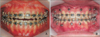
This retrospective study included patients who underwent orthognathic surgery and post-surgical orthodontic treatment between January 2012 and January 2013 at the Department of Oral and Maxillofacial Surgery, Pusan National University Dental Hospital. The patients consisted of 74 adults (39 male and 35 female patients; mean age, 21.7 ± 3.3 years) who had been diagnosed with skeletal Class III malocclusion and who subsequently underwent 2-jaw surgery (LeFort I osteotomy, bilateral sagittal split ramus osteotomy with rigid fixation). The patients were divided into two groups according to fixation type (Figure 1): conventional fixation using surgical archwires with hooks (group 1: wire fixation group, 20 male and 17 female patients; mean age, 21.6 ± 3.0 years) and miniscrew fixation using orthodontic miniscrews (group 2: screw fixation group, 19 male and 18 female patients; mean age, 22.0 ± 3.6 years). The inclusion criteria were as follows: healthy physical condition, lack of severe facial asymmetry (≤ 3 mm of Menton deviation from facial midline), no temporomandibular dysfunction symptoms or degenerative joint disease on examination, and skeletal Class III deformities. All the patients had undergone pre-surgical orthodontic treatment and now had well-aligned arches.
The orthodontic treatment entailed the use of a 022-slot preadjusted appliance: 0.019 × 0.025 stainless steel wire in group 1 and rigid rectangular stainless steel wire (minimum 0.017 × 0.025) in group 2. After 6 weeks, postoperative orthodontic treatment was resumed, according to protocols applied equally to both groups. This study was reviewed and approved by the Ethics Committee of Pusan National University Dental Hospital (PNUDH-2013-024).
Data acquisition
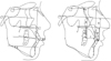
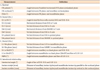
Lateral cephalograms were obtained immediately after surgery (T0), 3 months after surgery (T1), and 6 months after surgery (T2). Definitions of the landmarks, reference planes, and skeletodental variables are provided in Figure 2 and Table 1. The X axis has been defined as the Frankfort horizontal (FH) line and the Y axis as the line perpendicular to the X axis and passing through the Nasion. The intraskeletal reference planes were the palatal plane, the mandibular plane, and the maxillary occlusal plane.11
Assessment by lateral cephalogram
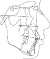
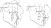
The angular and linear measurements are illustrated in Figure 3 and defined in Table 2. FH-palatal, FH-occlusal and FMA were measured to assess the extent of skeletal movement during post-surgical orthodontic treatment (Figure 3).
To assess the extent of dental changes to the maxilla and mandible during postoperative orthodontic treatment, the horizontal and vertical distances from the incisal edges of the maxillary central incisors were measured, as were the distances from the mesiobuccal cusp tips of the maxillary first molars to the vertical and horizontal reference planes (U1-X, U1-Y, U6-X, U6-Y, U1-palatal, U6-palatal). The perpendicular distances from mandibular central incisors and the mesiobuccal cusp tips of the mandibular first molars to the mandibular plane were also measured (Figure 4). The incisor inclination of the maxilla (U1 to SN, U1 to MxOP) and the mandible (L1 to Mn plane, L1 to MnOP) and the positional changes of the incisal edges (interincisal angle, incisor overjet, incisor overbite) were measured (Figure 5). The cephalograms were digitized by a single operator using the V-Ceph program (version 6.0; CyberMed, Seoul, Korea). To evaluate intraoperator variability, 10 variables for each patient were reassessed by the same operator after 3 weeks. Because no statistically significant differences (p > 0.05) were observed, the first set of measurements was accepted.
Statistical analysis
Statistical analyses were conducted using IBM SPSS Statistics software version 21.0 (IBM Co., Armonk, NY, USA); p-values less than 0.05 were considered statistically significant. Time-dependent changes in cephalometric measurement variables indicating dental and skeletal changes were compared with one-way analysis of variance (ANOVA) and Duncan's post-hoc test in each group. Independent sample t-tests were used to compare the differences between the groups.
The intraassessor reproducibility of the measurements was determined by evaluating the variables of 10 randomly selected patients twice, at 3-week intervals, according to the intraclass correlation coefficient (ICC). The mean ICC was 0.965 in the 95% confidence interval.
RESULTS
The mean and standard deviation values, as determined from all of the cephalometric measurements immediately after surgery (T0), 3 months after surgery (T1), and 6 months after surgery (T2), were determined. The differences between the T0 and T1 stages (ΔT1; T0-T1) and the T0 and T2 stages (ΔT2; T0-T2) were analyzed.
Skeletal changes
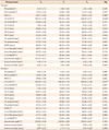
The FH-palatal plane, FH-occlusal plane, and FMA, as referenced in the measurement of skeletal changes during post-surgical orthodontic treatment, showed no significant time-dependent differences in either group (Table 2). There was no significant skeletal relapse 6 months after surgery (p > 0.05).
Dental changes in the wire-fixation group (group 1; Table 3)
Vertical changes
In group 1, U1 to SN increased significantly (p < 0.05) but U1 to MxOP tended to decrease. For both U1-Y and U1-palatal, there was no significant difference in the vertical position at each time point. Likewise, the U6-Y and U6-palatal values, indicating the vertical position of the maxillary first molar, did not change with time.
Horizontal changes
U1-X and U6-X, indicating the anteroposterior dental position, showed no significant differences over time (p > 0.05).
Changes in interdental relationship
The interincisal angle and incisor overjet tended to decrease but the decreases were not statistically significant. ANOVA analysis of time-course changes showed that the incisor overbite value increased at each measurement point and that the differences were significant (p < 0.05). The Duncan post-hoc test was used to determine the differences between T0 and T1.
Dental changes in screw fixation group (group 2)
Vertical changes
In the screw fixation group (group 2), U1 to SN increased significantly (p < 0.05) but U1 to MxOP did not. There were no significant differences in the U1-Y or U1-palatal values at any of the time points. Likewise, the U6-Y or U6-palatal values, indicating the vertical position of the maxillary first molar, showed no significant change.
Horizontal changes
U1-X, representative of the anterioposterior position of the maxillary incisor edge, showed no significant differences over time. U6-X tended to decrease, but the decrease was not statistically significant.
Changes in interdental relationship
The interincisal angle and incisor overjet showed a tendency to decrease but the decreases were not statistically significant. However, changes in the incisor overbite showed a statistically significant increase between T0 and T1 (p < 0.05), with the values reflecting those of the Duncan post-hoc test differences between T0 and T1, as was the case in the wire fixation group (group 1).
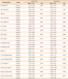
Dental-change differences for groups 1 and 2 (Table 4)
Vertical change differences
The results of an intergroup comparison are listed in Table 4. U1 to SN increased significantly in ΔT1 and the extent of change was similar. However, U1 to MxOP did not show statistically significant differences (p > 0.05). In group 1, U1-Y and U1-palatal, indicating the mean vertical position of the maxillary incisor, showed smaller differences than in group 2 (p < 0.05). However, this fact is not meaningful because an intragroup comparison (Table 4) showed no significant differences over time. Finally, the changes to U6-Y and U6-palatal (ΔT1 [T0-T1], ΔT2 [T0-T2]), indicating the mean vertical positional change of the maxillary first molar, did not show statistically significant differences.
Horizontal change differences
The differences in the horizontal changes (U1-X, U6-X in both groups) between the groups, as determined by independent samples t-test, were not statistically significant (p > 0.05).
Interdental-relationship change differences
Considering the interincisal angle and incisor overjet, there were no significant differences between the groups in ΔT1 or ΔT2. The changes to incisor overbite manifested as statistically significant time-dependent increases in both groups (Table 4), although the extent of increase differed according to the group (p < 0.05, Table 4). The incisor overbite in group 1 in ΔT1 (1.64 ± 1.48) was smaller than that in group 2 (0.87 ± 1.22); the difference was small but statistically significant (p < 0.05).
DISCUSSION
With the advent of the use of screws for IMF, many surgeons have shifted to that technique to avoid or reduce the disadvantages of using the traditional arch bar and wiring techniques. Certainly, the use of screws for IMF has provided many benefits to patients and surgeons. The dental-change differences according to the fixation method had not been evaluated.
Hovell12 reported maxillary incisor inclination increased at 3 months after surgery, although the inclination decreased significantly with movement of the maxilla immediately after surgery. In the present study, postoperative labial-inclination tendency of the maxillary incisor was seen in both groups, but there was no significant difference in this regard between the groups. Furthermore, there was no significant time-dependent change in any measurements of vertical dental position. The labioversion of the maxillary incisors during the post-surgical orthodontic treatment was caused by the Class III mechanics. Skeletal relapse and compensatory movement of dental structures take place simultaneously after surgical correction of mandibular prognathism.13,14 Furthermore, dental decompensation in skeletal Class III malocclusion had labioversed maxillary incisors due to skeletal and muscular limitations.12 Control of maxillary incisor inclination during postoperative orthodontic treatment is needed.15
Some studies have reported a limited correction in mandibular incisor inclination during surgical orthodontic treatment, as well as difficulty in changing mandibular incisor inclination by more than 5 degrees.12,16,17 In the present study, the measurements for the mandibular dentition did not show statistically significant change between the groups.
No significant time-course interdental-relationship differences were found between the groups for the cephalometric variables evaluated, except for incisor overbite. Incisor overbite increased significantly from T0 to T1 in both groups, and more so in the screw fixation group than in the wire fixation group. We suggest that maintenance of upper teeth position after surgery should continue for 3 months in order to stabilize the bone structure.
Wisth and Isaksen2 reported that all forces are transmitted to all dentitions; the vertical vector causes extrusion of anterior teeth and muscle forces are responsible for changes in incisor inclination. They showed that application of vertical forces during orthodontic treatment can result not only in incisor inclination but also in root resorption and consequent loss of marginal periodontal support for extruded teeth. Baek et al.11 suggested that the maxillary central incisor's direction and extent of relapse (from the vertical position) seem to be more influenced by postoperative orthodontic treatment than is the case for the maxillary first molar, and that post-surgical orthodontic treatment mechanics should be applied to maintain surgical results.
In the present study, increasing tendency of overbite during post-surgical treatment was greater in the wire fixation group than the screw fixation group. We suggest that the use of miniscrew fixation can be beneficial for maintaining surgical results and for minimizing maxillaryincisor extrusion.
We set the surgical occlusion on the basis of proper molar relations, canine relations, overjet and overbite, considering both pre-surgical dentition and the extent of post-surgical dental compensation. The overbite of the surgical occlusion at the surgical treatment objective can be made deeper or shallower according to IMF type. In a surgery-first approach, passive surgical wires can be bonded directly to the tooth surface or ligated into the brackets.15 In our comparison of the two types of fixation appliances there was no significant difference in any dental cephalometric measurement except incisor overbite. Furthermore, the overbite difference was too small to be clinically relevant. Therefore, miniscrew IMF can be viewed as a useful method for first-surgery approach or, in cases where the use of full-size rectangular surgical wire is problematic. On analyzing the use of miniscrews versus surgical archwire in IMF from the crucial perspective of both practical and clinical considerations, we believe that there were no significant differences in the post-surgical dental changes for the two methods.
The use of the miniscrews for IMF provides many benefits to patients and surgeons: 1) quick and easy insertion; 2) compatibility with any plating system; 3) ideal for use in cases of heavily restored teeth; 4) ease of maintenance of gingival health compared with that seen for arch bars and eyelet wires, and 5) easy and painless removal without anesthesia on an out-patient basis.1 Additionally, orthodontic miniscrews can be used as anchors for elastic traction. The most common complication associated with the use of miniscrews is iatrogenic injury to dental roots.19,20,21,22,23 Clinicians need to be aware of this potential complication and take appropriate steps to avoid it.24
CONCLUSION
In the present study, the changes in cephalometric values after 2-jaw orthognathic surgery were compared between two groups of patients, one with miniscrew fixation and the other with wire fixation. The results showed that there were no dental-change differences attributable to one or the other type of fixation, except for incisor overbite. Incisor overbite increased moderately in both groups during the post-surgical orthodontic treatment but the increase was slightly more in the wire fixation group than the miniscrew group. Overall, the results suggest that miniscrew fixation is no less effective clinically than wire fixation. This means that a clinician can choose the fixation type depending on the individual case, e.g., miniscrew fixation may be more useful in a surgery-first approach without orthodontic bracket bonding, or in cases where the use of surgical archwire would be difficult.


 XML Download