

 PDF
PDF ePub
ePub Citation
 Print
Print


INTRODUCTION
The pendulum device is one of the most commonly used conventional distalizing devices.12 However, despite its efficacy in molar distalization, premolar mesial movement and anterior anchorage loss34567 continue to represent an unpleasant problem and require additional treatment time for correction during fixed appliance therapy.
In order to reduce anchorage loss, the use of mini-implants or miniscrews was introduced.8 Based on their low cost, reduced invasiveness, ease of insertion and removal, possibility for immediate loading, and versatility, these devices have become increasingly popular, even in combination with distalizing devices. Paramedian and midpalatal insertions appear to be most suitable for this purpose,910 because placing miniscrews in interradicular sites may require additional radiographic examinations, complex surgical protocols to avoid root damage, and prevent spontaneous retraction of premolars during distalization until the screws are inserted.1112 A recent meta-analysis13 evaluated the efficacy of conventional versus bone-anchored anchorage, showing that both systems were effective for molar distalization but that there were differences in anchorage loss. Conventional and indirect skeletal anchorage showed a certain amount of anchorage loss at the premolars and incisors, whereas these side effects were not seen with direct skeletal anchorage.
For this reason, various modifications of distalizing appliances used in combination with paramedian miniscrews have been developed in recent years.111415161718192021 Many authors141516 have described the effects of a bone-anchored pendulum appliance (BAPA), a modified pendulum in which the palatal arms on the premolar are eliminated. The distal jet appliance was modified into a skeletonized distal Jet appliance,17 in which the Nance button was eliminated but the arms on the premolar were retained; it was later modified into the distal screw appliance (Micerium S.p.A., Avegno, Italy),1118 in which the metallic palatal arms on the premolar were eliminated. Similar devices were the skeletal frog,19 a modified frog appliance without arms on the premolar and without the acrylic palatal button; the intraoral miniscrew implant-supported distalization system (MISDS)20 and the Beneslider,21 which is characterized by midpalatal miniscrews and compressed palatal coils.
Dentoskeletal changes produced by these appliances have been described in the literature. However, there have been few comparisons between skeletal-anchored and conventional distalizing devices.22 Moreover, a comparison between distal jet and implant-supported distal jet devices has recently been published.23 Therefore, the aim of this study was to compare the dentoalveolar and skeletal changes produced by the pendulum appliance (conventional anchorage) and distal screw appliance (skeletal anchorage) in Class II patients. The null hypothesis was that the two appliances would produce similar dental and skeletal changes.
MATERIALS AND METHODS
A sample of 75 patients was retrospectively obtained from two orthodontic dental offices. All patients were selected according to the following criteria:
·Skeletal Class I or mild Class II malocclusion and a bilateral full cusp or end-to-end Class II molar relationship
·Absence of protrusive profile or mandibular retrusion24
·Nonextraction treatment
·Mandibular inclination (sella-nasion-gonion-gnathion [SN-GoGn] angle) less than 37°
·Good-quality radiographs with adequate landmark visualization and minimal or no rotation of the head
From the initial sample, the records of 5 patients in the pendulum group (PA) and 4 patients in the distal screw group (DS) were excluded because their mandibular plane was greater than 37°. An additional 3 patients in the PA group and 3 patients in the DS group were excluded because extraoral traction was used after the distalization phase, either as anchorage support or because of miniscrew failure. Nine patients in the PA group and 8 patients in the DS group were excluded due to poor radiograph quality or incomplete records (Table 1). Thus, the final sample consisted of 43 white patients divided in 2 groups: 24 (10 men, 14 women) with a mean age of 12.2 ± 1.5 years (range, 10 years 5 months to 14 years 2 months) in the PA group, and 19 (9 men, 10 women) with a mean age of 11.3 ± 1.9 years (range, 10 years 4 months to 13 years 7 months) in the DS group. Initial cephalometric characteristics of the patients in the two groups were considered comparable (Table 2). The average amount of Class II molar relationship was 3.5 mm in the PA group and 3.8 mm in the DS group, with mean overjets of 3.9 mm and 4.2 mm, respectively, at the beginning of treatment. Two serial cephalograms for all patients were available at 2 observation times: before treatment (T1) and after distalization (T2). Demographics of observation periods and observation intervals are reported in Table 3.
Clinical management
All patients underwent maxillary molar distalization therapy with either a pendulum or a distal screw appliance.
Dental and skeletal changes during fixed appliance therapy were not taken into consideration in order to highlight the differences between the two appliances during the distalization phase.
Pendulum appliance
The pendulum appliance used in this study was similar to that described by Hilgers.2 The 0.032-inches [in] titanium-molybdenum alloy (TMA) springs were bent parallel to the palatal midline and placed in the lingual sheaths on maxillary first molar bands, exerting approximately 230 g of distalizing force per side, as measured with a dynamometer (Correx; Dentaurum GmbH & Co., Ispringen, Germany). A Nance button was placed on the anterior vault of the palate as anchorage, adding arms on the first premolars and occlusal rests on the second premolars. Palatal TMA arms were removed from the palatal molar sheaths and intraorally reactivated every 6 weeks on average. As recommended by Byloff and Darendeliler,5 uprighting bends were added to the ends of the TMA wire to prevent excessive molar tipping. The appliance was left in situ until a super Class I molar relationship was achieved.
The mean treatment time for distalization was 7 ± 2 months (Table 3).
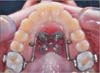
Distal screw appliance
The distal screw appliance is a modified distal jet in which the metallic palatal arms on the premolars, normally used for dental anchorage, are eliminated and the Nance button includes a moldable metal plaque which is used as a guide to insert two miniscrews (Titanium V, 11 mm long, 2.2 mm diameter; Micerium S.p.A., Avegno, Italy) in the palatal vault. The shank was 1.0 mm in diameter, the threaded part had a length of 8.0 mm, and the head featured a hexagonal slot to house the head of the screwdriver or contra-angle handpiece. The two miniscrews (one for each side) were placed in the paramedian region of the anterior palatal vault along a line connecting the first premolars. The head had a flat shape completely included in the Nance button, minimizing the possibility of tongue loadings and reducing the risk of compromising the primary stability. The arms on the first molars were activated as a distal jet, completely compressing the superelastic springs until a force of 240 g was obtained; reactivation was carried out at 4-week intervals.25 Distalization continued until the Class II molar relationship was corrected to a Class I molar relationship.
The mean treatment time for distalization was 9 ± 2 months (Table 3).
Cephalometric analysis
Lateral cephalograms for each patient at T1 and T2 in each treatment group were standardized at the same magnification factor (6% enlargement). Lateral cephalograms were hand traced in random order by one investigator with verification of anatomic outlines and landmark position by a second investigator. In cases of disagreement, the structures in question were retraced to the satisfaction of both. In instances of bilateral structures (e.g., gonial angle and teeth), a single averaged tracing was made. The centroid points of the maxillary first and second molars and the maxillary first premolars were obtained as the midpoint between the greatest mesial and distal convexity of the crowns, as seen on the cephalometric radiographs.3 Table 2 shows the measurements and summary statistics of the pooled sample before initiation of treatment.
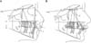
A conventional analysis, including soft tissue, skeletal, and dental measurements (Figure 3), was done.3 Sagittal skeletal measurements were added in order to complete the cephalometric analysis. The Frankfort-mandibular plane angle was not used because of the difficulty of detecting the Porion point; SN-GoGn was used as an indicator of vertical facial dimension. The SN plane and palatal plane (PP) were used as the horizontal reference planes, and the pterygoid vertical (PTV) line, as described by Enlow et al.,2627 was used as the vertical reference plane. The PTV line extends inferiorly from a point (the sphenoethmoidal) located by the intersection of the shadows of the great wings of the sphenoid with the floor of the anterior cranial fossa (representing the boundary between the anterior and posterior portions of the anterior cranial base as well as the boundary between the anterior and middle endocranial fossae) through the inferior point of the pterygomaxillary fissure (Figure 3).
The cephalometric analysis consisted of 25 landmarks, 10 angular measurements, and 15 linear measurements for each tracing (Figure 3). To assess the sagittal changes in jaw relationships, measurements were made from points A and B to the PTV. The PTV line was used to assess the amount of linear horizontal movement, whereas the linear vertical movement was measured on PP. The long axes of the premolar and molar teeth were obtained by drawing a perpendicular line to the midpoint of a line connecting the most convex points on the crowns of these teeth. Angular differences in tooth position were then determined by the inclination of the long axes to the SN plane.3
Statistical analysis
Statistical analysis was performed using SPSS software, version 13.0 (SPSS® Inc., Chicago, IL, USA). Descriptive statistics were calculated for age, duration of treatment, and cephalometric measurements at T1 for the 2 groups. Mean differences and standard deviations were also calculated for the treatment changes between T1 and T2. A Shapiro-Wilk test did not reveal a normal distribution of the tested variables, and nonparametric tests were used for the inferential statistics. In order to compare pre-treatment cephalometric data, an independent sample Mann-Whitney U test was performed between the two groups at T1. No significant differences were found. A paired-data Wilcoxon test was used to identify significant differences in each group between T1 and T2, and a Mann-Whitney U test was used to identify significant differences between groups (Δ = T2 - T1) for each cephalometric variable. Statistical significance was tested at p < 0.05, p < 0.01, and p < 0.001.
Method error
Fifteen randomly selected cephalograms were retraced by the same author after a period of 2 months. No significant differences between the 2 series of records were found using paired t-tests. Dahlberg's28 formula was used to establish the method error. A range of 0.5 to 0.8 mm for linear measurements and 0.6° to 0.9° for angular measurements was found. The reliability coefficient (r)29 ranged from 0.94 to 0.98 and from 0.92 to 0.97, respectively.
RESULTS
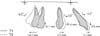
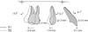
The mean, standard deviation, and statistical significance of the dentoskeletal changes relative to T1-T2 are summarized in Table 4. Graphic representations of maxillary superimposition for the PA and DS groups describing dentoalveolar changes during the distalization phase (T1-T2) are shown in Figures 4 and 5.
Pretreatment to post-distalization (T1-T2)
No significant sagittal or vertical skeletal change was detected between the two groups during the distalization phase. There was a slight opening of the mandibular plane angle in both groups (0.8° ± 3.0° in PA and 0.5° ± 2.1° in DS), with an increase of lower anterior facial height (1.8 ± 1.8 mm in PA and 1.7 ± 1.2 mm in DS). However, these differences were not statistically significant.
The maxillary first molar showed a mean distal movement of 4.7 ± 2.0 mm in the PA group and 4.2 ± 1.4 mm in the DS group, but these changes were not statistically significant. However, the maxillary first molar showed greater distal tipping in the PA group than in the DS group (U6 to SN: -9.0° ± 4.1° vs. -3.2° ± 3.0°; p < 0.01). Moreover, the first molar slightly intruded in the PA group (-0.1 ± 1.6 mm) and extruded in the DS group (0.3 ± 0.8 mm). The mean treatment time for distalization was 7 ± 2 months in the PA group and 9 ± 2 months in the DS group.
A significant difference was found in the anchorage loss. The first premolar showed a mesial movement (2.7 ± 3.3 mm) and a mesial tipping (3.6° ± 1.6°) in the PA group, whereas a slight distal movement (-1.9 ± 1.7 mm) and distal tipping (-5.1° ± 2.0°) was noted in the DS group. Accordingly, no significant change was described at the maxillary incisor in the DS group (0.1° ± 3.5° and -0.1 ± 1.5 mm), whereas a significant proclination (5.0° ± 3.6°) was reported in the PA group (p < 0.001). The first premolar and maxillary incisor also slightly extruded relative to the palatal plane in both groups (1.4 ± 1.9 mm and 0.5 ± 1.4 mm, respectively, in the PA group; 1.3 ± 2.0 mm and 0.5 ± 0.6 mm, respectively, in the DS group), but these changes were not statistically significant.
The overjet increased more in the PA group than in the DS group (1.3 ± 1.2 mm vs. 0.9 ± 1.1 mm) and the overbite decreased more in the PA group than in the DS group (-0.4 ± 1.9 mm vs. -0.1 ± 1.3 mm), but neither of these changes was statistically significant.
The soft tissues showed no significant change in either group. The upper lip moved slightly forward relative to the esthetic plane (E-plane) in both groups, whereas the lower lip was insignificantly protruded in the PA group and retruded in the DS group.
DISCUSSION
Cephalograms at the beginning of treatment revealed that patients in the 2 groups were in general not substantially different, confirming that this study had a low susceptibility bias (Table 3).
The appliances were equally effective in molar distalization (4.7 mm in the PA group and 4.2 mm in the DS group), even if the pendulum appliance required less distalization time (2 months less than distal screw). As previously reported by a recent meta-analysis,13 the amount of molar distalization did not seem to be the main difference between conventional and skeletal-anchored distalizing devices, because it seemed mainly related to the molar movement required before treatment to correct the Class II molar relationship. A confounding factor might be simply the tendency to treat patients with more severe Class II malocclusions using skeletal anchorage, where greater anchorage is desired.
Despite a slight molar intrusion in the PA group and extrusion in the DS group during the distalization phase, both appliances showed similar findings in the vertical facial dimension (SN-GoGn, 0.8° and 0.5°, respectively), but none of these differences was statistically and clinically significant.
However, differences were noted in molar distal tipping (9.0° in the PA group and 3.2° in the DS group), probably due to the intrinsic characteristics of the two appliances. Molar tipping could be related to the rigidity of the distalizing arms and the point of force application with respect to the center of resistance of the molar.3 The greater elasticity and flexibility of the TMA arms in addition to a coronal point of force application in the pendulum could cause greater distal molar tipping, which could be partially reduced by adding uprighting bends.5 Conversely, the distal screw acted as a distal jet,25 using telescopic rigid arms acting in proximity to the center of resistance of the first molar and providing more bodily distal movement.
Bodily movement could require longer treatment time during the distalization phase, probably because of the increased resistance to the movement. However, greater molar distal tipping could involve a longer treatment time after the distalization phase, requiring subsequent mechanics for root uprighting and producing an additional burden on molar anchorage.6
The anchorage loss at incisors and premolars was the main difference between the two appliances (Figures 4 and 5). In the PA group, the maxillary incisor showed a buccal inclination of 5.0°, similar to the finding of 4.0° described by Angelieri et al.30 The first premolar showed a mesial movement of 2.7 mm, and premolar anchorage loss was calculated at 36.5% in our study, in accordance with other studies in which mean percentage ranged between 24%4 to 43%.3 Mesial movement of the anchor unit was an unavoidable side effect that could occur with any conventional intraoral distalizing appliance. Unfortunately, the increase of anchoring teeth might not be sufficient to overcome this problem6; in addition, most of the mechanotherapy used to contrast such forces (e.g., headgear or Class II elastics) is compliance-dependent.1 Consequently, the combination between intraoral distalizing appliances and skeletal anchorage could be a reasonable approach to minimize anchorage loss during the distalization phase.13 In the present study, the maxillary incisor was almost stable in the distal screw (0.1°), and the first premolar showed distal movement of 1.9 mm.
A recent meta-analysis13 revealed that only with direct skeletal anchorage, a spontaneous distal premolar movement with no anchorage loss could be observed, probably due to the stretching of the interseptal fibers. Conversely, indirect skeletal-anchored devices showed a certain amount of anchorage loss at the premolar and incisor, even though they are smaller than conventional distalizing devices. This difference can be easily noted by comparing the findings relative to two modifications of the distal jet appliance in conjunction with paramedian miniscrews: the skeletonized distal jet (arms on the premolar) showed a premolar mesial movement of 0.72 mm, while in our study, the distal screw (no arm on the premolar) showed a premolar distal movement of 1.9 mm. This phenomenon had previously been described by Sar et al.16 and Polat-Ozsoy et al.,22 who reported premolar distalizations of 1.7 mm and 2.7 mm, respectively, using a BAPA; and by Cozzani et al.,23 who reported distalization of 2.1 mm using a distal screw appliance.
Moreover, our results revealed that the maxillary premolar showed a consistent distal tipping (5.1°) rather than a true bodily distal movement, possibly indicating that transseptal fibers acted on the premolar crown more than on the root (Figure 5). Our findings confirmed the results described by Cozzani et al.23 using the distal screw (-3°), Sar et al.16 using BAPA (-4.93°), and Polat-Oszoy22 using MISDS (-6.85°).
These phenomena might facilitate the retraction of premolars and canines, reduce round tripping of the anchor teeth, and decrease total treatment time by the early stages.11
The retrospective nature of this study and the analysis focusing primarily on the changes before and after the distalization phase could be considered weaknesses. However, all patients treated with these mechanics were included in the study, in order to avoid selection bias and the possibility of choosing some patients and screening out others. This reduced the risk of influencing the final outcomes.
Further long-term prospective studies should be conducted to confirm our results and report data at the end of orthodontic treatment or long-term follow-up.
CONCLUSION
·The null hypothesis that pendulum and distal screw appliances would produce similar dental and skeletal changes was disproved.
·Pendulum and distal screw appliances are equally effective in distalizing the maxillary molars, although treatment time was shorter in the pendulum group.
·Greater molar distal tipping, premolar mesial movement, and incisor proclination were noted with the pendulum appliance than with the distal screw during the distalization phase.
·The distal screw caused premolar distal movement during the distalization phase, possibly positively influencing the total treatment time.


 XML Download