

 PDF
PDF ePub
ePub Citation
 Print
Print


INTRODUCTION
Currently, a single-tooth implant has become a common treatment option for adults that has survival rates of about 90% after 10 years.1,2,3,4,5 Obtaining an esthetically satisfactory solution that mimics the adjacent natural teeth, however, represents a great challenge for clinicians, especially if an implant-supported crown is anticipated to be part of the treatment plan for a highly esthetically demanding area in a young adult who has a long life expectancy.5,6
The lack of an adequate recipient site secondary to alveolar bone resorption, as a result of a root fracture, does not create an ideal three-dimensional implant position and limits the predictability of a long-term esthetic result with an implant-supported crown because the gingival tissue follows the osseous crest, resulting in gingival recession and exposure of the crown margins.1,6 Progressive remodeling of the alveolar bone that was initiated by tooth extraction leads to a physiological reduction in thickness and height of the alveolar ridge, a concerning condition, which occurs at the buccal aspect of the anterior maxillary teeth where the cortical plate is anatomically thin and porous.7,8,9 Moreover, to remove an apical root-fractured fragment, surgical instrumentation can further increase resorption of the alveolar bone. In these situations, various surgical techniques such as guided tissue regeneration, grafting procedures, distraction osteogenesis, and ridge splitting have been proposed in order to correct local bone and soft tissue deficiencies6 that aim to provide a dimensionally adequate and potentially esthetic recipient site for an implant.
In order to improve a potential implant site, an alternative approach is the orthodontic extrusion of hopeless teeth. It is well known that extrusive movement produces forces on the periodontal fibers in the same direction as the movement, thus resulting in new bone apposition as the tooth moves coronally.10 At the same time, it also enhances soft tissue volume by increasing the amount of attached gingiva.11 Furthermore, to increase the success of this non-surgical technique, cooperation among a general dentist, a periodontist, and an orthodontist is a necessity. As compared with other surgical augmentation procedures, this technique is more time consuming but entails no risk of postoperative pain or inflammatory complications. Orthodontic extrusion has been proposed for periodontally compromised teeth at potential implant sites and also as an alternative to surgical crown lengthening in subgingivally fractured teeth prior to their restoration.6,9,10,11,12 However, few reports exist on the orthodontic extrusion of a root-fractured tooth that would undergo extraction, along with a lack of extended long-term clinical evaluations.13,14
This case report describes an interdisciplinary approach for the replacement of a maxillary central incisor that had a root fracture in the apical one-third of a young adult, in whom an orthodontic extrusion of the injured tooth was conducted in order to help maintain the volume of the osseous and gingival tissue for a subsequent esthetic implant placement. The results of a 12-year follow up are presented.
DIAGNOSIS AND ETIOLOGY
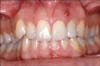
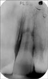
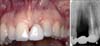
A 21-year-old woman in good general health, presented with discomfort of her left maxillary central and lateral incisors. The patient reported that her maxillary anterior teeth were injured 6 months before this visit. During an intraoral examination, a fistula between the roots of the left maxillary incisors was apparent at their buccal aspect, at the level of the mucogingival line (Figure 1). A periapical radiograph demonstrated a circular radiolucency around the apex of the lateral incisor, and a horizontal fracture was located in the apical one-third of the root of the adjacent central incisor (Figure 2). With the electric and cold pulp vitality tests, a delayed response was observed for the lateral incisor, as compared with that of the homologous contralateral tooth. An absence of tooth caries or defective restorations that could account for this response, together with evidence of a radiographic radiolucency, led to the diagnosis of an endodontic lesion, and the lateral incisor was endodontically treated (Figure 3).
The central incisor had the following pathological probing pocket depths: 7 mm mesiobuccally, 8 mm buccally, 5 mm distobuccally, 6 mm mesiolingually, 5 mm lingually, and 5 mm distolingually. In the maxillary anterior region, mesial and distal interdental papillae completely filled the embrasure spaces up to the contact points. A thick periodontal biotype was apparent on the basis of the negative visibility of an underlying periodontal probe through the gingival tissue,15 and the maxillary buccal and interdental soft tissues were not exposed while the patient smiled because of a low smile line. The patient was a non-smoker and did not take any medications that were known to interfere with periodontal healing.
TREATMENT OBJECTIVES AND ALTERNATIVES
In such a young adult, a clinician's primary concern is to provide an esthetically satisfactory and predictable outcome, especially from a long-term perspective.
The central incisor required extraction and replacement because of the apical location of the horizontal root fracture. The main treatment alternatives included the following: (1) conventional fixed partial dentures; (2) implant-supported restorations with surgical augmentation procedures at the time of implant placement; and (3) orthodontic extrusion of the fractured tooth followed by implant placement. For this patient, an implant-supported restoration was chosen in order to preserve the structure of the adjacent teeth. A team approach involving orthodontic extrusion of the injured tooth in order to help maintain the volume of the osseous and gingival tissues at the potential implant site was thought to be highly recommended, so as to compensate for the expected post-extraction tissue remodeling and avoid any surgical augmentation procedures, if possible.9,10,11,13,14
TREATMENT PROGRESS
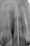
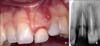
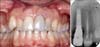
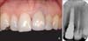
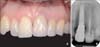
After provision of a careful explanation of the treatment plan to the patient, instrumentation of the central incisor root canal was conducted, and a hand file was cemented into it in order to provide an anchorage system so the fractured root could be orthodontically moved (Figure 3). Then, brackets were bonded in the maxillary arch. The bracket on the central incisor was positioned more apically than those on the adjacent teeth in order to allow for reduction of the incisal surface during extrusion. Further, an elastic chain was applied in order to extrude the fractured tooth (Figures 4A and 4B); a multiple-loop design orthodontic appliance was subsequently adopted in order to provide optimal control of the direction of traction until the desired extrusion level was obtained (Figures 4C and 4D). At that time, a great soft tissue volume was available, as the gingival margin of the extruded tooth was located at the height of the interdental papilla between the right maxillary incisors. The width of the keratinized gingiva increased by 4 mm with respect to baseline, and an immature red tissue (called a "red patch")16 appeared coronally to the original gingival margin.
After a 4-month extrusion period, as well as a 3-month retention phase, the orthodontic appliance was removed (Figure 5), and the tooth was extracted using a flapless, minimally invasive surgical procedure in order to preserve the integrity of all the socket walls. A Maryland bridge was cemented on the teeth adjacent to the extraction site that could serve as an interim restoration for esthetics, as well as space maintenance (Figure 6).
After a 3-month healing period, the temporary Maryland bridge was removed; a full thickness flap was raised; and a single 13-mm-long implant (3.3 mm in diameter) was placed with a platform collar (3.5 mm in diameter) that was positioned 2 mm beneath the bone peaks of the adjacent teeth. No dehiscence, fenestration, or fracture was observed in the socket walls at the time of implant insertion. No bone substitutes were employed. The Maryland bridge was carefully realigned in order to avoid any soft tissue compression or re-cementation.
Oral hygiene instructions were provided, and the patient was recommended to eat a soft diet for 8 weeks. Three months after implant placement, a provisional crown was placed in order to obtain proper conditioning of the peri-implant soft tissues. Final impressions were taken 6 months later by using a polyether impression material. The final metal-ceramic crown was placed about 12 months after the tooth extraction.
After having treatment, mesial and distal papillae completely filled the embrasure spaces up to the contact points (Figure 7A), and the height of the alveolar bone crest was well preserved at the interproximal areas (Figure 7B). A discrepancy existed between the level of the gingival margin of the restored tooth and that of the contralateral natural tooth (Figure 7A). Postoperative clinical and radiographic examinations revealed that the patient had good, esthetically pleasing results within 6 (Figure 8) and 12 years (Figure 9). The soft tissue color and texture at the buccal aspect of the implant site mimicked that of the adjacent natural teeth, including an adequate width of keratinized gingiva. As compared with the contralateral tooth, only a slight discrepancy persisted in the emergence line of the prosthetic crown from the soft tissues, probably secondary to passive eruption of the right central incisor. The interdental papillae in the maxillary anterior region were well preserved and filled the embrasure spaces up to the contact points. Accordingly, the height of the interproximal bone crest was well preserved and stable over time (Figures 8B and 9B). At 12 years, a cone-beam computed tomography (CBCT) follow-up also demonstrated the presence of adequate bone height and thickness at the buccal cortical plate (Figure 10).
DISCUSSION
After a tooth is extracted, the alveolar bone and soft tissues remodel with a resulting reduction in the horizontal and vertical dimensions of the future implant site.7,8 Moreover, marginal bone loss has been reported to occur over time at the buccal cortical plate of an implant-supported crown, a factor that predisposes a patient to gingival recession especially in the presence of a thin gingival biotype and vigorous toothbrushing.5,17 Accordingly, a recent study that used cross-sectional CBCT reconstructions demonstrated almost no buccal bone was detected in 36% of implants after 7 years, while the gingival margin was located 1 mm more apically than those in which the buccal bone plate was covering the entire implant surface.18 A lack of interdental papilla fill can also be expected in implant-supported single tooth crowns secondary to progressive interproximal bone loss.5,19
Many surgical procedures have been proposed in order to regenerate missing bone volume around single-tooth implants in esthetic sites,1,3,20 but these techniques are not without risk of postoperative inflammatory complications.21 An alternative approach to help maintain bone and soft tissue volume at the potential implant site before extraction is an orthodontic extrusion.6,9,11,13,14,22
In the present case report, a 4-month orthodontic extrusion followed by a 3-month retention phase created a greater volume of alveolar bone tissue without any surgical augmentation procedures. Despite having an increased treatment period, this procedure entails no risk of postoperative inflammatory complications21 and has a lower biological cost to patients. Orthodontic extrusion enabled the removal of the fractured root with an atraumatic and conservative procedure without the need for extended and invasive surgical instrumentation. A greater amount of bone was spared around the extraction socket; after extraction, reduction of the alveolar bone was minimized in the horizontal and vertical dimensions.
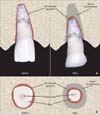
At the end of the orthodontic tooth extrusion, the bone volume had increased vertically (Figure 11A). Moreover, the portion of the root that had a smaller cross section was located coronally within the alveolus at the end of the extrusion (Figure 11B). For this reason, it can be hypothesized that bone volume also horizontally increases after extrusion. Such a well-represented osseous support helps retain stability of the gingival tissues around a single-tooth implant restoration, without any gingival recession or exposure of the crown margin. This is somewhat similar to what happens to the labial gingival dimensions in the horizontal and vertical dimensions if a lingual orthodontic movement is performed in order to place a root into a more proper position within the alveolar bone limits.23,24 Accordingly, in the present case report, an adequate buccal cortical plate was still present during a 12-year CBCT examination.
After 12 years, at the facial aspect of the implant site, the soft tissue color and texture were harmoniously integrated with the adjacent natural teeth and interdental papillae in the maxillary anterior region that had completely filled the embrasure spaces up to the contact points. Previous study reports exist regarding the augmentation of the coronal soft and hard tissues around an implant-supported crown of a root-fractured maxillary incisor that had a 10-week extrusion and 10-week stabilization period; however, a lack of long-term follow-up evaluations exists regarding these techniques.13
Waiting a period of 3 months prior to implant placement was also important, as achievement of the goal of complete healing and hard and soft tissue maturation over the extraction socket were possible.1,3,8,25,26 At the time of implant surgery, having an increased volume of healed soft tissues probably helped with respect to the improved predictability of a complication-free osseointegration of an implant and enhanced the site in order for a more esthetically pleasing gingival architecture around the final restoration.
CONCLUSION
Orthodontic extrusion of hopelessly fractured teeth seems to help maintain bone and soft tissue volume without the use of any surgical augmentation procedures and thus can be utilized as an esthetically satisfactory solution that mimics the neighboring natural teeth. Although modern dentistry has been shifting toward simplified clinical procedures and shorter treatment times, both general dentists and orthodontists should be aware of the possible long-term esthetic advantages of this interdisciplinary approach in highly esthetically demanding areas and should educate and motivate patients regarding the choice of this treatment solution, if necessary.


 XML Download