

 PDF
PDF ePub
ePub Citation
 Print
Print


INTRODUCTION
In orthodontics, patient noncompliance with treatment has been a concern for over 40 years,1 and several recent publications attest to this phenomenon.1,2,3,4,5 Therefore, much interest is presently shown in fixed appliances requiring minimal patient compliance. The Herbst appliance,6,7 mandibular anterior repositioning appliance (MARA),8,9 Jasper Jumper,10,11 and Eureka Spring12 have been proposed to manage noncompliance by patients with Class II malocclusion.
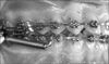
One fixed appliance for correcting Class II malocclusion is the Forsus™ fatigue resistant device (Forsus; 3M Unitek, Monrovia, CA, USA). It is a three- (L pin module) or two-piece (EZ module), semirigid telescoping system incorporating a stainless steel coil spring that can be assembled at the chair side and is compatible with complete fixed orthodontic appliances. Forsus is attached to the maxillary first molar and mandibular archwire, distal to either the canine or the first premolar bracket. As the coil is compressed, opposing forces are transmitted to the sites of attachment (Figure 1). The appliance is relatively well accepted by patients, although some may experience initial discomfort and functional limitations, which generally diminish with time.13 Jones et al.14 compared the therapeutic changes induced by Forsus with those induced by Class II elastics: they found no significant differences between the appliances with the exception of compliance. Franchi et al.15 evaluated the dental, skeletal, and soft-tissue changes following comprehensive treatment with fixed appliances including Forsus in patients with Class II malocclusion and reported that Forsus is effective in correcting Class II malocclusion via skeletal (mainly maxillary) and dentoalveolar (mainly mandibular) modifications. Further, Aras et al.16 compared the dentoskeletal changes and alterations of the mandibular condyle-disc-fossa relationship in patients at the peak and end of the pubertal growth period treated with Forsus. According to them, Forsus treatment is not a risk factor for the development of temporomandibular dysfunction in subjects without previous signs and clinical symptoms of dysfunction. Recently, Gunay et al.17 analyzed the effects of Forsus from its insertion to its removal (6 months) by comparing 15 late-adolescent patients with 12 subjects having untreated Class II malocclusion and reported that Forsus corrected the Class II discrepancy through dentoalveolar changes. The main limitation of their study was its small sample size. No previous study evaluated the contribution of active treatment with Forsus to the overall effects of comprehensive treatment.
The aim of the present study was to evaluate the active-treatment effects of Forsus during comprehensive correction of Class II malocclusion in growing patients.
MATERIALS AND METHODS
Study design
Fifty-four patients (27 boys, 27 girls) with Class II division 1 malocclusion were included in this study. The inclusion criteria were overjet longer than 5 mm, ANB angle larger than 3°, and full Class II or Class II tendency molar relationship. All the patients were in the permanent dentition phase at the start of the treatment. They consecutively underwent a specific nonextraction therapeutic protocol with 0.022-inch slot preadjusted fixed appliances in combination with Forsus at a single private practice of one author (L.A.) The amount of mandibular crowding ranged from mild (<3 mm) to moderate (3 mm).18 Forsus was used at the end of the leveling and alignment phase of the treatment, when a 0.019 × 0.025-inch stainless steel archwire was applied on both the arches. The mandibular archwire was consistently cinched distal to the molars. Brackets on the mandibular incisors had a torque of -6° to limit the buccal inclination of these teeth. The Forsus rods were placed on the mandibular archwire distal to the first premolars. Class II elastics were not used throughout the beginning of the fixed treatment (T1) till the removal of the appliance (T3) where Forsus was the only mean of Class II correction. The breakage rate of Forsus in this sample was 1% (2/54 cases). No transpalatal arches were applied during the treatment.
The active phase with Forsus was undertaken until Class II occlusion was overcorrected to an edge-to-edge incisal relationship. Thereafter, the fixed appliances were retained to finalize the occlusion. Ten patients (5.4%) wore Class II elastics during the final therapeutic phase from T3 to end of the comprehensive treatment (T4). Lateral cephalograms were taken at T1 (mean age = 12.5 ± 1.2 years), Forsus insertion (T2; mean age = 13.6 ± 1.1 years), T3 (mean age = 14.1 ± 1.1 years), and T4 (mean age = 14.8 ± 1.1 years). The duration of the comprehensive treatment was 2.3 ± 0.4 years. At T2, 15%, 70%, and 15% of the patients were in the prepubertal, pubertal, and postpubertal periods, respectively, as assessed by the cervical vertebral maturation method.19
Cephalometric analysis
Evaluation of dentoskeletal relationships
A customized digitization regimen and cephalometric software (Viewbox 3.0; dHAL Software, Kifissia, Greece) were utilized for the cephalometric evaluation. All the cephalograms were taken with the same equipment and magnification factor of 8%. Cephalometric variables from many other analyses20,21,22,23 were used to generate 28 (10 angular and 18 linear) measurements per tracing.
Both horizontal and vertical movements of upper and lower central incisors (U1 and L1) and first permanent molars (U6 and L6) were measured with respect to fiducial markers that were placed in the maxilla and mandible on the first tracing and then transferred to second, third, and fourth tracings in each patient's cephalometric series, based on superimposition of internal maxillary or mandibular structures.9 Molar relationship was measured as the distance between the projections of the mesial contact points of the upper and lower first permanent molars on the functional occlusal plane.
Error of the method
All the cephalograms were traced and superimposed by the same operator (G.C.) and checked by a second operator (L.H.) to verify anatomical outlines, landmark placement, and tracing superimpositions. Any disagreements were resolved by the consensus of both the observers.
Twenty randomly selected cephalograms were redigitized by the same operator (G.C.) and the measurements were recalculated to determine the method error with the intraclass correlation coefficient (ICC). The ICCs ranged from 0.93 to 0.99 for the linear measurements and from 0.94 to 0.98 for the angular measurements. All the recalculated measurements were within 1 mm or 1° of the original values.
Statistical analysis
Descriptive statistics (mean and standard deviation) were calculated at the four therapeutic time points. Statistical comparisons between the therapeutic intervals were performed by repeated-measures ANOVA with the Tukey's post-hoc test using statistical software (SigmaStat® 3.5; Systat Software Inc., San Jose, CA, USA).
The power of the study for repeated-measures ANOVA was calculated on the basis of the sample size of 54 subjects, an alpha level of 0.05, and an effect size for the Wits appraisal of 0.83.15 The power of the study was 0.99.
RESULTS
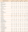
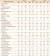
The results of the descriptive statistics and statistical comparisons are shown in Tables 1 and 2, respectively.
During the leveling and alignment phase (T1-T2; mean duration = 1.1 ± 0.4 years), significant increases occurred in the total mandibular length (Condylion [Co]-Gnathion [Gn] = +3.3 mm), mandibular ramus height (Co-Gonion [Go] = +2.4 mm), and lower anterior facial height (Anterior nasal spine [ANS] - Menton [Me] = +2.5 mm). As for the dentoalveolar changes, the overbite and interincisal angle decreased significantly (-1.7 mm and -5.9°, respectively) due to the significant proclination of the maxillary incisors in relation to the Frankfort horizontal plane (FH plane; +3.3°). The maxillary and mandibular molars showed significant mesialization and extrusion ranging between +1.1 and +1.7 mm. The occlusal plane showed significant anterior rotation in relation to FH plane (-1.4°).
In the active phase of Forsus (T2-T3; mean duration = 0.5 ± 0.1 years), no significant differences occurred in the sagittal maxillary skeletal measurements, whereas all the analyzed mandibular skeletal parameters showed significant increases (Sella-nasion-B point [SNB] = +0.6°; Pogonion [PG] - Nasion perpendicular [N per] = +1.4 mm; Co-Gn = +1.8 mm; Co-Go = +1.2 mm). The statistical comparisons revealed a significant decrease in the A point-Nasion-B point (ANB) angle (-1.0°) and Wits appraisal (-3.5 mm). With regard to the vertical skeletal measurements, a significant decrease was noted in the intermaxillary divergence (palatal plane to mandibular plane [PP-MP] = -0.9°). Basically, Forsus treatment produced dentoalveolar changes, especially in the mandibular arch. The overjet and overbite decreased significantly (-3.5 and -1.5 mm, respectively) and the molar relationship improved by 4.3 mm. These changes were associated with significant retroclination of the maxillary incisors (U1 to FH = -3.1°), proclination and intrusion of the mandibular incisors (L1 to MP = +5.0°; L1 vertical = -1.5 mm), and mesialization of the mandibular molars (L6 horizontal = 2.0 mm). The occlusal plane showed significant posterior rotation in relation to FH plane (+2.8°). All the other significant dentoalveolar changes were within 1.0 mm.
During the final therapeutic phase (T3-T4; mean duration = 0.7 ± 0.3 years), the dentoalveolar changes remained quite stable, with significant extrusion of the mandibular incisors (L1 vertical = +1.1 mm) and anterior rotation in relation to FH plane (-1.4°). At the skeletal level, significant increases were observed in the Wits appraisal (+1.0 mm), total mandibular length (Co-Gn = +1.6 mm), mandibular ramus height (Co-Go = +1.5 mm), and lower anterior facial height (ANS-Me = +1.3 mm).
DISCUSSION
In this study, the dentoskeletal changes induced by Forsus from its insertion to its removal were examined during comprehensive fixed appliance treatment in growing patients with Class II malocclusion. The patients could not be compared with a control group having untreated Class II malocclusion because of the short observational time points. Moreover, the use of a control sample would have raised ethical issues, as it would have required no treatment in subjects with Class II malocclusion during the circumpubertal growth phase, a biological period associated with the most favorable therapeutic effects in such patients.19 The lack of a control sample did not allow assessment of whether the skeletal changes in each interval occurred due to growth or the treatment. For the discussion, only the clinically significant changes (>1.5 mm or 1.5°) are considered.
Significant dentoalveolar changes during the leveling and alignment phase of the treatment (~1 year) occurred mainly in the anterior region, with a reduction in both the overbite and interincisal angle due to significant proclination of the maxillary incisors. These effects are probably related to the typical labial tipping of the incisors in this phase of treatment with a preadjusted appliance. Moreover, significant mesialization of the maxillary molars and extrusion of the mandibular molars were detected. These side effects can occur during this therapeutic phase depending on the geometrical relationships among the brackets.24
During the active phase of treatment with Forsus (~6 months), no significant changes were found in the maxillary skeletal measurements, as shown by Gunay et al.17 in a postpubertal sample treated with Forsus. This outcome however disagrees with those of studies that showed restriction of maxillary growth ("headgear effect") with the Jasper Jumper,11 Herbst appliance in combination with fixed appliances,6 and Forsus with controls.15 In the mandible, a significant increase in the total mandibular length was found, which accounted for 27% of the overall increase in the total mandibular length (6.7 mm). As the active treatment constituted 22% of the total therapeutic time (2.3 years), the impact of Forsus on mandibular growth was minimal. This outcome may also be correlated with the short duration of active treatment25 and early "burning" of the overjet due to significant proclination of the mandibular incisors (+5.0°, 80% of the total value). During the application of Forsus, significant improvement in the Wits appraisal was recorded (-3.5 mm). This change may be related primarily to the significant posterior rotation of the occlusal plane (+2.8°) rather than to the sagittal effects in both the jaws. This significant rotation of the occlusal plane, however, was transitory as the overall change was 0.0°. Treatment with Forsus did not induce significant changes in the vertical skeletal relationship.
With regard to the interdental changes, the active treatment with Forsus reduced both the overjet and the overbite, and improved the molar relationship. With respect to the overall outcome, this therapeutic phase contributed to 85%, 50%, and 123% of the changes in the overjet, overbite, and molar relationship, respectively. Slight relapse occurred in the molar relationship during the T3-T4 interval. The T2-T3 interdental findings were mainly related to the dentoalveolar changes. In particular, the decrease in overjet was associated with significant retroclination of the maxillary incisors and proclination of the mandibular incisors. The decrease in overbite was attributable to significant intrusion of the mandibular incisors, while the correction of the molar relationship was associated with significant mesialization of the mandibular molars. The mesialization of the mandibular dentition accounted for about 80% of the overall effects and occurred despite the cinching of the mandibular archwire distal to the molars and torque of -6° in the brackets on the mandibular incisors. Therefore, all procedures that can counteract the proclination and protrusion of the mandibular incisors during treatment should be applied (e.g., use of mandibular rectangular archwires of greater size and addition of a negative torque on the archwire in the mandibular incisor region). Recently, Aslan et al.26 showed that unfavorable labial tipping of the mandibular incisors can be effectively minimized by using miniscrews inserted between the mandibular canine and the first premolar roots bilaterally. Interestingly, the significant amount of relapse at the dentoalveolar level during T3-T4 occurred on the teeth more directly bearing the forces produced by Forsus. In particular, the maxillary molars and mandibular incisors showed significant extrusion after the removal of Forsus. Similar effects have been shown with the Herbst appliance.27 Further studies on the long-term stability of the dentoskeletal effects produced by the Forsus are required.
 XML Download