

 PDF
PDF ePub
ePub Citation
 Print
Print


INTRODUCTION
Maxillary canines are the most frequently impacted teeth with the exception of the third molars.1 Since they have great functional and esthetic value, surgical exposure followed by guided eruption with orthodontic treatment is commonly used. In this procedure, the orthodontist bonds an attachment to the surgically exposed area of the impacted tooth. Unfortunately, there is a risk of contamination of the bonding area with blood. Studies have revealed that contamination of the bonding site with blood during the bonding procedure results in a significant and drastic drop in the shear bond strength (SBS) of orthodontic brackets.2,3 To circumvent this problem, a blood stopper agent can be used to prevent the leakage of blood into the bonding site. Whether blood stopper agents also interfere with the SBS of orthodontic brackets is unclear; to our knowledge, there is only 1 previous study that addresses this issue.
Ankaferd blood stopper (Ankaferd Drug Inc., Istanbul, Turkey) is a plant extract that has been used in medicine as a hemostatic agent. This product comprises a standardized mixture of the plants Thymus vulgaris , Glycyrrhiza glabra, Vitis vinifera, Alpinia officinarum, and Urtica dioica .4 Trakyali and Oztoprak4 evaluated the effect of Ankaferd blood stopper on the SBS of orthodontic brackets bonded to bovine teeth. They concluded that "Ankaferd blood stopper could be used as a blood stopping agent during application of direct bonding brackets to surgically exposed canines to prevent bond failure due to blood contamination."
The aim of the present study was to evaluate the effects of blood contamination and a hemostatic agent on the SBS of orthodontic buttons bonded to impacted human teeth. The null hypothesis tested was that contamination of the bonding area by blood or a hemostatic agent would not decrease the SBS of orthodontic buttons.
MATERIALS AND METHODS
The minimum sample size for 0.75 power in this study was calculated as 15; therefore, 45 freshly-extracted, non-carious, impacted third molars without visible defects were used in this study. After extraction, any residual tissue attached to the root surface was removed mechanically. The teeth were washed under running tap water and stored in distilled water until use. Each tooth was individually embedded in an auto-polymerizing acrylic resin (Meliodent; Heraus Kulzer, Hanau, Germany). The facial surfaces of the teeth were cleaned with a mixture of water and pumice. The teeth were rinsed thoroughly with water and dried with compressed air.
Each tooth was etched with 37% phosphoric acid gel for 30 s, rinsed with a water/spray combination for 30 s, and dried until a characteristic frosty white etched area was observed. All teeth were randomly assigned to 3 groups of 15 each and treated as follows: group I, human blood was applied to the tooth surface and airdried; group II, blood stopper (patent number 2007-0-1-114485; Ankaferd Drug Inc.) was applied to the surface and air-dried; and group III, neither blood stopper nor blood was applied (control).
Blood and the blood stopper agent were applied according to the methods previously described by Trakyali and Oztoprak.4 In group I, all tooth surfaces were covered with fresh human blood from a male donor. The blood was applied to the labial surfaces of the teeth using a brush. In group II, 1 drop of Ankaferd blood stopper solution was applied directly onto the conditioned enamel surface.
Orthodontic buttons (9.6 mm2 surface area; G & H Wire Company, Greenwood, IN, USA) were used. Light bond (Reliance Orthodontic Products Inc., Itasca, IL, USA) was used as an orthodontic adhesive. A thin, uniform layer of sealant was applied to the etched enamel with a microbrush and cured for 20 s. A thin coat of sealant was also painted onto the metal button base and cured for 10 s before the paste was applied. A syringe tip was used for applying the paste to the button base. The button was then positioned onto the tooth and pressed lightly into the desired position. Excess adhesive was removed with a sharp scaler, and the adhesive was cured using an LED light curing unit (Ortholux 3M Unitek, Monrovia, CA, USA) for 20 s.
Each specimen was loaded into a Universal testing machine (Instron Universal test machine; Elista, Istanbul, Turkey), with the long axis of the specimen kept perpendicular to the direction of the applied force. The standard knife edge was positioned in the occlusogingival direction and in contact with the bonded specimen. Bond strength was determined in the shear mode at a crosshead speed of 0.5 mm/min until fracture occurred. The values of failure loads (N) were recorded and converted into megapascals (MPa) by dividing the failure load (N) by the surface area of the button base (9.6 mm2).
After debonding, all teeth and buttons in the test groups were examined under 10× magnification. Any adhesive remaining after debonding was assessed and scored according to the modified adhesive remnant index (ARI).5 The scoring criteria are as follows: 1, all of the composite, along with an impression of the button base, remained on the tooth; 2, more than 90% of the composite remained on the tooth; 3, more than 10% but less than 90% of the composite remained on the tooth; 4, less than 10% of the composite remained on the tooth; 5, no composite remained on the tooth.
Statistical analysis
The Kolmogorov-Smirnov test was used to check the normality of the SBS distribution. The values indicated that the data were normally distributed (p = 0.772). Therefore, parametric tests were used. Descriptive statistics, including the mean, standard deviation, and minimum and maximum values, were calculated for each of the groups tested. One-way ANOVA and Tukey's multiple comparison tests were used to compare the SBS values between the groups. The Fisher's exact test was used to determine significant differences in ARI scores between groups. Significance for all statistical tests was predetermined at p < 0.05. All statistics were performed using PASW Statistics 18.0 (IBM Co., Armonk, NY, USA).
RESULTS
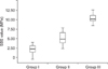
Descriptive statistics for the SBS (MPa) of all groups are presented as boxplots in Figure 1. ANOVA indicated a significant difference between groups (p < 0.001) (Table 1). The highest SBS values were measured in controls (10.73 ± 0.96 MPa). The SBS in the "blood" and "blood stopper" groups (4.17 ± 1.11 and 6.59 ± 1.5 MPa, respectively) were significantly lower than that of controls (p < 0.001). The lowest SBS values were measured in the "blood" group (4.17 ± 1.11 MPa; p < 0.001).
The frequency distribution of the ARI scores is presented in Table 2. Chi-square comparison revealed significant differences between the groups. There was a higher frequency of ARI scores of 2, 3, and 4 in the "blood stopper" and control groups, and ARI scores of 4 and 5 were more frequently observed in the "blood" group.
DISCUSSION
We observed that the SBS of orthodontic buttons was significantly decreased by the contamination of tooth surfaces with either blood or blood stopper. Several studies4,6,7 have shown that blood contamination significantly reduces the SBS of orthodontic brackets. The authors of these studies suggested that the pores of the acid-etched surface could become plugged due to wetting of the surface, resulting in insufficient numbers and lengths of resin tags.6 They also concluded that contamination by blood adversely affects bond strength by depositing an organic adhesive coating on the tooth.6 Similar to these results, we found a significant decrease in the SBS of orthodontic buttons due to contamination by blood.
Extrinsic factors such as bacteria, food, sugar and abrasive materials change the enamel surface composition. Therefore, the enamel may differ between erupted and unerupted teeth. In the present study, freshly extracted unerupted human third molars were used.
Oztürk et al.8 evaluated the influence of different tooth types on the bond strength of orthodontic adhesives, finding no significant difference between canines and molars. Several authors9-11 used molars instead of premolars to evaluate the SBS of orthodontic brackets. Therefore, impacted molars were used in the current study.
Since clinicians commonly use orthodontic buttons rather than brackets on impacted teeth, buttons were used in this study to better simulate the actual clinical conditions. Orthodontic buttons are usually smaller than brackets and the SBS of the buttons could be lower than that of brackets. Although different attachments were used in this study, our results are comparable to those of other studies. The failure loads were recorded in Newton (N) and converted into MPa by dividing the failure load (N) by the surface area of the bracket base.
Trakyali and Oztoprak4 evaluated the effect of Ankaferd blood stopper and blood on the SBS of orthodontic brackets bonded to bovine teeth. They found significant differences in the SBS between the three bonding procedures. The authors found that bonding was strongest in the control group, followed by the "blood stopper" group. Brackets bonded to teeth in the "blood" group had the lowest bonding strength. Similarly, we observed the highest SBS values in the control group, followed by the "blood stopper" and "blood" groups. The SBS in the "blood stopper" group may be lower due to the prevention of contact between the tooth surface enamel and the bonding agent and/or the obstruction of resin tags on the etched enamel surface.
Successful orthodontic treatment depends on adequate bonding of the brackets to the enamel.12 The minimum bond strength required to withstand normal orthodontic forces is believed to be 6 - 8 MPa.13 In the present study, the SBS was 4.12 ± 1.16 MPa in the "blood" group and 6.59 ± 1.5 MPa in the "blood stopper" group. These results indicate that the SBS of the "blood" group was inadequate, while the SBS of the "blood stopper" group is adequate for orthodontic treatment. Although contamination with the blood stopper agent significantly decreased the SBS of the orthodontic buttons, the resulting bond strength was still preferable to that obtained under conditions of contamination by blood. Therefore, our null hypothesis was rejected. Contamination of either blood or the blood stopper agent significantly decreased the SBS of orthodontic buttons.
CONCLUSION
Contamination of tooth surfaces with either blood or blood stopper significantly decreased the SBS of orthodontic buttons. Etching and bonding procedures should therefore be delayed until complete removal of the contaminating blood. The blood stopper agent is recommended for bonding orthodontic buttons to impacted teeth where the risk of contamination by blood is high. Remnants of blood and blood stopper agents should be completely removed from the bonding area to enable successful bonding.


 XML Download