

 PDF
PDF ePub
ePub Citation
 Print
Print


INTRODUCTION
Among orthognathic surgery procedures that can induce condylar position changes, sagittal split ramus osteotomy (SSRO) is one of the most frequently performed. This procedure offers several advantages, including an improved bony interface between the segments, rigid fixation, and rapid recovery of oral function. However, rigid fixation by SSRO, in comparison with non-rigid fixation, can induce greater changes in the condylar position and the axis of the condyle as well as higher incidences of temporomandibular disorder (TMD).1,2 Several studies have shown condylar displacement after mandibular set-back SSRO and elucidated the influence of mandibular set-back or lengthening osteotomy on the post-treatment osseous morphology of the temporomandibular joint (TMJ).3-5 However, these displacements and their accompanying morphologic changes did not lead to TMJ dysfunction because they occurred within the range of the natural adaptive mechanisms actuating after orthognathic surgery, such as glenoid fossa remodeling, condylar remodeling, and condylar repositioning within the fossa, which can attenuate the incidence of TMJ dysfunction.6,7 A double contour of the condylar head and/or the mandibular fossa has been reported to be a specific radiographic indication of remodeling activity, and this distinct double contour, specifically in the posterior area of the condylar head, was found in most of the joints studied in that report.8 Condylar remodeling has also been reported in other long-term post-operative radiographic examinations.9 However, only a few studies have utilized cone-beam computed tomography (CBCT) to investigate physiologic condylar head remodeling after orthognathic surgery in cases involving skeletal class III deformity.5
For accurate and effective CBCT-based comparison of remodeled areas, precise superimposition of CBCT images is essential, because the sliced multiplanar reformation (MPR) images vary according to the reference plane. For successful CBCT volume superimposition under these conditions, 3-dimensional (3D) medical imaging software programs using mutual information registration can be employed.10 The aim of this study was to evaluate condylar head remodeling after mandibular setback by bilateral SSRO with rigid fixation in skeletal class III deformities by using CBCT superimposition and to determine the correlation between the condylar head remodeling and changes in the condylar axis.
MATERIALS AND METHODS
Subjects and data acquisition
The study protocol was reviewed and approved by the ethics committee at Pusan National University Hospital (#2009062). The subjects were 22 adults (9 men and 13 women; mean age, 22.8 ± 2.9 years) diagnosed with skeletal class III malocclusion. These subjects underwent modified Hunsuck SSRO with rigid fixation, which was performed by 2 oral surgeons in the Department of Oral and Maxillofacial Surgery, Pusan National University Dental Hospital. The inclusion criteria were as follows: healthy physical condition; no severe facial asymmetry; no TMD symptoms and degenerative joint disease on examination; and skeletal angle class III. In SSRO, rigid intermaxillary fixation was carried out with bicortical screws. The skeletal fixation was followed by release of maxillo-mandibular fixation (MMF), after which the condylar and occlusal positions were checked. One week after MMF, patients received physiotherapy.
CBCT (Pax-Zenith3D; Vatech Co., Seoul, Korea) was used to evaluate the condylar remodeling and skeletal changes before and after surgery (average interval between examinations, 15.8 ± 3.7 months). The maxillofacial regions were scanned using a CBCT machine with a field of view of 20 × 19 cm, a tube voltage of 110 kVp, a tube current of 4.0 mA, voxel size of 0.3 mm, and a scan time of 24 seconds. The CBCT data were reformatted into Digital Imaging and Communications in Medicine (DICOM) format and reconstructed with 3D image software.
Superimposition method for skeletal and condylar axis changes and head remodeling
The anterior cranial base was used as the registration area for skeletal and condylar axis changes. The condylar neck and the posterior ramal area above the lingula of the mandible were registered for condylar head remodeling using OnDemand3D™ software (Cybermed Technology Co., Seoul, Korea). In this study, the superimposition method allows accurate, rapid, and automatic superimposition without segmentation or landmark definition. To superimpose the CBCT data, the investigator designated the registration areas, such as anterior cranial base and ramus region, on the MPR images. The software automatically calculated the best registration areas using the mutual information algorithm. After superimposition, data analysis was performed.10 The reference planes and axes for skeletal and condylar axis changes were defined in a previous study.11 The measurements were obtained under the same scanning conditions (grayscale: bit depth 14 bit, window width: 4,000, window level: 1,000).
Measurements of the condylar head
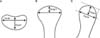
For analysis of the condylar head remodeling, the superimposed condylar head images were compared with regard to their width and depth on the axial plane, width and height on the coronal plane, and height and depth on the sagittal plane; the measurements were obtained according to the method reported by Hoppenreijs et al.3 (Figure 1).
Assessment of remodeling areas
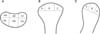
On the basis of the signs of condylar head remodeling, the superimposed axial, coronal, and sagittal pre- and postoperative images were classified as "bone resorption; loss or reduction of the shape and size of the bone," "bone formation; increase of the shape and size of the bone," or "unchanged; no significant change in the shape and size of the bone." Three areas (medial, superior, lateral) on the coronal plane, 3 (anterior, superior, posterior) on the sagittal plane, and 6 (anteromedial, anterior-middle, anterolateral, posteromedial, posterior-middle, posterolateral) on the axial plane were examined for each condyle (Figure 2).
Statistical analysis
The data were statistically analyzed using PASW Statistics version 18.0 for Windows (IBM Co., Armonk, NY, USA). Pre- and postoperative comparisons of the changes in the condylar axis and the measurements of the condylar heads were made using a paired t-test. Pearson correlation coefficients were calculated to analyze the correlation between the magnitude of mandibular set-back and condylar axis changes. The condylar remodeling sign distribution data for the right and left sides were compared using the Wilcoxon signed rank test (p < 0.05). To isolate the distribution of the condylar head remodeling signs, chi-square tests were performed in each predefined area between the pre- and postoperative stages. The Shapiro-Wilks normality test was applied to the data, which were found to be normally distributed. The reproducibility for the superimposition of the condylar remodeling signs was assessed using Cohen's kappa index (κ). In this study, the reproducibility for the superimposition of the condylar remodeling signs showed substantial agreement: Cohen's kappa index for intra-investigator agreement was 0.72, and that for inter-investigator agreement was 0.67.
RESULTS
The mean amount of mandibular set-back was 6.8 ± 2.2 mm. The mean vertical change of the mandible (FH plane to Me) was 1.7 ± 6.6 mm, which was not significant (p > 0.05).
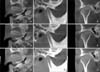
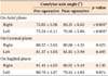
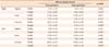
The pre- and postoperative right and left condylar axis angles (°) on the axial planes were significantly different (p < 0.05; Table 1). There was no significant correlation between the amount of mandibular set-back and condylar axis changes (p > 0.05). Measurements of the condylar head showed no changes on the axial plane. However, the values obtained on the sagittal and coronal planes indicated a statistically significant postoperative diminution in the condylar height (p < 0.05; Table 2 and Figure 3).
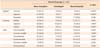
The distribution of condylar remodeling signs showed no significant difference between the right and left condyles (p > 0.05). However, a chi-square test comparison of the distribution showed that bone resorption predominantly occurred on the anterior and superior areas on the sagittal planes (p < 0.05; Table 3). On the coronal planes, resorption was predominantly observed on the superior and lateral areas, as in the case of the anterior-middle and anterior-lateral areas on the axial planes (p < 0.05).
DISCUSSION
In a radiographic study on condylar remodeling, Hollender and Ridell8 found a distinct double contour in the posterior area of the condylar head. They insisted that such a manifestation, whether of the condylar head or the glenoid fossa, indicates remodeling activity.8 Similar findings were reported in other studies.6,12,13 However, direct evidence of postoperative TMJ remodeling is difficult to obtain in 2-dimensional radiographic analysis. To resolve this limitation, a fully automated system with mutual information registration was used in this study. Structures that remain stable after orthognathic surgery, i.e., the cranial bases, the condylar neck, and the posterior ramal area above the lingula of the mandible, were referenced to compare the skeletal and condylar axis changes and remodeling.10,11
Several lateral cephalography studies have reported condylar remodeling and resorption in the anterosuperior aspects of the condyles after orthognathic surgery.3,14-16 The results of the present study were consistent with those findings: the condylar heights on the sagittal and coronal planes reduced after surgery (p < 0.05; Table 2) and the anterior and superior areas on the sagittal plane showed prominent bone resorption (p < 0.05; Table 3). Park et al.11 also reported the same results in two-jaw surgery cases. The extent of increase in the condylar height was not significant with reference to the voxel size of CBCT machine. These clinically insignificant changes could not produce clinical signs and symptoms of TMD.
Condylar remodeling during repositioning of the mandible may be attributed to mechanical force, specifically overloading.7 Hoppenreijs et al.3 reported that rigid fixation is also an important factor influencing condylar morphologic changes and that a rigid-fixation group showed condylar remodeling. Other studies suggested that inward rotation of the condylar head on the axial plane following mandibular set-back surgery could be an important factor inducing remodeling (p < 0.005; Table 1).17,18 The present study evaluated the effect of the amount of mandibular set-back on the condylar axis changes and remodeling and their correlation; however, no statistically significant correlation was noted (p > 0.05).
In the present study, most areas of the condylar heads showed various degrees of change. The anterior and superior areas on the sagittal plane, the superior and lateral areas on the coronal plane, and the anterior-middle and anterior-lateral areas on the axial plane predominantly showed signs of resorption (p < 0.05; Table 3). However, on the axial plane, these signs were predominantly observed in the anterior-middle and anterolateral areas. Park et al.11 showed similar results in cases involving 2-jaw surgery, although in their study, these signs were predominantly observed in the anteromedial, anterolateral, and posterolateral areas on the axial plane. These differences in the distribution of the regions showing prominent changes may have resulted from differences in the sample size rather than differences in the type of orthognathic surgery. The anterolateral area on the axial plane was a common resorption area in both studies. Resorption in this area might be influenced by the inward rotation of the axial condylar axis (Table 3). Further studies with larger sample sizes and control subjects should be performed to elucidate these findings. Several computed tomography studies suggested that the degree of postoperative condylar long-axis alteration might be a predictive factor of condylar remodeling.5,8,9,19,20 The movement associated with this alteration imparts compressive loading to the condyle and glenoid fossa, which could be one of factors related to condylar remodeling. However, in the present study, remodeling occurred in most areas of the condylar head. Among the different signs of remodeling, resorptive remodeling was predominant only in limited areas: the anterior and superior areas on the sagittal plane; the superior and lateral areas on the coronal plane; and the anterior-middle and anterior-lateral areas on the axial plane. These results differed from those of the previous study, which reported bone formation in the posterior region of condylar head.21 However, our results were consistent with those obtained in studies involving mandibular advancement surgery, which showed that condylar resorption usually occurred on the anterior-superior surface of the condyle, and not the posterior surface.3,14,16 The posteriorly directed force after mandibular advancement does not appear to have a direct influence on the development of condylar resorption. In our study, condylar resorption was predominantly observed on the anterosuperior surface of the condyle, and not the posterior surface as observed in the previous reports with mandibular set-back movement. The condylar remodeling in the sagittal and coronal aspects might be related to the complex mechanical vector mechanisms between the musculature and proximal segment rotation and the less dense surface of the condyle.22 As mentioned previously, a double contour of the condylar head and/or mandibular fossa is noted as the specific radiographic indication of remodeling activity.8 Moreover, Liu et al.23 also reported that this distinct double contour in the posterior area of the condylar head. However in this study, we did not find a double contour of the condylar head. Further studies would be needed with a large sample size and long-term data to clarify this discrepancy.
Several studies have found that individual adaptive capacity is related to systemic factors such as age, autoimmune system function, endocrine system, and metabolic disorders. These factors might influence fibrocartilage metabolism and blood supply, thereby contributing to resorptive changes.24 O'Ryan and Epker22 found a difference in the trabecular pattern of condyles between high- and low-angle patients. Condyles in patients with anterior open bite appear to be very sensitive to functional loading and their adaptive capacity probably is smaller than in patients with deep bites.21,22 Patients presenting with predispositional factors relating to condylar adaptive capacities, therefore, should be informed about the possibility of condylar resorption and its possible effects on stability.
CONCLUSION
Even though many patients can adapt to occlusion or condylar positions that are not considered ideal, in orthognathic surgery patients, the range of the physiologic adaptation should be studied further because the surgery changes the stomatognathic environment. It should also be considered that patients with risk factors for imbalance of the adaptive ability and masticatory system would experience difficulty in physiologic adaptation.
 XML Download