

 PDF
PDF ePub
ePub Citation
 Print
Print


INTRODUCTION
Cleidocranial dysplasia (CCD), also known as cleidocranial dysostosis or Marie-Sainton syndrome, is an autosomal dominant disorder with a prevalence of 1 in 1,000,000 individuals. It is characterized by delayed or absent fontanel closure, parietal and frontal bossing, hypoplastic clavicles, and narrow thorax; it may also present deafness, hypertelorism, and low nasal bridge. Young individuals with CCD show relatively normal jaw proportions and mandibular morphology, whereas older affected individuals tend to have short lower facial height, acute gonial angle, anterior inclination of the mandible, and mandibular prognathism. These differences are attributed to pronounced horizontal mandibular growth resulting from impaired vertical maxillary growth and eruption of permanent teeth.1 The dental manifestations include delayed exfoliation of primary teeth, delayed eruption or noneruption of permanent teeth, supernumerary teeth, retention cysts, and enamel hypoplasia (OMIM #119600).
Heterogeneous mutations in core-binding factor subunit alpha-1 (CBFA1) are associated with CCD.2 CBFA1, also known as runt-related transcription factor 2 (RUNX2), is a transcription factor of the runt family, located on chromosome 6p21.3 RUNX2 binds to and regulates multiple genes in osteoblasts; it has been identified as an osteoblast-specific transcription factor and is also regulated by bone morphogenetic proteins (BMPs) and vitamin D3.4,5 Along with its role in bone development, RUNX2 participates in tooth development by mediating interactions between epithelial and mesenchymal cells during tooth morphogenesis.6
Studies of the role of RUNX2 in normal tooth development have provided some molecular explanations for the abnormal dental features in CCD. In the Lossdörfer et al.7 study, an individual with CCD having a missense mutation in RUNX2 showed reduced basal expression of mRNA for receptor activator of nuclear factor kappa-B ligand (RANKL), which is normally expressed on the cell membrane of osteoblasts. Given that RANKL is responsible for osteoclastic differentiation and activation, insufficient resorption of alveolar bone and failure of root resorption of primary teeth could lead to delayed exfoliation of primary teeth in affected individuals with an RUNX2 mutation.
One explanation for the noneruption of permanent and supernumerary teeth is the lack of cellular cementum in the apical region of impacted teeth. Manjunath et al.8 found that all the examined teeth from unaffected individuals had cellular cementum at the apices, whereas only 8% of the teeth from a 60-year-old patient with CCD had cellular cementum at the apices. Because the patient's teeth had spontaneously erupted, the authors did not correlate the absence of cellular cementum with the failure of tooth eruption. Further, Counts et al.9 studied two teeth from a 22-year-old patient with CCD and found no significant correlation between cementum in this patient and that in a patient without CCD. These incongruent results indicate the need for further research on this topic. The issue becomes more perplexing because 40% of the CCD cases do not have any apparent genetic cause.10 A possible role of environmental and other nongenetic factors in the variation of CCD symptoms, especially in the formation of supernumerary teeth, has been suggested by Suda et al.,11 who studied a case of monozygotic twins with an identical mutation in RUNX2 showing different numbers and positions of supernumerary teeth.
The most recognizable dental features of CCD are supernumerary and retained primary teeth. The cranial defects may not be noticeable at birth. The physical symptoms are not progressive or debilitating. However, retention of primary teeth, midfacial hypoplasia, overclosure, and skeletal class III malocclusion cause both functional and aesthetic problems.12
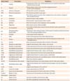
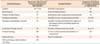
The dental management of individuals with CCD is challenging and involves comprehensive orthodontic and surgical treatments. The four main therapeutic approaches published in the literature are the Toronto-Melbourne, Belfast-Hamburg, Jerusalem, and Bronx methods.13 Here, we report two cases of CCD that had been treated surgically and orthodontically at the University of California, San Francisco (UCSF) School of Dentistry by using modifications of the Bronx (case 1) and Belfast-Hamburg (case 2) approaches. We also summarize the clinical features of 17 individuals who had been diagnosed with CCD at UCSF. Refer to Table 1 for details of the cephalometric points and measurements.
CASE REPORT
Case 1
Features
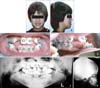
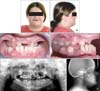
A 12-year-old Asian boy diagnosed with CCD presented with his father, who had the same condition. The patient's chief complaint was described by his father as "anting normal eruption of his permanent teeth and better alignment of his upper and lower arches." His medical and surgical histories included recurrent otitis media with tubes placed in the tympanic membranes and bilateral femoral osteotomies to correct coxa vara. The boy showed the classical craniofacial features of CCD: broad forehead, hypertelorism, and short upper third of the face (Figure 1A and 1B). The initial extraoral examination revealed a retrognathic and vertically deficient maxilla (Figure 1A and 1B), resulting in a concave facial profile.
The initial intraoral examination revealed a skeletal class III pattern with 9-mm anterior crossbite and 3-mm open bite, moderate-to-severe maxillary crowding, and mild mandibular crowding (Figure 1C and 1D). The panoramic radiograph showed nine retained primary teeth, seven supernumerary teeth, four impacted permanent teeth, and no congenitally missing permanent teeth (Figure 1E). The initial lateral cephalogram confirmed the skeletal class III relationship due to maxillary underdevelopment (Figure 1F).
Treatment
The treatment involved five steps and was expected to continue until skeletal maturity, when the underdeveloped maxilla was to be surgically corrected. The first step was to treat multiple primary and newly erupted permanent teeth for caries due to poor oral hygiene.
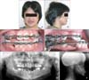
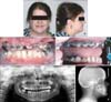
At age 14, nine retained primary teeth (i.e., 53, 62, 63, 74, 73, 72, 82, 83, and 84) were extracted and seven supernumerary teeth were surgically removed under general anesthesia (Figure 2E). All the permanent canines were also removed, because they were determined to be nonretrievable due to severe impaction. The permanent teeth were observed for possible spontaneous eruption over 9 months.
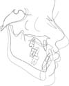
The mandibular lateral incisors and maxillary right lateral incisor failed to erupt spontaneously. These teeth were surgically exposed and bonded with brackets and gold chains tied to maxillary and mandibular lingual arches for guided eruption (Figure 2E). Simultaneously, a mesiodens and the maxillary right primary second molar were extracted. Shortly thereafter, full maxillary and mandibular fixed orthodontic appliances were placed for alignment and coordination of the dental arches (Figure 2C and 2D). The maxillary right lateral incisor was repositioned in the canine space (Figure 3E).
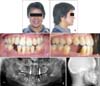
To correct the transverse maxillary discrepancy, a transpalatal arch was used. At skeletal maturity (age 19), the maxilla was advanced and downfractured to correct 6 mm of the sagittal maxillary discrepancy and vertical deficiency (Figure 3B and 3F). The postsurgical healing was good with excellent patient compliance and the orthodontic treatment was completed (Figure 3A, 3C, and 3D).
The final step was implant placement and prosthodontic rehabilitation. Implants were placed for the maxillary right lateral incisor and left canine and mandibular canines (Figure 3E). The mandibular right canine-premolar space required two separate implants. Because the second molars were the last teeth to develop and the patient had been in treatment for a long time, these teeth were not retrieved. Treatment was completed with the delivery of the final crowns for the implants at age 24.
Outcome
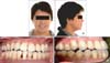
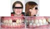
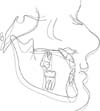
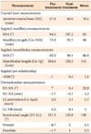
The final extraoral and intraoral photographs are shown in Figure 4. Fifty-three hard-tissue and soft-tissue landmarks were digitized by an orthodontist using Dolphin software (version 11.5; Dolphin Imaging and Management Solutions, Chatsworth, CA, USA). The pretreatment and posttreatment lateral cephalograms were superimposed to illustrate treatment-induced changes (Figure 5). Comparison of the ANB, overbite, and overjet values between these time points (Table 2) showed a dramatic improvement in the sagittal jaw relationship after the maxillary advancement surgery and orthodontic treatment.
Case 2
Features
A 14-year-old Caucasian girl diagnosed with CCD presented for orthodontic treatment with a desire to have greater number of teeth to be visible. Her mother and younger sister also had CCD. Her medical and surgical histories revealed mitral valve prolapse, tethered spinal cord, arthritis, Crohn's disease, thyroid reduction surgery, tonsillectomy, and Z-plasty of the lingual frenulum. The initial evaluation revealed jaw pain, bilateral temporomandibular joint clicking, and facial muscle pain. Mouth breathing, tongue thrusting, and clenching habits were also noted.
The patient had a concave facial profile and decreased facial height due to maxillary underdevelopment. Her lips were retrusive, resulting in an edentulous appearance (Figure 6A and 6B). The intraoral examination showed that she had five retained primary teeth (i.e., 55, 63, 75, 74, and 73) and 15 fully erupted permanent teeth (Figure 6C and 6D). She had a negative sagittal jaw relationship with class III malocclusion due to the hypoplastic maxilla (Figure 6D). On the panoramic radiograph, 14 impacted permanent teeth were identified and the left mandibular second molar was determined to be congenitally missing (Figure 6E). The lateral cephalogram confirmed maxillary hypoplasia (Figure 6F). Multiple impacted supernumerary teeth had been extracted 2 years earlier along with the mandibular right first molar, due to extensive caries.
Treatment
Orthodontic treatment was initiated with the following objectives: maxillary expansion, removal of the retained primary and supernumerary teeth, guided eruption or extrusion of the impacted permanent teeth, and correction of the class III jaw relationship by maxillary advancement surgery.
A hyrax expander was placed for 2 months, which achieved 11 mm of expansion (Figure 7C). Heavy upper and lower wires were placed; the retained primary teeth were removed; and impacted permanent teeth 11, 15, 21, 22, 23, 35, and 47 were surgically exposed and bonded with gold chains placed close to the cusp tips for guided eruption (Figure 7C and 7D). All the bonded teeth except 21 erupted after approximately 11 months. The maxillary left central incisor was extracted because of exposure of the root, which was very short.
Orthodontic alignment and coordination of the dental arches were complete in 14 months (Figure 7E). Le Fort I osteotomy was performed at age 18 (Figure 8D). With the removal of the remaining orthodontic appliances and placement of an implant and a fixed prosthesis for the maxillary left central incisor, the treatment was completed (Figure 8A-8D). However, the patient's poor oral hygiene during the retention stage led to complete loss of the maxillary dentition.
Outcome
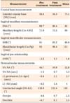
Although no post treatment extraoral and intraoral photographs were obtainable, because of the loss of the maxillary teeth, the Dolphin software was used to digitize and superimpose the pretreatment and postsurgical lateral cephalograms (Figure 9). Comparison of the ANB and overjet values between these time points indicated improvement in the sagittal jaw relationship after the maxillary advancement surgery and orthodontic treatment (Table 3). The overbite also improved significantly, from -6.7 mm to 0.1 mm
Clinical features of individuals with CCD
We reviewed our database (FileMaker Pro 10.0 V3; FileMaker Inc., Santa Clara, CA, USA) of all the individuals with CCD who had been diagnosed at the UCSF. Table 4 summarizes their clinical features.
Seventeen (nine female and eight male) affected individuals were identified. Four of 15 individuals (the height of two individuals was not recorded) had short stature. Further, 57% had a family history of CCD, with one-to-three generations of CCD passed down in the family. Eight of the 17 individuals had vertebral changes such as scoliosis. The characteristic features of this condition, such as missing or hypoplastic clavicles, persistent metopic sutures, frontal bossing, and hypertelorism, were observed in 93%, 94%, 88%, and 69% of these subjects, respectively. A high incidence of otitis media that required surgery to place pressure-equalization tubes was noted. Seven patients had speech problems and received speech therapy (Table 4).
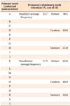
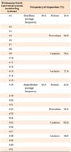
Regarding the dental features, congenital hypodontia was observed in 12% of this population. The number of retained primary teeth ranged from 0 to 20, with the mandibular canine being the most frequently retained primary tooth (Table 5). On average, the mandibular primary teeth were slightly more likely to be retained than the maxillary ones. The number of impacted permanent teeth ranged from 3 to 27, with the incisors and canines being the most frequently impacted in the maxillary and mandibular arches, respectively (Table 6). Zero to 18 supernumerary teeth were observed.
Noteworthily, 14 of the 17 subjects had maxillary hypoplasia, 88% had class III malocclusion due to the hypoplastic maxilla, and 10 patients required Le Fort I maxillary advancement surgery.
DISCUSSION
Management of the orofacial manifestations of CCD is lengthy and challenging, and requires careful planning and execution by an interdisciplinary team. The length of treatment ranges widely and is generally not completed until growth has seized. By reviewing these cases, we appreciate the importance of interdisciplinary teamwork for a successful outcome. We have also learned that it is critical to maintain the patient's motivation and keep the treatment within the patient's tolerance limit for a good outcome. Failure to meet these criteria may lead to deleterious results, as seen in case 2.
The UCSF approach in case 1 is similar to the Bronx approach. Extraction of all the retained primary and supernumerary teeth was followed by an observation period and a second surgical phase of exposing and bonding the impacted permanent teeth. After orthodontic alignment and coordination, patient 1 underwent a third surgical phase to correct the skeletal class III malocclusion. The treatment was completed with the restorative phase using implants and fixed prostheses.
Case 2 was approached by using a method similar to the Belfast-Hamburg approach. In a single surgical operation, all the retained and supernumerary teeth were extracted and surgical exposure and bonding of the impacted permanent teeth were completed. However, as with 14 out of 17 patients described in our summary of clinical features of CCD, case 2 showed maxillary hypoplasia and required a second surgical phase to correct the skeletal class III malocclusion.
We have summarized the clinical features of CCD observed in 17 individuals to aid in early diagnosis and, thereby, better management of this rare condition. A greater number of genetic and molecular studies are needed to explain the features, patterns, and variation in the severity of the syndrome.
Recent advances in dentistry have yielded better therapeutic options and outcomes for individuals with CCD. For example, dental implants can be successfully used not only for orthodontic tooth movement with reduced side effects but also for replacing teeth that cannot be guided into the arch orthodontically, as presented in cases 1 and 2.
Although the cases presented here were completed before the introduction of cone-beam computed tomography (CBCT), this technology is now routinely used to provide a three-dimensional image of the dentition, reducing guesswork and ensuring better anatomical localization of supernumerary and impacted teeth. CBCT can provide pertinent information such as the precise location of a supernumerary tooth in relation to important structures such as the cortex of the nasal floor, labial cortex of the nasal ridge, nasopalatine duct, and adjacent root apices.14 Such developments will continue to improve the management of complex dental cases.
CONCLUSION
Individuals with CCD require extensive diagnostic work-up and a long-term treatment plan. The UCSF treatment protocol incorporates the Bronx and the Belfast-Hamburg approaches and can be organized into five steps. It involves the appropriate timing of removal of supernumerary and retained primary teeth, surgical exposure of impacted permanent teeth, orthodontic extrusion and alignment, followed by Le fort I advancement and implant retained prosthesis. The two cases reported prove the efficacy of this protocol in resolving the clinical dentofacial features of CCD.
 XML Download