

 PDF
PDF ePub
ePub Citation
 Print
Print


INTRODUCTION
Patients who undergo teeth-bleaching procedures may also opt for further enhancement of their smile with orthodontic treatment. However, the bleaching procedures and the methods used for enamel etching may affect the bonding forces of the brackets to enamel surfaces. The procedures for vital bleaching are thought to decrease bond strength by altering enamel surface topography.1,2 Bleaching of vital teeth can be performed in the dental office or at home; the 2 approaches differ in terms of the chemicals and concentrations used and the treatment durations. In a study wherein the bonding strengths of metal brackets were tested after bleaching, the authors have concluded that home bleaching with 10% carbamide peroxide did not change the bonding strength; however, in-office bleaching with 35% hydrogen peroxide decreased the bonding strength considerably.3
This reduction in bond strength may be due to loss of prismatic formation,2 loss of calcium and decreased micro-hardness,4,5 residual oxygen interference with resin penetration into the etched enamel, or inhibition of polymerization.6-9 Methods proposed for recovering the reduced bond strengths include removal of a layer of the enamel surface,10 delaying the bonding procedure,11 antioxidant treatment of the surface,12 use of alcohol-based bonding agents,13 or pretreatment of the surface with alcohol.14
Etching the surface of enamel can be carried out by acid, laser, or sandblasting. Whereas some studies suggest that laser etching can be as effective as acid etching,15-17 other studies have concluded that laser-etching results in considerably lower bonding forces.18-20 Because there is no study on bonding to laser-etched surfaces of bleached teeth, the aim of this study was to investigate the bonding strength and characteristics of orthodontic brackets attached to laser-etched surfaces of in-office bleached teeth, and compare them with the corresponding characteristics of acid-etched surfaces.
MATERIALS AND METHODS
Seventy-eight, sound, human, upper premolar teeth were used in this study. The teeth were scaled with a periodontal scaler to remove organic debris. After scaling, they were disinfected in 0.5% chloramine solution and stored in distilled water at 4℃ until the beginning of the test. Each tooth was examined under a stereomicroscope (Leica/Meyer Instruments, Houston, TX, USA) to eliminate teeth with cracks or hypoplastic defects. The criteria for tooth selection included no pretreatment with a chemical agent such as alcohol, formalin, or hydrogen peroxide, or any other bleaching material. The buccal enamel surfaces of the teeth were polished with non-fluoridated pumice and rubber prophylactic cups at a low speed (3,000 rpm) and then washed and dried before any procedure was performed.
Specimens were then randomly divided into a control group (n = 26) and a bleaching group (n = 52). In the control group, surface treatments of acid etching or laser application prior to bracket bonding were performed immediately after enamel-surface cleaning in two subgroups (group A and B, n = 13 for each subgroup). In the bleaching group, the surface treatments were done 1 day (n = 26) or 3 weeks (n = 26) after bleaching. Each of these bleaching groups was then divided into 2 subgroups (yielding groups C, D, E, and F, n = 13 for each subgroup) according to the surface treatments. The detail procedures employed for each subgroup are provided below:
Group A: A 38% phosphoric acid gel (Etch-Rite; PulpDent, Watertown, MA, USA) was used to etch specimens. After 30 s, buccal surfaces were washed for 10 s with distilled water and dried with oil-free compressed air until the chalky white appearance of acid-etched enamel was obtained.
Stainless steel, upper premolar, standard edgewise brackets (Standard Edgewise Dynalock; 3M Unitek, Monrovia, CA, USA) with a base surface of 13.44 mm2 were used for this study. After surface treatments, the liquid primer Transbond XT (3M Unitek) was applied to the etched surfaces, and the brackets were bonded on the teeth with Transbond XT adhesive. Excess resin was removed with an explorer before polymerization. Then, a light-emitting diode (Starlights; Mectron S.p.a., Carasca, Italy) was used for curing the specimens for 20 s.
Group B: The specimens were irradiated with Er:YAG (VersaWave; HOYA Photonics Inc., Fremont, CA, USA) laser with a wavelength of 2.94 µm at 300 mJ/pulse, 10 pulse per second. The laser was operated for 10 s in a focused, noncontact mode under water spray. The beam was aligned perpendicular to enamel at a 1-mm distance and was moved in a sweeping fashion by hand over an approximately 4 × 4-mm area during exposure. After laser application, upper premolar brackets were bonded by using the method described in group A.
Group C: For groups C, D, E, and F, bleaching with 38% hydrogen peroxide (Opalescence Boost; Ultradent, South Jordan, UT, USA) was performed as follows: The enamel surfaces were dried with cotton pellets, and tooth shades of the specimens were determined and recorded before bleaching using Opalescence Boost's own shade scale. Bleaching material was mixed according to the manufacturer's recommendations. A 1-mm thick layer of 38% hydrogen peroxide was applied to buccal enamel surfaces for a total of 45 min in the first session. After 15 min of application, bleaching gel was removed with a soft brush under running tap water and the procedure was repeated 3 times. Two sessions of office bleaching were performed, with a 3-day interval between sessions. During the test procedures, the specimens were stored in distilled water at 37℃ and the distilled water was changed daily. Post-bleaching changes in the shades of the specimens were determined and recorded by using Opalescence Boost's shade scale. Bleaching treatments were applied in an environment of 100% humidity at 37℃. For groups C and D, acid or laser etching and bracket bonding procedures were performed 1 day after bleaching as described previously. In groups E and F, acid/laser etching and bracket bonding procedures were performed 3 weeks after bleaching.
After bracket bonding, crowns and roots of the teeth were separated with a diamond bur under water coolant and the crowns were embedded in acrylic resin blocks by using a mounting jig to align the buccal surface of the crowns and the base of brackets parallel to the base of the mold.
Specimens were then stored in distilled water at 37℃ for 24 h and thermocycled for 500 cycles between 5℃ and 55℃ with a dwell time of 30 s each. After thermocycling, debonding was performed with a shearing force using a testing machine (Instron 3345; Instron, Canton, MA, USA). Each specimen was oriented such that the buccal surface of the crown was parallel to the force during the shear strength test. A 50-kg tension cell was used at a crosshead speed of 1 mm/min. The force required to cause bond failure was recorded electronically, measured in Newton (N), and converted into megapascal (MPa) with the following equation:
Shear bond strength (SBS; MPa) = Debonding force (N)/Bracket base area (mm2) and 1 MPa = 1 N/mm2
After debonding, all crowns and brackets were examined under ×40 microscopy and classified according to the adhesive remnant index (ARI). The ARI scores range from 0 to 3 (score 0 = no adhesive left on the tooth; score 1 = less than half of the adhesive left on the tooth; score 2 = more than half of the adhesive left on the tooth; and score 3 = all adhesive left on the tooth with a distinct impression of the bracket mesh).
Statistical analysis
Statistical calculations were carried out with NCSS 2007 software (NCSS LLC, Kaysville, UT, USA) for Windows. In addition to descriptive statistics (mean, standard deviation), to assess normal distribution of the SBS values, the Shapiro-Wilk test was used. Because the SBS values were normally distributed, and because the Levene test did not show homogeneity in intergroup variance, the Welch test was used to evaluate SBS of the groups. Dunnet's T3 test was used for subgroup comparisons. For the evaluation of qualitative data, Fisher's exact test was used. The results were evaluated at the p < 0.05 significance level, with a 95% confidence interval.
Ethical approval was obtained from the institutional board of Yeditepe University (approval no:107).
RESULTS
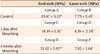
The bleached teeth showed an average change of 3 shades. The mean and standard deviation values for both acid- and laser-etched groups are presented in Table 1. In evaluations with the Welch test, there was a statistically significant difference in bond strength between acid- and laser-etched groups (p < 0.001). In both the acid- and laser-etched groups, the SBS was significantly lower 1 day after bleaching (p < 0.05). When the mean and standard deviation values of bond strengths for the acid-etched groups were evaluated, the teeth bonded 3 weeks after bleaching showed slightly lower bond strengths than the control; however, this difference was not statistically significant (p > 0.05).
A comparison of SBSs for the acid- and laser-etched groups revealed that the laser group showed significantly lower values (p < 0.001). The mean shear strength of the laser-etched teeth bonded 1 day after bleaching was 4.59 ± 2.1 MPa. In this subgroup (Group D), only 4 of the 13 samples (31%) had values above 6 Mpa. In the laser-etched groups, the bonding strength after 3 weeks was the same as in the control group (p > 0.05).
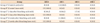
Table 2 shows the bonding failure sites and percentages. In the 3 laser groups, bond failure occurred at the adhesive-enamel interface in all specimens, with no statistically significant difference between groups (p > 0.05). In the nonbleached acid-etching group, all 13 samples had an ARI score of 1, indicating that less than half of the adhesive was left on the tooth. For the group in which the acid etch and bonding procedures were carried out just after bleaching, the ARI scores were as follows: in 6 specimens, there was no adhesive left on the enamel surface; in another 6, there was less than half of the adhesive on the enamel surface; and in 1 specimen, more than half of the adhesive was found on the enamel. In the group that underwent bonding 3 weeks after bleaching, two types of bond failure (ARI 1 and 2) were observed.
DISCUSSION
The results of this study indicate a significant reduction in the SBS of orthodontic brackets bonded to enamel surfaces prebleached with 38% hydrogen peroxide. The ARI scores for these bleached groups showed that the failure generally occurred between enamel and adhesive. The bond strengths of brackets bonded to bleached surfaces after waiting for 3 weeks showed a statistically nonsignificant increase; however, the increase was not enough to reach the unbleached control values. The reason for choosing a duration of 3 weeks to monitor changes in this study lies in the results of the clinical study by Mullins et al.,21 who stated that teeth bleached 2 - 3 weeks before bonding had a higher bond-survival rate than those bleached less than 24 hours before bonding. In the present study, the ARI scores for the group in which bonding was carried out 3 weeks later indicated a tendency to show breaks within the adhesive, similar to the findings in the unbleached control group. Similar patterns of decreased bond strength were noted in studies that used hydrogen peroxide in-office bleaching.3,9,22-26 Contradictory results are seen only in one study, in which the authors concluded there were no significant differences in bond strengths between a control group (nonbleached), an immediate-bonding group (bleaching with 35% hydrogen peroxide), and a 30-day delay group (bleaching with 35% hydrogen peroxide).27 However, in their study, the use of 2 acid etchings before and after the bleaching procedures could have masked the enamel changes created by the bleaching agent. The ARI scores in the study by Uysal et al.,27 however, were very similar to those obtained in the present study and other studies,3,9,22-26 showing that the failure site moves to the enamel-adhesive interface when the bleached surfaces are bonded immediately.
Similar to the findings of the present study, others have reported recovery of bond strengths with different waiting periods after bleaching.9,24,26 One of these studies used 25% hydrogen peroxide for bleaching and reported recovery of bond strengths even after 1 day, and this result was consistent until 1 month, wherein a slight decrease was noted.9 The authors have stated that scanning electron microscopy revealed an apparent decrease in the number of resin tags present in the enamel-composite interface. Polymerization inhibition of the resin bonding agent was proposed to cause the decrease in bond strength. This conclusion was supported by microscopic observation, which suggested that interaction between the resin and residual peroxide in the enamel influenced the bond-strength reduction.8 The results of a study by Abe et al.26 were very similar to those of our study, but the authors chose a 1-week recovery period. After 1 week, SBSs were similar to, but slightly lower than, those in the nonbleached group.
The SBSs of Er:YAG laser-etched groups showed a similar trend to acid-etched teeth in the case of bleached surfaces; however, the mean bond strengths were much lower and standard deviations much higher than in the groups receiving acid etch. Laser etch has been reported to yield a surface suitable for bonding and also resistant to carious attack.15,28 While some studies comparing acid- or self-etching to laser etching have recommended the use of laser etching for orthodontic bonding,15,16,29,30 some have raised doubts over the usefulness of laser etching because of large standard deviations and coefficients of variation and the irregular surface topography caused by the procedure.20 Similar to our results, another study has shown that laser etching led to failure mainly at the enamel-adhesive interface.20 In another study, Martínez-Insua et al.19 compared differences in bonding to acid-etched or Er:YAG laser-treated enamel and dentin surfaces using bond strength tests and scanning electron microscopy. The authors stated that adhesion forces to laser-etched enamel were weaker than those with acid etching, and the bond failure was due to microcohesive fracture of tooth enamel. They explained this cohesive tooth fracture and lower bond strength values by the weakening of enamel by microexplosions. They also found extensive subsurface fissuring after laser etching, which creates less regular and less homogeneous surface patterns that were unsuitable for bonding. In our study, ARI scores for the laser-etched groups showed 100% failure at the enamel-adhesive interface, which demonstrated the inadequacy of bonding to the enamel surface. However, because we did not perform scanning electron microscopy, it is not possible to state if there were enamel microfractures. Less adhesive left on the enamel surface is regarded as less time spent cleaning the teeth after debonding.31 However when it is evaluated together with the SBSs (Table 1) of laser groups recorded in this study, it is evident that the enamel surface had not been adequately prepared for the bonding of the adhesive. The adequate bond strength for orthodontic attachments is reported to be 8 MPa, which is higher than the mean values of laser-etched groups in the present study.32
It is so far impossible to duplicate the oral environment in vitro, because of the complexity of the system and changing stress, humidity, and acidity of the medium. In vitro studies are performed to overcome limitations by simplifying and controlling the conditions affecting the system; however, these limitations should be kept in mind while interpreting the results. The inconsistencies in study results can arise from variations in testing procedures, such as different kinds of specimens used (bovine/human enamel), the sequence of applications (e.g., etch procedure before and/or after bleaching), fluoridation differences in the study design, treatment chemicals and their concentrations, treatment durations and application frequency (cycling vs. single application), and the use of different technical devices. Besides these variations and limitations, some studies report different results with the same material and methods. It has been explicated that coordinated bleaching-bonding is both a technique-sensitive and system-specific treatment.26
Even though thermocycling is one of the recommendations of International Organization for Standardization for in vitro studies, reviewing the literature on this subject reveals that most studies have omitted this procedure. Another important concern is that in most studies, the authors have neglected to report on effectiveness of the bleaching agent they used.
CONCLUSION
In-office bleaching with 38% hydrogen peroxide resulted in dramatic reduction of shear bond strength of orthodontic attachments, with ARI scores indicating that the failure site switched from within the adhesive to the enamel-adhesive interface in the acid-atched teeth.
Er:YAG laser etching of bleached teeth resulted in clinically unacceptable low bonding strength.
Waiting for 3 weeks before bonding attachments to bleached surfaces resulted in similar, but not exactly the same, bond strength values as those obtained in nonbleached surfaces with acid etching; however, in laser-etched groups, the bonding strength after 3 weeks was the same as that in the nonbleached group.
When teeth bleached with 38% hydrogen peroxide are meant to be bonded immediately, acid etching is preferable.
 XML Download