

 PDF
PDF ePub
ePub Citation
 Print
Print


INTRODUCTION
The majority of researchers recognize that there is variability in the size and shape of the human arch form. Several classification schemes have been suggested, but the three basic arch forms that are commonly described by clinicians are tapered, ovoid, and square arch forms.1 Rudge2 believed that occlusion and arch shape are determined by the interplay between genetic factors and a wide variety of external environmental factors. Cassidy et al.3 suggested that arch size and shape are determined more by environmental influences than by genetic inheritance. In a study of teenage twins, however, researchers found a high genetic contribution to variation in dental arch dimensions.4 Determination of arch form is vital in clinical orthodontics for esthetics and for long-term occlusal stability through the maintenance of the original mandibular inter-canine width and preservation of the original arch form.5
Lavelle et al.6, measured the dental arches of adult men in four major ethnic groups: Caucasoid, Mongoloid, Negroid, and Australoid. They concluded that there were some basic differences in dental arch size and shape between the different populations. Studies of other populations have further supported these findings.7-11
Ethnic Malays make up the largest ethnic group in Malaysia. The Malaysian Aborigines (subsequently referred as Orang Asli) are the indigenous people of Peninsular Malaysia and are the descendants of the earliest inhabitants of the area.12 Although some of the Orang Asli still live in isolation from other communities and preserve their traditional way of life, many of them have integrated with urban communities to achieve a better quality of life.13 The Orang Asli may seek orthodontic treatment following urbanization. Given the anthropological and ethnographical similarities between the Orang Asli and ethnic Malays, a comparison of the dental arch forms in these ethnic groups may help clinical orthodonists to determine the esthetic requirements and to ensure the long-term occlusal stability of these patients.
There have been very few studies of dental arch dimensions among indigenous people throughout the world. In Malaysia, three studies have been conducted to describe and analyze the dental arch characteristics of ethnic Malays.14-16 However, none of these studies investigated the relationship between dental arch characteristics and orthodontics. We therefore investigated the morphological differences between ethnic Malay and Orang Asli arch forms in Peninsular Malaysia and determined the frequency distribution of arch shapes in these two groups. The findings from this study may serve as population study data and a database for future comparisons.
MATERIALS AND METHODS
We examined 249 study models. Of these, 120 (60 maxillary, 60 mandibular) were from 60 ethnic Malays (30 males, 30 females) that were obtained from the Orthodontic Department, Faculty of Dentistry, University of Malaya, and 129 (66 maxillary, 63 mandibular) were from 129 Malaysian Aborigines (35 males, 94 females) that were obtained from the Centre of Malaysia Pribumi Studies Databank. The models that were selected for study were of Malay or Orang Asli ethnicity (up to second generation). They were from non-growing, postpubertal adults aged 18 - 50 years, had intact permanent dentition from the left first permanent molar to the right first permanent molar, had normal tooth size and shape, and had a ≤3-mm arch length discrepancy. Models were excluded if they had a history of orthodontic treatment, retained any primary teeth, were damaged or fractured, had active pathology, or had restoration extending to contact areas, cusp tips, or incisal edges. Ethical approval was obtained from the Research Ethics Committee, Faculty of Dentistry, University of Malaya.
Cast preparation
Eighteen buccal cusp tips and incisor line angles were marked using 2B pencils on each maxillary and mandibular model. The occlusal sufaces of the maxillary and mandibular models were scanned using a Panasonic KX-MB772 all-in-one scanner (Panasonic, Osaka, Japan). A ruler was included in the scans to permit magnification correction. The scan ned images were digitized using Engauge version 4.1 (Free Software Foundation Inc., Boston, MA, USA). The software automatically defined each point in a 2-dimensional coordinate system with associated x and y values.
Arch dimension measurements
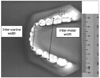
Using the computer software, we measured inter-canine width, defined as the distance between canine cusp tips. We also measured inter-molar width, defined as the distance between the first molar mesio-buccal cusp tips (Figure 1): Marked landmarks on scanned study models were selected on the computer by one investigator, and the measurements were calculated by the software. We also calculated the ratio of inter-canine width to inter-molar width.
Statistical analysis
Data were analyzed using the SPSS version 12.0 (SPSS Inc,. Chicago, IL, USA). We examined the normality of data for each variable, and calculated descriptive statistics. Independent t-tests were used to examine differences in arch measurement dimensions between genders and ethnic groups. A chi-square test was used to examine the statistical significance of any association between ethnic group and arch shape. The level of significance used in this study was p < 0.05.
Methodological error
We took duplicate measurements of all variables to determine the degree of error associated with the measurement method. We used the method described above to re-scan and digitize 10 randomly selected study models. One week after the first set of measurements were taken, the inter-canine width and inter-molar width measurements were measured by the same investigator (E.S.X.) using the same protocol. The investigator was blind to the first measurements when measuring the models for the second time. The intra-examiner reliability was measured using the Intra-Class Correlation Coefficient (ICC) and paired t-tests. The ICC value was 0.99 for both inter-canine and inter-molar widths, indicating that the measurements were highly consistent. The first and second measurements were not significantly different for either inter-canine or inter-molar width (p = 0.963, 0.946). This also indicates that the measurements were reliable.
Similarly, the arch shape measurement error was determined by re-measuring a set of randomly selected models using the same equipment and method as described above. The investigator (L.S.Y.) was blind to the first results when examining the scans for the second time. The intra-examiner reliability was measured using a Kappa test to determine agreement between measurements and a McNemar test to determine the difference between the first and second measurements. Both tests showed that the superimposition and selection of best fit arch shape was consistent and reliable. The Kappa value was high at 0.787 and the McNemar test revealed no significant differences between the two readings (p = 0.083).
RESULTS
The arch dimension measurements and independent t-test results for the ethnic Malay males and females are shown in Table 1 and the measurements and t-test results for Orang Asli males and females are shown in Table 2. We found no significant sexual dimorphism for any of the variables for either the maxillary or mandibular arches in either group.
The maxilla and mandible arch dimension measurements are compared between ethnic Malays and Orang Asli in Table 3. None of the measured variables were significantly different between the ethnic Malay and Orang Asli groups.
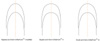
The frequency distribution of maxillary and mandibular arch shapes in ethnic Malays and Orang Asli groups are presented in Table 4. Overall, approximately 60% of the samples had ovoid maxillary arches and included 65% of the ethnic Malay samples and 55% of the Orang Asli samples. None of the ethnic Malays and only 1.5% of the Orang Asli had square maxillary arches. The remaining samples (35% of the ethnic Malay samples and 44% of the Orang Asli samples), had tapered maxillary arches. Approximately 56% of all samples had tapered mandibular arches, including 50% of the ethnic Malays and 62% of the Orang Asli. The next most common mandibular arch shape was ovoid (37%). Only 6.5% of all samples had square mandibular arches. The samples with square mandibular arches included 1.7% of the ethnic Malays and 11% of the Orang Asli.
The square arch shape was omitted from further analysis, due to the very small number of samples with this shape. There were no significant differences observed in the maxillary arch shape between genders either among Malays or among Orang Asli. However, among Malays, approximately 66% of males had tapered mandibular arches, while approximately 66% of females had ovoid mandibular arches (p = 0.027). No such difference was observed among the Orang Asli. There was no significant association found between maxillary arch shape and ethnicity either in males, females, or in both genders combined. However, more female Orang Asli had tapered mandibular arches than did female Malays (p = 0.022). Similarly, when both genders were considered together, more Orang Asli had tapered arches than did Malays (p = 0.040).
DISCUSSION
The ethnic Malay study models we used in this study were equally distributed between males and females, but the Orang Asli dental study models were not. Only 35 (27.13%) of the 129 Orang Asli study models were from male subjects. This was largely due to difficulty in recruiting male Orang Asli subjects as study models were gathered during the daytime when most of the Orang Asli males had gone to work. Additionally, some of the men who were present in the settlements at the time that the study was conducted refused to participate in the study.
Different methods have been used to measure dental arch dimension and shape. The most widely used method for measuring dental arch dimensions is direct measurement using a calliper on dental study models, as in the studies conducted by Ling and Wong,9 Lara-Carrillo et al.10, Barrett et al.17, Tibana et al.18 and Radzi et al.19 The measurement method we used in this study was based on the digital method used by Burris and Harris.7 We used computer digitizing software to reduce the time needed to measure arch dimensions. However, we could not take three-dimensional measurements using this method, and ommision of the third dimension mayhave been a shortcoming.
In studies of dental arch shape, a variety of different land marks have been used. The most commonly used landmarks are the incisal edges and cusp tips that we used in this study , and that were used in studies carried out by Burris and Harris7 and Ling and Wong.9 Nojima et al.5 and Kook et al.8, however, used clinical bracket points as landmarks in their studies. These bracket points corresponded to bracket slot points that were mathematically estimated from the most facial portion of the proximal contact area of each tooth. Kook et al.8 argued that using clinical bracket points as landmarks for measurement of dental arch shapes was of greater value for modern orthodontic treatment than the conventional incisal-edge and cusp-tip landmarks, because preformed superelastic archwires are frequently used for clinical treatment.
Because aborigines have limited access to dental services, e.g., dental scaling, supra-gingival calculus frequently forms on the buccal surfaces of the first molar in the maxilla and on the lingual surfaces of mandibular anterior teeth.20 We used incisal edges and cusp tips as landmarks in this study, because the calculus frequently seen on aboriginal teeth might introduce error into estimates of clinical bracket points made from Orang Asli study models. The Orang Asli study models showed severe tooth wear in comparison to those of ethnic Malays. Nonetheless, anatomical cusp tips were used as landmarks on all models, even if abrasion had occurred. Significant occlusal tooth wear from mastication can result either from a large amount of abrasive substances in food boluses or from acid softening of enamel and dentine that results in altered cusp morphology, such as rounding or cupping of cusps and grooves on incisal edges.21,22 Caglar et al.21 found that 20% of central incisors, 62.5% of lateral incisors, 78.5% of canines, 83.3% of first premolars, 89.4% of second premolars, 89.7% of first molars, 82.1% of second molars, and 85% of third molars are worn. Nishijima et al.23 carried out a study of occlusal tooth wear in female rats and found out that tooth wear increases with age. Therefore, there is a certain degree of discrepancy between the original and estimated locations of cusp tips.
Many recent studies have favored the use of mathematical formulae to represent human dental arch forms, as in Braun et al.24 We used the method described by Nojima et al.5 and Kook et al.8 The determination of arch shape using this overlay method is subjective. However, in this study, there was 78.7% agreement between first and second arch shape selection, indicating consistency in the superimposition and selection of best-fit arch shape. This method is also limited by the small range of available arch-shape template sizes in comparison to the wide range of sizes of ethnic Malay and Orang Asli dental arches. The three-dimensional imaging technology would have been better to identify the clinical arch form25-27 and worth considering in future research.
As people of different genders and ethnic groups may present with varying dental arch widths, sizes, and shapes, clinicians must recognize the pre-treatment arch form of patients in order to determine the most suitable form of arch wire before commencing orthodontic treatment. The size and shape of dental arches have considerable implications for orthodontic diagnosis and treatment planning, and affect the space available, dental esthetics, and post-treatment dentition stability.28
Inter-canine width
Inter-canine width can serve as a basis for estimation of the total width of the maxillary and mandibular anterior teeth.29 The results from this study showed that sexual dimorphism and ethnic differences do not exist among ethnic Malays and Orang Asli. This differs from results reported by Burris and Harris,7 Ling and Wong9 and Lara-Carrillo et al.10, indicating significant differences in inter-canine width between ethnic groups, and from studies by Kook et al.8, Ling and Wong,9 Lara-Carrillo et al.10, Khin et al.14, Hussein et al.16, and Barrett et al.17, indicating significant differences between genders in inter-canine width. However, Nojima et al.5 also found no significant differences between ethnic groups in terms of dental arch dimensions. In addition, the mean difference between the inter-canine widths of males and females, or of Malays and Orang Asli, observed in this study were too small (<2 mm) to have any obvious significance for orthodontic treatment planning. Mean inter-canine width generally increases in both dental arches during treatment of all types of malocclusion, and tend to return close to or narrower than the original width after orthodontic treatment.29 Therefore, the data collected in this study may serve as a guide for planning the eventual arch dimensions for Malay and Orang Asli patients so that the arches are not expanded beyond their established dimensions.
Inter-molar width
We did not find any statistically significant sexual dimorphism or ethnic differences in inter-molar width among ethnic Malays and Orang Asli. This is contrary to results obtained by Burris and Harris,7 Ling and Wong,9 and Lara-Carrillo et al.10, who all found significant differences in inter-molar width between ethnic groups, and to the results of Kook et al.8, Ling and Wong9, Lara-Carrillo et al.10, Khin et al.14, Hussein et al.16, and Barrett et al.17, who found significant differences in inter-molar width between genders. The variability in dental landmarks used for measurement in these studies may have contributed to the differences in results. In this study, the mesiobuccal cusp tips of the first permanent molar were used as the reference points. However, there have been no studies comparing measurements made using various dental land marks. Our results agreed with those of Nojima et al.5, who did not find dental arch dimensions to be different between ethnic groups. In addition, the mean difference between the inter-molar widths of males and females, or of Malays and Orang Asli, that we observed in this study were too small (<2 mm in most cases) to be useful for orthodontic treatment planning. While most dif ferences between males and females were less than 2 mm in this study, ethnic Malays had an inter-molar width difference of slightly more than 2 mm (2.33 mm) between genders (Table 1). A difference of this size may be significant for selection of arch wire for orthodontic treatment. Therefore, care must be taken during treatment planning to avoid over-generalizing cases.
Inter-canine width/inter-molar width ratio (ICW/IMW ratio)
While there have been no studies directly comparing inter-canine width to inter-molar width, Harris30 concluded that inter-molar distance widens with age, but intercanine width remains unchanged. This results in the arch shape becoming more tapered with age. Kook et al.8 reported that there were significant decreases in inter-canine and inter-molar width as the mandibular arches changed in form from square to ovoid to tapered. Similarly, Nie and Lin31 found that posterior arch width contributed to the observed shape difference between males and females. It can be inferred from these findings that smaller ICW/IMW ratios are associated with broader arches.
We compared the ICW/IMW ratios between gender and ethnic groups and found no statistically significant differences in the maxillary and mandibular ICW/IMW ratios between ethnic Malays and Orang Asli, or between genders in either ethnic group. The obvious close resemblance in ICW/IMW ratios of ethnic Malays and Orang Asli imply that dental arch shape distribution between the two ethnic groups are similar.
Dental arch shape
Ovoid arches were the most common maxillary arches among both ethnic Malays and Orang Asli in this study, followed by tapered arches and square arches. On the other hand, the most frequent mandibular arch shape in both ethnic groups was tapered, followed by ovoid and square. The only statistically significant ethnic difference in dental arch shape frequency was found in the mandible generally (p = 0.040) and in the mandibular arches of females, specifically (p = 0.022). This finding is not in accordance with those of Burris and Harris,7 that indicated African Americans have squarer maxillary dental arches than Caucasians. Our results validated our prediction of a similar dental arch shape distribution between ethnic Malays and Orang Asli, based on the strong correspondence of ICW/IMW ratios between these two ethnic groups.
We found no correlation between dental arch shape and gender in either ethnic Malays or Orang Asli, except in the frequency distribution of mandibular arch shape (tapered and ovoid) among ethnic Malay males and females (p = 0.027). Burris and Harris7 similarly found that the shape of the dental arches was similar between genders within American ethnic groups. The lack of disparity in dental arch shape distribution between the sexes we observed in this study is not consistent with the results obtained by Nie and Lin31 that indicate dental arch shape is very different in males and females (p < 0.001). Therefore, further research involving a larger sample size with a more balanced gender distribution should be conducted to verify the association between gender and dental arch shape.
CONCLUSION
Ovoid shape was the most common maxillary arch shape in both ethnic Malays and Orang Asli groups and the most common mandibular arch shape in females, whereas square arch shape was the rarest in all groups. There were no significant differences between the dental arch dimensions and shape distributions of the ethnic Malays and Orang Asli. Therefore, arch wire selection for orthodontic treatment of these ethnic groups may not be affected by these factors.


 XML Download