

 PDF
PDF ePub
ePub Citation
 Print
Print


INTRODUCTION
Orthodontic brackets should be manufactured precisely,1 because they must be 3-dimensionally suitable for the form and characteristics of each tooth and have adequate strength and solidity to enable definite correction with orthodontic wires.2,3 The frictional resistance of a bracket increases as the orthodontic wire becomes thicker; such resistance, which acts against tooth movement, must be optimally regulated so that more ideal, low orthodontic forces can be applied.4,5
Although metallic materials, such as stainless steel, possess an excellent level of anti-corrosion, metal orthodontic brackets can corrode upon exposure to potentially harmful physical and chemical substances in the oral cavity for several months or even several years.6,7 Corrosion can be clinically defined as loss of metallic products or conversion to an oxide. In general, the formation of a strong oxide layer on a metallic surface is considered beneficial, but the formation of a weak oxide film is harmful because of rusting. This corrosion reduces the volume of orthodontic brackets, subsequently decreasing orthodontic forces, and causing cracking in areas of stress concentration.8 Ion release secondary to corrosion also results in fracture of orthodontic brackets, poor clinical outcomes, local hypersensitive reactions, and general deterioration of health when toxic products are absorbed locally or systemically.9-12
Permanent discoloration of enamel secondary to corrosion at the base of orthodontic brackets has been reported, 13,14 and attempts have been made to improve the corrosion and discoloration resistance of metal orthodontic brackets by enabling the formation of a TiN film using the ion plating method.6,15
Orthodontic brackets were introduced in the mid-1980s in Korea and nowadays, expensive products manufactured in the United States, Japan, and Germany are commonly used in most dental clinics. Domestic manufacturers started to produce and sell orthodontic brackets in the early 2000s. Moreover, brackets manufactured overseas that are not yet verified are being imported and distributed in the country. Despite these developments, national standards for such products have not been established and international standards explain only the related terms but do not regulate detailed requirements.
This in vitro study was performed to evaluate the physical, chemical, and biological properties of commercially available metal orthodontic brackets in Korea.
MATERIALS AND METHODS
Brackets
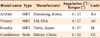
Four brands of commonly used metal orthodontic brackets manufactured in different countries were tested: Archist (Daeseung Medical, Seoul, Korea; KA), Victory (3M Unitek, Monrovia, CA, USA; AV), Kosaka (Tomy, Tokyo, Japan; JK), and Confidence (Shinye Odontology Materials, Hangzhou, China; CC). Table 1 lists the brackets analyzed in this study.
Dimensional accuracy test
Dimensional accuracy was tested to determine whether the brackets meet the criteria stated by the manufacturers. Five upper central incisor brackets (#11) per brand (n = 20) were randomly selected and measured for angulation, torque, and manufacturing errors in these parameters.
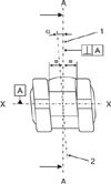
To evaluate angulation, the faces of the brackets were photographed by optical microscopy at a magnifying power of 25 and their angulations were measured with a computer-based measuring tool (I-Solution, Olympus, Tokyo, Japan), as shown in Figure 1 [ISO 27020: 2010].16 To evaluate torque, the samples were embedded in epoxy resin to minimize measurement errors due to the curvature of the bracket base, and the profiles were obtained by microgrinding (EXAKT 310 CP Macro Band, EXAKT Technologies, Oklahoma, OK, USA). Then, torque was measured as shown in Figure 2, and the absolute values were analyzed.
Furthermore, the differences between the measured and the standard values were divided by the standard values to determine manufacturing errors in angulation and torque, and the resultant values were charted. All the measurements were performed twice to reduce photographic errors.
Cytotoxicity analysis
Cytotoxicity was evaluated through an agar overlay test. Mouse fibroblasts (L929, 2.5 × 10⁵ cells/mL) were cultivated at 37℃ in 5% CO2 for 24 hours after suspension in liquid medium and adhered by dropping onto 100-mL dishes. Agar and the medium were mixed (1:1) and condensed when the cells covered more than 80% of each dish, and the mixture was dyed with 0.3% neutral red solution.
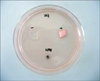
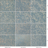
Two brackets (#11 and #13) from each brand (n = 8) were used after ethylene oxide gas sterilization. The samples were placed on the prepared dishes and latex and glass slide pieces were used as the positive and negative controls, respectively (Figure 3). After the samples and controls were incubated at 37℃ in 5% CO2 for 24 hours, the diameter of discoloration around the specimens was measured and the level of cellular destruction within the discolored area was observed under microscopy.
Cytotoxicity was assessed with the measured values obtained only when clear destruction was observed in the positive control group and no discoloration or cellular destruction was noted in the negative control group. A level above grade 2 was considered indicative of cytotoxicity [ISO 10993-5:2009].17
Compositional analysis
The composition of the brackets from the 4 brands was analyzed by inductively coupled plasma atomic emission spectrometry (Optima 3000, PerkinElmer, Wellesley, MA, USA).
Elution test
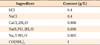
The elution of metal ions from the brackets over time was analyzed in artificial saliva (pH 6.5) produced by the Fusayama-Meyer method (Table 2).
Five brackets per brand (n = 20) were immersed in 15-mL tubes containing 2-mL artificial saliva and incubated at 37℃ in 5% CO2 for 24 hours. The solution in the tubes was collected by using pipettes, refrigerated, and replaced with fresh artificial saliva. This procedure was repeated on days 7, 14, and 28.
For analysis, 2 mL of artificial saliva was diluted with 48 mL of fresh artificial saliva, and a part of the diluted solution was dried and dissolved in 3 mL of HCl and HNO3. The dissolved solution (n = 80) was analyzed by inductively coupled plasma mass spectrometry (Elan 6100, PerkinElmer, Houston, TX, USA).
Corrosion analysis
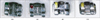
Corrosion of the brackets was analyzed by potentiodynamic polarization. Five samples from each brand (n = 20) were prepared for this experiment. In brief, an electric wire was attached to the solder of each bracket, and the brackets were embedded in epoxy resin. They were then ground to expose their base (Figure 4).
A potentiodynamic polarization device was connected to the corrosion cells. Reduction was forced for 10 minutes at -1,000 mV and the electric potential was activated for 10 minutes to stabilize the cells. The electric potential for corrosion was started at -100 mV and increased at a rate of 1 mV/second up to 2,000 mV to observe the changes in the electric currents. Ez (zero current potential), Ec (active peak potentials), Ep (breakdown potential), and I300 (current density at potential of Ez + 300 mV) were assessed from the polarization curves [ISO 10271: 2011].18
Statistical analysis
Data are expressed as means (standard deviation). SPSS for Windows (version 12.0; SPSS Inc., Chicago, IL, USA) was used for statistical analysis. The Kruskal-Wallis nonparametric test was used to assess differences in the experimental results and the Duncan's multiple comparison test was used to identify individual differences. A p-value less than 0.05 was considered significant.
RESULTS
Dimensional accuracy
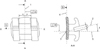
Figures 5 and 6 show the frontal and cross-sectional views of the tested brackets, respectively. Most of the sectioned slots were even and clearly formed, but the CC slots appeared to be uneven and their angles were not clear.
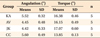
Angulation was not significantly different among the products (Table 3), because the standard values of the manufacturers were similar. CC brackets showed a difference in torque; however, this difference was not interpreted as significant because the standard torque value of every manufacturer was different.
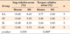
Table 4 shows the deviation (manufacturing error) of the measured values from the standard values. AV and CC brackets showed the largest deviation in angulation and torque, respectively. Although the difference in torque was significant (p < 0.05), the difference in angulation was not.
Cytotoxicity
Discoloration was not clearly observed under the samples or in the surrounding areas. Further, the cells and their density around the samples did not appear different from those in the negative control group. In other words, obvious cytotoxicity was not identified in any specimen (Figure 7).
Composition
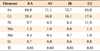
Fe, Cr, and Ni were the predominant metal ions, but small volumes of Mn, Mo, Si, and Ti were also identified. JK, CC, and KA brackets had relatively higher amounts of Fe, Ni, and Cr, respectively (Table 5).
Elution
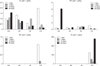
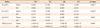
As shown in Figure 8, the eluted components did not have a regular increasing or decreasing trend over time. The volume of eluted Cr, Mn, and Mo was significantly different at times, but the total eluted volume over 28 days was less than 10 ppb, indicating low risk of elution. However, the volume of eluted Ni from CC brackets was about 20 - 30 times higher than that from the other products (p < 0.05).
Table 6 shows the volume of the total eluted metal ions. The volume of eluted metal ions from CC brackets was significantly larger than that from the others (p < 0.05), possibly due to excessive Ni elution. Otherwise, KA brackets showed the greatest volume of eluted metal ions, but this finding was not significant.
Corrosion resistance
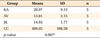
CC brackets had the lowest Ez (-0.368 mV) and Ec (-0.014 mV) values (p < 0.05). Further, they had the lowest Ep value, but this measurement was not significantly different. The I300 value of KA brackets was the smallest, but no significant difference was detected with this measurement either (Table 7).
DISCUSSION
Applying accurate force through orthodontic brackets and wires is a decisive factor in the success or failure of orthodontic treatment. However, orthodontic treatment with precise angulation and torque is challenging because the tooth morphology and reactions to orthodontic forces can vary.
Precise regulation of brackets is necessary for accurate manufacturing of such products. Therefore, dimensional accuracy is extremely important, but research regarding this matter is lacking. Oh et al.19 evaluated various physical and chemical properties of custom-made bracket and commercially available brackets, but their analysis of dimensional accuracy was limited to the slot size and horizontal and vertical dimensions of the bracket wing, which are relatively easy to measure. Angulation and torque of the bracket slot, which are complicated parameters, were measured in the present study. About 10.4 - 13% of the brackets showed manufacturing errors in angulation, and AV brackets appeared to have the largest, deviation from the standard values, though this finding was not significant. Furthermore, about 2.6 - 15.4% of the brackets showed manufacturing errors in torque, and CC brackets showed the largest deviation, which was significant. Nonetheless, inaccuracy of the tangential, perpendicular, and median lines drawn for the measurements and distortion of facets due to 2-dimensional photography should be considered and accounted for while interpreting the results.
All dental materials must be biocompatible, and orthodontic brackets, which are in direct contact with teeth and are exposed to saliva, should not contribute to any toxicity due to metal ion release from their surfaces. 11,12,19-22 In the present study, orthodontic brackets were placed on a cultivated cellular medium under simulated oral conditions for 24 hours; however, no clear evidence of cytotoxicity was noted. Oh and Kim23 researched the impact of heating and cooling on the amount of metal ions released from stainless steel wires and their cytotoxicity: wires that did not undergo heat treatment (control group) showed minimal toxicity. Freitas et al.24 used stainless steel orthodontic wires as a negative control group (nontoxic group) to assess the cytotoxic impact of silver solder on fibroblasts. Although various metallic materials may not initially display any cytotoxicity in the form of finished products, they may eventually become cytotoxic when exposed to the oral environment for extended periods and metal ions are released.21,25 Therefore, cellular reactions should have been observed in the present study. This proved difficult, however, because the fibroblasts underwent apoptosis when cultivated for longer than 24 hours due to the high density of cells, even in the absence of toxins. This may have led to an inaccurate result, and the method used in this study may be inadequate for cytotoxicity testing. Moreover, previous studies11,20-22 studying the cytotoxicity of orthodontic materials used human gingival fibroblasts collected during premolar extraction in pedodontic patients undergoing orthodontic treatment, as opposed to mouse fibroblasts. Further research using human gingival cells and longer cultivation periods is needed for greater clinical relevance.
The compositions of the brackets were investigated to assess their risk of corrosion and secondary products. Fe was detected in the range of 65.8 to 72.7 wt% in all of the products. In particular, Ni was detected in the 6.4 - 11.9 wt% range. Ni in stainless steel alloys is thought to improve processing and increase ductility of products, but eluted Ni ions cause allergic reactions in vivo.10,26,27 Ni ions are highly related to the corrosion resistance of metals; however, metal ions are not easily eluted in vitro when brackets have a high level of solid solubility or dense oxide film on the surface.28 Cr is primarily used to prevent corrosion as it forms a dense protective oxide film, such as Cr2O3 on the surface, once it is distributed evenly on the crystal grain, comes into contact with air and as long as the appropriate concentration is maintained in the alloy.21,22,28 Cr was present from 16.1 wt% to 20.4 wt% in this study. Mn, Mo, and Si were also found in small amounts.
The characteristics of eluted reactive products should be observed under various conditions and over time, because dental materials present in the oral cavity for a long time can react with environmental factors. Dental implants induce less reaction in connective tissues due to the formation of protective films, although they are more invasive than orthodontic devices. On the other hand, orthodontic brackets or wires induce various and persistent reactions.7,25 In this study, the eluted metal ions in artificial saliva did not show clear patterns of persistent increase or decrease over time, but CC products showed the largest amount of Ni elution. The release of Ni seems to be more closely related to the structure and manufacturing methods of brackets rather than to the concentration of Ni in the brackets, considering that all the products contained Ni to varyingregardless of its concentrations.27,29 Small amounts of Cr were eluted from most of the products initially, and the volume decreased or increased over time. Less that 10 ppb of Mn and Mo was also eluted during the 28-day period. However, the levels were judged to carry low risk compared with the recommended dietary allowances (Cr, 35 µg; Mn, 2.3 mg; Mo, 45 µg).30 Moreover, according to the international standards for metallic materials used in prosthodontics, the volume of the total eluted metal ions should not exceed 200 µg/cm2 when the metallic materials are exposed to corrosion test solutions for 7 days. All the products in this study satisfied this criterion. However, CC products appeared to have a significantly larger total elution volume possibly due to excessive elution of Ni. Metallic corrosion is influenced by various factors such as intraoral pH,20,27 temperature,28 internal stress, friction of brackets and wires due to constant movements,29 dental plaque and its secondary products, and oral flora.25 However, in this study, metal ion release was analyzed in artificial saliva for only 28 days, without accounting for such influential factors. Considering that brackets, as opposed to other orthodontic materials, are exposed to intraoral conditions for several months or years, an experiment with a harsher condition for corrosion, such as lower pH of the immersion medium or abrupt change in temperature, is warranted.
In corrosion testing, a negative electric current will pass through the brackets if the voltage increases from -0.1 V and a reduction reaction will occur on the surface of the brackets until it reaches the Ez. The increasing voltage causes an oxidation reaction on the surface of the brackets, increasing the positive electric current on the surface, and a passivation film is formed (the state at which the increased voltage does not increase the electric current) when it reaches the threshold of electric current (I300). This passivation film can resistan corrosion, and the corrosion resistance is higher and lasts longer when the passivation film is formed early.8 In other words, the corrosion resistance is considered to be high when the threshold of electric current to form the passivation film is lower and the threshold of voltage at which the passivation film is destroyed (Ep) is higher. Although KA products had the lowest I300 and highest Ep, indicating the best corrosion resistance, these values were not significant. This result can be explained by the fact that KA products contain the highest Cr concentration, increasing the corrosion resistance.21,22,28 On the other hand, CC products showed significantly low Ez and Ec values, indicating low corrosion resistance and that corrosion started early and that the passivation film became unstable earlier. Potentiodynamic polarization facilitated objective assessment by allowing numeric measurement of the level of corrosion on the metallic surface. However, the reliability of the experiment was low, because the standard deviation of each measured index was too large in this study. The large deviations could have occurred because the smaller bracket base was polished and used despite the fact that the surface of the sample should be at least 10 mm2, according to ISO 10271.18
Newly developed orthodontic materials and methods will lead to a continuous evolution of orthodontic treatment, just as the development of direct bonding systems has incomparably extended the level and range of fixed orthodontic treatments. However, the biostability of orthodontic materials in the oral cavity should be continuously maintained and guaranteed by establishing thorough testing methods and criteria for newly developed materials.
CONCLUSION
Through dimensional accuracy measurements, no differences were found between the products in manufacturing errors of angulation, but CC showed a significant difference in manufacturing errors of torque (p < 0.05).
None of the products showed cytotoxicity in the cytotoxicity assessment through the agar overlay test.
In the elution test, the volume of eluted metallic components did not show regular patterns over time, but there was an increase in final volume.
CC showed the highest volume of eluted metallic components due to excessive Ni elution (p < 0.05).
CC showed significantly low values of Ez and Ec in potentiodynamic polarization, indicating that the corrosion resistance of CC was low (p < 0.05).
KA, AV, and JK showed relatively favorable results when compared to CC. The present results can be applied to establish national standards for orthodontic brackets and to evaluate commercially available products. When compared to such standards, brackets with smaller manufacturing errors, less leaching of metal components and greater corrosion resistance are needed.


 XML Download