

 PDF
PDF ePub
ePub Citation
 Print
Print


INTRODUCTION
Orthodontically induced root resorption (OIRR) is a sequela of orthodontic treatment that affects nearly 100% of treated patients.1 OIRR can induce cementum and dentin damage, depending on its severity. Although the normal root structure might later be partially repaired by cementum, severe resorption can destroy it, leading to the failure of orthodontic treatment. Because of its harm to dental health, great efforts have been made to identify mechanotherapies that might reduce or eliminate root resorption.
OIRR is related to total apical displacement, treatment duration, age, gender, malocclusion type, root shape, type of appliance, treatment modality, and force regimen.2 However, a hypofunctional periodontium may also contribute to OIRR.3 The primary cells responsible for this process are the odontoclasts.4 A group of proinflammatory factors and enzymes are involved in the differentiation, maturation, and activation of these cells.5 Increased knowledge of this process has caused investigators to focus on certain inhibitors as potential treatments. Fangchinoline (C37H40N2O6) is an alkaloid that is extracted from bisbenzylisoquinoline and is widely used in Asian countries as an anti-inflammatory drug. Fangchinoline acts through more mechanisms, and is safer, than other anti-inflammatory agents.
Fangchinoline inhibits 90% of interleukin (IL)-1 and tumor necrosis factor-alpha (TNF-α) production at a dose of 10 µg/mL.6 Fangchinoline also partially inhibits cyclooxygenase and human IL-6,7 is a nonspecific calcium channel inhibitor,8,9 and has an antithrombosis function.10 At the same time, fangchinoline has effective antioxidant and radical-scavenging activity.11 Our laboratory recently found fangchinoline to be a potential cathepsin K (CK) inhibitor. Fangchinoline is an excellent candidate for inhibiting root resorption because it is safe and has multiple anti-inflammatory effects.
The aim of this study was to examine the effects of fangchinoline on root resorption during tooth movement. We hypothesized that fangchinoline would limit root resorption during orthodontic tooth movement.
MATERIALS AND METHODS
Animals
All experimental procedures were approved by the Institutional Animal Care and Use Committee of the local district government and the Animal Care Commissioner of Jilin University.
We used an online sample-size calculator for clinical trials and scientific experiments (hedwig.mgh.harvard. edu) to determine the appropriate sample size for a study with a statistical power of 0.8. Twenty-four 8 week old male Wistar rats, with an average weight of 200 g, were purchased from the Animal Center of Jilin University. Rats were randomly allocated to 1 of 6 control or treatment groups. There were 4 rats in each group. Rats in the treatment groups had either 50 g or 100 g of orthodontic force applied to their maxillary right first molars, while control rats were not subjected to orthodontic force (0 g). Treatment and control groups were further divided into 2 subgroups, based on whether they received fangchinoline injections. Rats in the 0 g(+), 50 g(+), and 100 g(+) groups received 40 µL of 5 µg/µL fangchinoline every 2 days. Rats in the 0 g(-), 50 g(-), and 100 g(-) groups received no fangchinoline.
We based the dose of fangchinoline on a study by Onai et al.6 and the unpublished results of our earlier study. All of the rats were observed for 7 days prior to beginning the experiment, to ensure that they were healthy. Rats were fed a soft diet during the 14 days experiment.
Experimental protocol
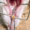
Orthodontic appliances were installed on the rats while under anesthesia with SuMianXin (Institute of Military Medicine Science, Changchun, China) (0.2 mL/kg) IM. The appliances consisted of a nickel-titanium coil spring (ShengMaTe Co., Shanghai, China) tied between the maxillary incisors and right first molar (Figure 1). The force of the springs was measured using an ergometer before installing them. We covered the end of the ligature wire with a piece of resin in order to prevent appliance loosening.
Fangchinoline injection
In our experiment, 200 µg fangchinoline was given every 2 days. Twenty milligrams fangchinoline (YuanYe Co., Shanghai, China) was added to 4 mL distilled water, forming an injectable suspension with a concentration of 5 µg/µL. We injected 40 µL fangchinoline every 2 days into the subperiosteum adjacent to the maxillary first molar of the rats in the 0 g(+), 50 g(+), and 100 g(+) groups. The suspension was stored at 4℃ and stirred before each injection.
X-ray and scanning electron microscope (SEM) observation
All rats were killed by decapitation at the end of the experiment. After 14 days of force application, the amount of tooth movement was measured on cephalometric radiographs, as previously reported.12 The maxillary right first molar and the adjacent alveolar bone was extracted in toto for further examination. All tissues were soaked in 5% sodium hypochlorite solution for 12 hours, at which time the alveolar bone was delicately removed, exposing the 5 roots of the first molar. The periodontal ligament residue of the distobuccal and distopalatal roots were then removed and carefully cleaned to reveal clear root surfaces. All teeth were dried and observed under an SEM (JSM-6390A; JEOL Ltd., Tokyo, Japan) at the same distance and orientation. Images of the mesial surfaces of both distal roots were saved as digital photographs.
Evaluation and statistical procedure
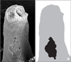
The distance between the contact points of the first and second molar in each rat was measured using ImageJ software (version 1.44; National Institutes of Health, USA). The resorption area and area of the entire mesial surface of the 2 distal roots were measured separately using the same software. We obtained the resorption-area ratio by dividing the crater area by the total surface area (Figure 2). Each measurement was made 3 times by the same examiner, and the mean of these values was used in further analyses. We performed a 2-way ANOVA to compare the paired treatment groups using SPSS version 17 (SPSS Inc., Chicago, IL, USA).
RESULTS
Tooth movement
None of the rats had any distance between the first and second molar crown prior to the experiment. Tooth movement occurred in all of the rats in the treatment groups by the end of the experiment (Table 1). No force was loaded on the rats in either control group and no movement had occurred. Statistical analysis indicated that the distance of tooth movement in the 50 g and 100 g groups was significantly greater than that in the 0 g group (p < 0.05). However, there was no significant difference in the distance of tooth movement between the 50 g and 100 g groups, irrespective of fangchinoline injection (p > 0.05).
Root resorption-area ratio
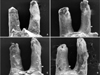
Root resorption was not detected in the control group, and smooth cementum could be seen on the mesial surface of the distobuccal and distopalatal roots. However, root resorption was observed among the treatment group rats, mainly in the cervical and middle one-third of the root (Figure 3). There was no significant difference between the 50 g(-) group and the 100 g(-) group in resorption area ratio. However, the resorption area ratio was less severe in the fangchinoline treatment groups than in the non-fangchinoline treatment groups, irrespective of force magnitude (p < 0.05) (Table 2).
DISCUSSION
In this study, we used SEM and X-ray to quantify root resorption and tooth movement. After 14 days, the maxillary first molar moved no further in the 50 g(+) and 100 g(+) groups than it did in the 50 g(-) and 100 g(-) groups. These results agree with those of Gonzales et al.,12 indicating that there is no significant difference in tooth movement after application of force at these magnitudes. Rat tooth movement is divided into 3 phases: an initial phase lasting 2 days, a lag phase, and a final linear increment of tooth movement.13,14 In our study, tooth movement over the 14 days was relatively slow and was the result of the teeth shifting in the periodontal ligament space. Because odontoclasts and osteoclasts are similar, we expected that when the same amount of force was applied, there would be more tooth movement in the fangchinoline group than in the non-fangchinoline group. However, we found no difference between the tooth movement of groups that received fangchinoline and those that did not. This is may be partially due to a lag in tooth movement caused by hyalinization. Additionally, there is still no consensus regarding the relationship between root resorption and bone resorption; therefore, further research is necessary in this regard.15 Experience with human patients indicates that molars have a tendency to move toward open spaces along the arch. We assumed this would also apply to rats: the second molar would move forward after the movement of the first molar. Because this process takes time to occur, we did not expect any movement of the second molar within the 14 day experimental period. However, this effect should be taken into account in studies with longer treatment periods.
Several researchers have attempted to investigate the precise mechanisms of root resorption. Orthodontic movement is dependent on bone resorption on the pressure side and odontoclasts are now considered to be responsible for root resorption. Odontoclasts are thought to originate from circulating progenitor cells and have characteristics that are similar to osteoclasts, such as expression of CK, cathepsin D, matrix metalloproteinase-9, H+-ATPase, and others. The activation of odontoclasts is regulated by the nuclear factor kappa B/nuclear factor kappa B ligand/osteoprotegerin (RANK/RANKL/OPG) pathways.16,17
In the present study, there was no significant difference in the resorption area ratio of 50 g(-) rats and 100 g(-) rats. This result parallels those of Zhuang et al.,18 indicating that force level is not the sole contributor to root resorption. Resorption areas in the 50 g(+) and 100 g(+) groups were smaller than in the 50 g(-) and 100 g(-) groups, respectively, supporting the hypothesis that fangchinoline inhibits root resorption. CK is a cysteine protease that is responsible for the degradation of collagen and matrix proteins.19 CK inhibitor has been investigated as a potential target for osteoporosis treatment.20 Our past research indicated that fangchinoline is a CK inhibitor; therefore, we assumed that this inhibitory effect would also apply to resorption. Moreover, fangchinoline partly inhibits IL-1 and TNF-α, which may also explain its inhibitory effect on root resorption. A study by Jäger et al.21 indicated that systemic application of soluble receptors to IL-1 and TNF-α reduces the number of odontoclasts, lending further support to our hypothesis that fangchinoline would limit root resorption during orthodontic tooth movement.
Because root resorption craters are usually 3-dimensional, further investigation is needed to determine the effect of fangchinoline on crater depth, as well as its influence on the various phases of tooth movement.


 XML Download