

 PDF
PDF ePub
ePub Citation
 Print
Print


INTRODUCTION
Colorectal carcinoma is tumor of adults peaking at 65 years [1]. It is rare in children and adolescents and therefore frequently overlooked in the differential diagnosis of abdominal pain, weight loss, and anemia [1]. Literature suggests that unlike adults, children are more likely to have advanced-stage disease at presentation, unfavorable (mucinous) tumor histology and coupled with delayed diagnosis, a poor outcome [1]. We present a case of a 12-year-old girl with rectal carcinoma.
CASE REPORT
A 12-year-old female child presented with complaints of constipation and bleeding per rectum since one and half month and abdominal distention since ten days. She had been treated earlier for anal fissures with serial dilatation elsewhere. Family history was non-contributory. At admission, she was cachectic with a grossly distended abdomen. Per rectal examination revealed tight anal stenosis (Fig. 1). Abdominal X-ray suggested intestinal obstruction.
Under anaesthesia, severe anorectal stenosis was confirmed and biopsied. In view of the massive abdominal distension, a transverse colostomy was done. The biopsy revealed inflammatory lesion. Carcinoembryonic antigen (CEA) and CA 19-9 were within normal limits. Contrast computed tomography (CT) abdomen showed circumferential thickening of rectum with preserved peri-rectal fat planes suggestive of inflammatory pathology. Colonoscopic evaluation revealed linear ulcers in distal colon. Biopsy from these lesions was also non-specific. Patient was taken home at persistence of parents.
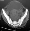
After three months, she was admitted with rectal bleeding. Tumor markers were (CEA and CA 19-9) were raised. CT abdomen showed circumferential thickened rectum with frozen pelvis suggestive of malignancy (Fig. 2). Repeat colonoscopic biopsy from the rectum revealed highly undifferentiated mucinous adenocarcinoma of rectum (Fig. 3). Patient was referred to the oncology team. She was advised radiotherapy in view of inoperability of the lesion. She succumbed within 6 months.
DISCUSSION
Colorectal carcinoma is a common gastrointestinal cancer in adults and second most common in Indian population [123]. However, it is rare in pediatric age group (<19 years) with an incidence of 1:1,000,000 and represents a small fraction of neoplasms encountered in children [234]. Adenocarcinoma of the colon in children is often seen in the setting of hereditary cancer syndromes which account for 5-10% of all cancers [5]. The incidence in the general population is 6% vs. 60-80% in Lynch syndrome and >90% in familial adenomatous polyposis (FAP) [678]. There is a significant association between acanthosis nigricans and gastrointestinal malignancy.
Paediatric colorectal carcinoma being rare, the literature is limited to studies from single institutions [29]. There is no data suggesting its incidence in paediatric Indian population [5]. In an 18-year experience from Rohtak, Nair et al. [5] have reported nine patients of colorectal malignancy. An Indian study reports four children with rectal carcinoma [6].
The age of diagnosis is usually in the second decade of life [67]. Males are usually affected more in the ratio of 2:1 [678910]. However, Nair et al. [5] found more females affected by rectal carcinoma. Predisposing factors such as familial polyposis of the colon and other polyposis syndromes, ulcerative colitis, familial multiple cancer syndromes are usually noted in 10% of the bowel cancers arising during childhood; however, studies from Indian populations did not have any children with these known predisposing factors [56]. A review by a large population based study done recently, indicates that the occurrence of colorectal malignancy in children and adolescents has similar natural history as seen in adult patients [21112].
The duration of symptoms, primary site, pathologic findings, stage and prognosis between the adults and the children are different [678]. Children usually present with vague complaints like abdominal pain, vomiting, change of bowel habits, bleeding per rectum, constipation and weight loss [678]. However, the diagnosis of colorectal malignancy is usually missed in the differential diagnosis whereas in adults the same history would warrant a colonoscopy to rule out malignancy [21112].
In adults, most of the colonic cancers are located in left side within 25 cm of the anus including the recto- sigmoid [678]. However, in children the site of involvement is varying and has been found to be equally distributed in all parts of the colon, transverse colon, right and transverse colon and recto-sigmoid [6913].
Children are known to present with advanced stage of the disease at diagnosis and have a higher occurrence of aggressive tumours [211]. Adults are usually affected with moderately differentiated adenocarcinoma; on the other hand, children have predominantly mucinous adenocarcinoma [61314]. Mucinous adenocarcinoma is rare in adults [69]. The mucin is known to absorb water, swell and invade the nearby tissues, thereby promoting spread of malignant cells. The tumours has the potential to grow to huge size because of the pooling mucin and also interferes with the immune recognition of carcinoma cells caused by mucopolysaccharide coating [691315]. The signet ring subtype is known to grow very rapidly. There is regional lymph node involvement and diffuse peritoneal seeding at the time of presentation, which leads to the worst prognosis [69]. The signet ring subtype was reported in series by Pandey et al. [6] in 2008. In another series from Rohtak reported by Nair et al. [5], poorly differentiated mucinous adenocarcinoma was the main histopathological subtype and the predominant growth was constrictive in nature.
The diagnosis is based on a high index of suspicion which can be confirmed by sigmoidoscopy or colonoscopy and biopsy [6]. CT and positron emission tomography (PET) are commonly used to detect the extent of the disease [6]. The role of CEA in the diagnosis and follow-up of colorectal carcinoma in children is controversial and not established at present [678]. The genetic screening of family members should be done to rule out the various hereditary polyposis syndromes.
In patients with resectable mass, complete tumour resection that includes the lymphatic basin of the affected colon and/or rectum has the greatest impact on the overall survival [6]. With the advance disease stage at presentation, such surgical resection may not always be possible.
The role of chemotherapy in paediatric colorectal cancer (CRC) is controversial [5]. In adults, the FOLinic acid, 5-Fluorouracil and OXaliplatin (FOLFLOX) regimen of chemotherapy has been reported to demonstrate a clear benefit in patients with stage III and IV disease and is actively evolving also [212]. A variety of new agents like irinotecan, oxaliplatin and leucovorin are also being considered to be used in association with 5-fluorouracil (5-FU) [615]. Advancements in DNA sequencing and molecular biology are being explored to look for potential biological markers of colorectal carcinoma that may reveal the differences between pediatric CRC and adult CRC [210].
However, in a developing country like India, the choice of chemotherapeutic agent is usually guided by the socioeconomic scenario [5]. This has led us to the use of a cheaper chemotherapeutic regimen [5]. In patients with a poorer performance score, palliative radiation therapy is given locally [5].
Colorectal carcinoma in children is usually a fatal disease [6]. The 5-year survival rate for children ranges from 7% to 12% and is similar in both developed and developing countries [6]. Delay in diagnosis, greater virulence, advanced stage of disease at the time of diagnosis and the mucinous type of histology—all lead to poor prognosis in children [6].
Any child who present with abdominal pain along with a history of constipation and rectal bleeding should be examined and investigated carefully to rule out malignancy [6].


 XML Download