

 PDF
PDF ePub
ePub Citation
 Print
Print


INTRODUCTION
Abdominal cysts are rare in children. Gastrointestinal (GI) tract cysts account for about 15% of abnormalities [1]. Common causes include duplication cysts, mesenteric and omental cysts, as well as meconium pseudocysts. We describe fourteen cases of GI tract cysts in children highlighting their varied presentation and diagnoses.
MATERIALS AND METHODS
Records of fourteen paediatric patients with cysts of GI origin were reviewed and analyzed on the basis of age, sex, presenting symptoms and signs, radiological investigation, type and site of cyst, management, outcome and follow-up.
Patients with acute presentation were kept nil by mouth, started on intravenous fluids and antibiotics. X-ray and ultrasound (USG) abdomen was done. A computerized tomography (CT) scan was done for all patients except patients presenting as acute intestinal obstruction. The cysts were deroofed or excised with or without bowel resection. Diagnosis was confirmed on histopathology.
RESULTS
A total of 14 patients with cysts of GI origin were retrospectively analyzed. The mean age was 4 years; the youngest patient was 4 days and the eldest 11 years at presentation. There were six males (42.8%) and eight females (57.1%). Abdominal pain was the most common presenting symptom seen in 10 patients. The other symptoms were lump (8), abdominal distention (8), vomiting (7), constipation (7) and fever (4).
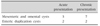
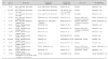
There were 7 patients with mesenteric cysts, 3 with omental cysts and 4 patients with enteric duplication cysts (Tables 1 and 2). Five patients had an acute presentation. Three patients presented in emergency with symptoms of acute abdominal pain, distention, bilious vomiting and constipation. On examination, abdomen was distended and tender but there was no palpable lump. Erect abdominal X-ray and USG suggested small bowel obstruction. At emergency exploration, all three had distal ileal mesenteric cysts; with one patient having two cysts. The cysts were excised in two patients and deroofed in one patient.
Two patients had a palpable abdominal lump along with acute abdominal pain, distention, nonbilious vomiting and constipation. The cysts were diagnosed pre-operatively on CT scan. Both of these patients had ileal duplication cyst sharing a common wall with ileum. Volvulus and gangrene of ileum was present in one of them. Excision of cyst with ileal resection-anastomosis was done.
Six patients presented with chronic abdominal pain and lump. They had symptoms of intermittent abdominal pain over months. Cysts were diagnosed on USG and CT scan. At exploration, three patients had omental cysts and three had mesenteric cysts– two of these in distal ileum and one in sigmoid colon. All were excised. The large mesenteric cyst of sigmoid required excision and resection anastomosis of the bowel.
Two patients presented with antenatally diagnosed palpable abdominal lump. Both patients were asymptomatic. Multilocular cyst was confirmed on post-natal CT scan. Intra-operatively, the neonate had ileal mesenteric cyst which was excised laparoscopically. The other had a distal ileal duplication cyst which required excision with resection and anastomosis. The diagnoses were confirmed on histopathology.
One patient had an atypical presentation. He was a known case of sickle cell trait and had presented with vague abdominal pain, recurrent cough and multiple episodes of haemoptysis since a period of one year. Multiple chest X-rays were suggestive of persistent consolidation in lower lobe of the left lung. CT chest and abdomen showed left lower lobe consolidation suggestive of sequestration. Magnetic resonance cholangiopancreaticography revealed a normal pancreas with a cyst in the lesser sac. At laparotomy, gastric duplication cyst was found which was excised completely. Histopathology confirmed the diagnosis.
There were no complications in any of these fourteen patients.
DISCUSSION
The differential diagnosis of abdominal cysts includes mesenteric cysts, omental cysts, cystic lymphangioma, cystic teratoma, ovarian cyst, enteric duplication cyst, hydatid cyst, etc. [2345]. Abdominal cysts of GI origin are usually mesenteric cysts, enteric duplication cysts, omental cysts and meconium cysts.
Mesenteric cysts
Mesenteric cysts are rare benign intra-abdominal masses with an incidence of about 1:100,000 in adults and 1:20,000 in pediatric age group [3456]. They may occur anywhere in the mesentery of the GI tract from the duodenum to the rectum, but most commonly are localized in the mesentery of the ileum followed by the mesentery of the large intestine and retroperitoneum [345789]. Though commonly solitary, multiple positions within the peritoneal cavity have also been reported [4]. Approximately, one third of the mesenteric cysts occur in the children younger than 15 years of age and are slightly more common in males [457].
The exact etiology for the development of the mesenteric cysts is unknown. The most commonly accepted theory as proposed by Gross states that it is the result of benign proliferation of ectopic lymphatics in the mesentery which lack communication with remainder of the lymphatic system [45]. The classification proposed by Beahrs et al. [10] in 1950 divides mesenteric cysts into four types: developmental, traumatic, infective and neoplastic [39]. de Perrot et al. [11] proposed a classification based on histopathological features and their origin dividing mesenteric cysts into six groups: lymphatic origin, mesothelial origin, enteric origin, urogenital origin, dermoid cyst and non pancreatic pseudocyst [11].
Clinical presentation of mesenteric cysts is variable. They may present as an asymptomatic abdominal mass, incidental finding during laparotomy for other abdominal conditions, or even as acute abdomen [34]. Acute presentation usually results due to the cyst rupture, infection, hemorrhage, intestinal obstruction and volvulus [3412]. Abdominal USG and CT are the investigations of choice [3459]; however, it may be not possible to confirm this diagnosis in all cases [3410]. Their sizes usually vary from 4 cm to 30 cm [34812]. Mesenteric cysts are most commonly single and multilocular and the fluid is generally serous when the cyst involves the distal small bowel or colonic mesentery and chylous when it is located in the proximal small bowel mesentery [345]. Histopathological examination is confirmatory [345].
Complete surgical excision is the preferred treatment for mesenteric cysts and the results are excellent [3457891213]. This can be accomplished laparoscopically [34]. Bowel resection and anastomosis may be needed in up to 50% to 60% of cases [34591213]. Partial excision with marsupialization of the remaining cyst into the abdominal cavity is recommended when the above is not possible [34]. This form of treatment is required in approximately 10% of patients [34]. The cyst lining should be sclerosed with 10% glucose solution, electrocautery, tincture iodine or OK432 after marsupialization to prevent recurrence [34].
Enteric duplication cysts
Calder [14] first reported a case of enteric duplication cyst in 1733. These are rare congenital anomalies with an incidence of 1 in 4,500 autopsy series [1315]. They are present in both genders with slight male predominance [1516]. Their presentation is variable and usually is confused with other GI pathologies. Abdominal examination may be unremarkable, but a mobile mass may be palpable in 50% of cases. Associated malformations are reported in 50% of patients, the most common being vertebral defects [151718].
Though there is no consensus on the exact embryological origin of enteric duplication cysts, the most widely accepted theory is the split notochord theory [17]. Other theories suggested are incomplete twinning, phylogenetic reversal, persistent embryonic diverticula, entrapment of endodermal cells or persistent epithelial buds within the body wall and dysvacuolation [17]. As per Ladd, the term ‘enteric duplication cyst’ will be applied if a congenital lesion has (1) coat of smooth muscle, (2) GI type epithelial lining, and (3) intimate anatomical location with some part of gastro-intestinal tract (GIT) [13]. These can arise from any part of GIT from oropharynx to anus. They can be classified into foregut, midgut and hindgut, depending on the site of origin [1318]. Enteric duplications may be cystic, tubular or mixed [13]. These may share blood supply with adjacent intestine by residing in leaves of its mesentery, posing a difficulty for safe resection [1319].
Laboratory investigations are usually non-specific however, they may show anaemia, which may be due to bleeding from heterotopic gastric mucosa present in the cyst wall [13]. USG and CT scan are the imaging modalities used [13].
Excision should be considered in all cases wherever possible. The surgical approach varies with location and type of the cysts [13]. Resection and anastomosis may be required.
Omental cysts
Omental cysts are rare cysts of the abdomen with only about 150 cases recognized till now, 25% of which presented in children less than 10 years of age [2021]. However, the true incidence of omental cyst may be much higher than reported cases in literature because usually only cysts of clinical importance are reported [2022]. The typical presentation of omental cyst is low grade partial intestinal obstruction with palpable freely movable abdominal mass [2022]; however, the lump is palpable in 25-50% of cases only [2023]. The cysts are incidental findings in approximately 40% of the cases [20].
The preferred treatment of omental cyst is total excision. Bowel resection is rarely necessary and recurrence is rare [2024]. Excision is usually indicated because of the possibility of torsion, rupture, bleeding and infection, even if the patient has no symptoms [2025]. Malignant degeneration of omental cyst is rare; only a small number of cases of sarcoma and adenocarcinoma transformation have been reported [2026].
Cysts of GI origin are rare and have varied presentation. They may have typical presentation of abdominal pain and lump; however atypical presentation in the form of obstruction, malena or haemoptysis may also be present. Pre-operative diagnosis may be difficult in cases with atypical presentation. Surgical excision is the mainstay of treatment. The results and prognosis are good.
 XML Download