

 PDF
PDF ePub
ePub Citation
 Print
Print


INTRODUCTION
Crohn's disease (CD) is a chronic inflammatory bowel disease of unknown cause. It occurs primarily in the terminal ileum, but can involve any area of the gastrointestinal tract (GIT), from the mouth to the anus [1]. To date, much research has been conducted to investigate the involvement of the lower GIT, but the involvement of the upper GIT in CD has not been studied sufficiently.
In studies involving Italian and American adults, the prevalence of upper gastrointestinal (GI) CD is reported to be 16–34% [23]. In pediatric patients with CD, studies have reported different findings; for example, the upper GIT was involved in 71% of cases in a study conducted in Canada [4], whereas, in a study conducted in the UK, esophagitis involvement was noted in 72%, gastritis in 92%, and duodenitis in 33% [5]. Moreover, in a study conducted in the US, 36% of patients showed symptoms suggestive of upper GI involvement, and 12% of pediatric patients demonstrated noncaseating granulomas [6]. In a study involving Korean adults who underwent upper GIT biopsy, 59% of cases had upper GI symptoms. Erosive gastritis was the most common endoscopic finding, and was noted in 66% of patients. In another study, approximately 70% of patients had an abnormal histological finding. However, these studies were based on only a few cases, and moreover, the focus was on abnormal findings in the stomach or Helicobacter pylori [7]. To date, data has been scarce regarding pediatric patients in Korea. In Asians, CD is more male-predominant (1.67:1 to 2.9:1) compared to in other races. Moreover, CD of an isolated colonic type is most common in Europeans, whereas CD involving both the small and large bowels is most common among Asians [8910].
Accordingly, this study aimed to examine whether the presence or absence of upper GI symptoms was related to upper GIT lesions in pediatric patients with CD and to investigate whether the relationship would help determine the need for testing. An additional objective of the study was to examine the clinical features, endoscopic and histological findings, and the prevalence of H. pylori in CD with upper GIT involvement to help create a guideline for upper GIT testing in pediatric CD patients in Korea.
MATERIALS AND METHODS
Study design and subjects
Data from the medical records of pediatric patients (age <18 years) with CD who underwent upper GIT endoscopy and biopsy between January 2001 and August 2016 were collected from 4 Korean tertiary hospitals. The 4 medical centers included Gachon University Gil Medical Center located in Incheon, Chungbuk National University Hospital in the central region, Kangwon National University School of Medicine in the north region, and Chung-Ang University Hospital in Seoul, South Korea.
Of 64 subjects, 52 were finally enrolled after meeting the following criteria: (1) patients who underwent biopsy for abnormal gastric lesions and/or normal-looking gastric mucosa and (2) patients who had no known chronic medical illnesses. Our diagnostic criteria for CD has been previously described [811].
We retrospectively reviewed the patients' medical records, upper GI endoscopic findings, and histopathological findings from the time of CD diagnosis. The following demographic and clinical information was collected: gender, GI symptoms (diarrhea, lower abdominal pain, weight loss, hematochezia, and poor oral intake), upper GI symptoms (epigastric pain, nausea, vomiting, dyspepsia, burning sensation, epigastric fullness, belching, and melena), perianal lesions (skin tag, fissure, abscess, and fistula), and medications ever used for CD (anti-tumor necrosis factor-α therapy [infliximab, adalimumab], immunosuppressive therapy [steroid], immunomodulators [6-mercaptopurine, azathioprine], 5-aminosalicylic acid [mesalamine, sulfasalazine], and antibiotics). Laboratory findings such as erythrocyte sedimentation rate (ESR) and C-reactive protein (CRP) were identified. Disease locations were evaluated according to the Montreal classification. Disease activity was evaluated with the pediatric Crohn's disease activity index (PCDAI) and was classified as remission (PCDAI <10), mild activity (PCDAI 10-27.5), moderate activity (PCDAI 30-37.5), and severe activity (PCDAI >40) [12]. The study protocol was approved by the institutional review boards of Gachon University Gil Medical Center (IRB no. 2016-368).
Upper GIT endoscopy, histologic evaluation, and H. pylori infection assessment
All upper GI endoscopic evaluations were performed by expert GIT endoscopists of the participating institutions. Biopsy specimens were obtained for pathologic evaluation from abnormal gastric lesions and normal-looking gastric mucosa at the endoscopists' discretion. Additional biopsies were performed at both the gastric antrum and corpus for the rapid urease test. Standard hematoxylin and eosin staining was performed on biopsy specimens. The diagnosis of H. pylori infection was based on either a positive histopathology plus a positive rapid urease test, or a positive culture. Gastric biopsies were obtained for histopathology [13].
The diagnosis of “upper GI involvement of CD” was based on a combination of compatible endoscopic (ulcerations, erosions, strictures, and aphthous lesions) and histologic findings (chronic inflammation, erosion, ulceration, granuloma, and gastric intestinal metaplasia) as described previously [1415]. Some nonspecific inflammation or inflammatory changes explained by other conditions (reflux esophagitis or H. pylori gastritis) were excluded.
After dividing the participants into 2 groups according to the presence or absence of upper GI symptoms, we evaluated the differences in demographics, H. pylori infection, treatment, surgery, PCDAI, perianal lesions, laboratory findings (ESR, CRP), and endoscopic and histologic findings.
Statistical analysis
Continuous variables were expressed as means with ranges. Discrete data were expressed as numbers or percentages or both. Demographics, clinical symptoms, and endoscopic and histologic GIT lesions of subjects with and without upper GI CD symptoms were compared. For comparative analyses, the Mann-Whitney U-test and Student's t-test were used for continuous variables, while the chi-squared test or Fisher's exact test were used for categorical variables, where appropriate. All statistical analyses were performed using IBM SPSS for Windows ver. 21.0 (IBM Co., Armonk, NY, USA). p<0.05 was considered statistically significant.
RESULTS
Demographics and disease characteristics
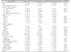
Of the total 64 patients, 2 patients did not undergo upper GI endoscopy and 10 patients were excluded from the study because of inadequate data. A total of 52 patients were enrolled. The subjects included 34 men (65.4%), and the mean age at CD diagnosis was 14.1±2.1 years. According to the Montreal classification, the dominant phenotypes were listed as follows: the sites of disease location were L3 (ileocolon, 33/52, 63.5%), L4 (isolated upper GI, 0/52, 0%), L2 (colon, 16/52, 30.8%), and L1 (terminal ileum, 3/52, 5.8%).
Perianal lesions were found in 59.6% of the patients. Of the lesions, the skin tag was the most common (21.2%, 11/52), followed by fistula (9.6%, 5/52). Overall, a mixed-type lesion was noted in 21.2% (11/52) of patients.
According to PCDAI, severe activity was the most common (51.9%, 27/52). Moderate activity was found in 32.7% (17/52) and mild activity in 15.4% (8/52), and remission did not occur in any patients. H. pylori test was positive in 3 patients (5.8%).
The mean CRP value at the time of upper GIT endoscopy was 9.7±19.6 mg/dL and the mean ESR level was 48.5±28.4 mm/h (Table 1).
GI symptoms
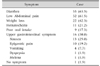
The most common GI symptom was diarrhea (63.5%, 33/52), followed by lower abdominal pain (61.5%, 32/52) and weight loss (42.3%, 22/52). Hematochezia (21.2%, 11/52) and poor oral intake (17.3%, 9/52) were rare relative to other symptoms.
Upper GI symptoms were noted in 30.8% (16/52). Of those, nausea was the most common symptom (25.0%, 13/52), followed by epigastric pain (19.2%, 10/52) and vomiting (7.7%, 4/52). There were no complaints of epigastric fullness, belching, or burning sensation (Table 2).
Upper GI endoscopic and pathologic findings
The upper GI endoscopic findings were examined according to the following sites: esophagus, stomach, and duodenum.
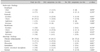
Gastric ulcer was the most common (19.2%) lesion found on upper GIT endoscopy, followed by duodenal ulcers (15.4%). In the group with upper GI symptoms, esophageal ulcer was observed in 12.5% (2/16) of patients, while in the group without upper GI symptoms, esophageal erosion was noted in 11.1% (4/36) of patients. The difference was not statistically significant.
Regarding gastric findings, gastric ulcer was the most common (19.2%, 10/52). In the group with upper GI symptoms, gastric erosion was noted in 25.0% (4/16), while in the group without GI symptoms, gastric ulcer was observed in 19.4% (7/36) of patients. The difference was not statistically significant.
Regarding duodenal findings, duodenal ulcer was the most common (15.4%, 8/52). There was no difference between the groups with and without upper GI symptoms.
Chronic inflammation was most common histopathologic feature (n=39, 75.0%) followed by gastric erosion (n=9, 17.3%). Chronic inflammation was the most common both in the group with (81.3%, 13/16) and without (72.2%, 26/36) upper GI symptoms. In 5 patients (9.6%), granuloma had previously been identified from biopsy specimens. In addition, erosion, ulceration, gastric intestinal metaplasia, etc. showed non-significant differences based on the presence or absence of upper GI symptoms (Table 3).
Differences between patients with and without upper GI symptoms
In the group with upper GI symptoms, the mean age at CD diagnosis was 14.2±1.7 years and 43.8% (7/16) were female. In the group without upper GI symptoms, the mean age at CD diagnosis was 14.1±2.3 years and 30.6% (11/36) were female. No between-group differences were statistically significant.
When the association between upper GI symptoms and H. pylori infection, CD medications, PCDAI, perianal lesions, etc., was analyzed, no variables showed an association with upper GI symptoms except for the mean values of ESR (60.7±27.1 vs. 43.0±27.6 mm/h, p=0.037) and CRP (16.5±28.2 vs. 6.62±13.4 mg/dL, p=0.014) (Table 1).
DISCUSSION
Upper GI symptoms were not correlated with upper GI lesions, and H. pylori infections were relatively uncommon in Korean pediatric CD. However, the extent of inflammation suggested the presence of upper GI symptoms. These results can aid the establishment of guidelines for upper GIT examination.
According to studies involving adults in Western countries, the prevalence of upper GI CD ranges from 16–34% [23]. However, the correlation between upper GI symptoms and true endoscopically and pathologically proven disease has not been fully established, even in pediatric patients. In the present study, upper GI involvement in pediatric CD was 50.0% (26/52), which is higher compared to results in Western countries. We believe the differences are due to dissimilar subject baseline characteristics such as age, race, and disease severity, and differences in diagnostic criteria.
A previous study involving adult Korean patients with CD reported that 59.6% demonstrated upper GI symptoms [7]. In the present study, 16 (30.8%) patients showed upper GI symptoms, which is consistent with other studies, and there was no significant difference based on the presence or absence of symptoms. Concerning individual symptoms, nausea (25.0%) was the most common, followed by epigastric pain (19.2%), vomiting (7.7%), dyspepsia (1.9%), and melena (1.9%).
Regarding endoscopic findings, gastric ulcer was the most common (19.2%) lesion found on upper GIT endoscopy, followed by duodenal ulcers (15.4%). In the presence of upper GI symptoms, gastric erosion (25.0%) and duodenal ulcer (25.0%) were the most common, and gastric ulcer (18.8%) was the most common in the absence of upper GI symptoms. The difference in endoscopic findings based on the presence and absence of upper GI symptoms was not statistically significant. A study involving adults found gastric erosion in 9.6%, duodenal ulcer in 5.3%, duodenal erythema or erosion in 5.1% [2]. There were fewer abnormal findings in that study compared to the present one [2]. It is speculated that the discrepancy may be due to inter-observer differences and the presence or absence of overlapping findings.
According to a meta-analysis conducted on studies with adult patients with CD in Western countries, nonspecific gastric inflammation was the most common histological finding (32%) [2]. Gastric granuloma was noted in 7.9% and focal gastritis in 30.9% [2]. Regarding inflammatory findings by site, 84% occurred in the stomach, 28.2% in the duodenum, and 23.2% in the gastric granuloma [2]. In the present study, the most common histological finding in all pediatric patients with CD, regardless of the anatomical site, was chronic inflammation (75.0%), followed by erosion (17.3%). Granuloma was observed in 9.6% of patients, which was higher than in adults. Moreover, there was no association between the presence or absence of upper GI symptoms and histological findings (Table 3).
In Western countries, one study reported that in small bowel or colonic surgical specimens of patients with CD, perianal fistula increased significantly in the presence of noncaseating granuloma [16]. Other studies reported that gastric granuloma was found in 5–83% of gastric biopsy specimens from patients with CD and suggested an association between gastric noncaseating granuloma and perianal abscess/fistula [1718]. In the present study of pediatric patients, gastric granuloma occurred in 9.6% (5 patients) and perianal abscess/fistula co-occurred in none of the 5 patients with gastric granuloma. We believe this finding shows that in pediatric patients with CD, unlike in adult patients, there is no association between gastric granuloma and perianal abscess/fistula. This finding should be clarified with studies including a larger number of cases.
H. pylori infections are acquired early in life by young children and adolescents. In one study involving South Korea children, the prevalence of H. pylori infection was 22% [19]. Another study reported that H. pylori infections were found in 7.4% of South Korean children with recurrent abdominal pain [20]. A previous study reported a lower H. pylori infection rate in patients with CD than patients without CD [21]. Additionally, another study based on a meta-analysis showed a significant negative association between H. pylori infection and irritable bowel syndrome (IBD) and suggested a possible protective effect of H. pylori infection against IBD [22]. In the present study on pediatric patients with CD, H. pylori infection was identified in 5.8% of patients, which was lower than in pediatric patients without CD. We believe this finding shows that in pediatric patients with CD, there is negative association between H. pylori infection and pediatric patients with CD in South Korea. This finding should be clarified with studies including a larger number of cases.
CD is commonly complicated by perianal manifestations. In studies of adult patients with CD in Western countries, the reported incidence of perianal CD varied from 3.8% [23] to 80% [24]. In a study involving children [25], perianal disease co-occurred in 21% of pediatric patients with CD and was also more common among black people (26%) compared to white people (20%, p=0.017). In the present study, 59.6% (31/52) of the pediatric patients showed more than one perianal lesion such as skin tag, fissure, fistula, and abscess. It seems that racial characteristics are important, suggesting that if an Asian pediatric patient with CD presents with a perianal lesion, more attention should be paid to the CD.
According to previous research, increased CRP in adult patients with CD increases the risk of CD-related hospitalization and CD-related intestinal resection. CRP levels >1 mg/dL have been found to be correlated with granulomatous CD in pediatric patients in the United States [26272829]. In the present study, we found that ESR and CRP significantly increased in the presence of upper GI symptoms. CRP testing was conducted at slightly different time points across different studies, but care should be taken if CRP and ESR are elevated in a patient with CD even when there is no difference in PCDAI or histological or endoscopic findings.
According to the European Society of Pediatric Gastroenterology, Hepatology and Nutrition (ESPGHN) guidelines, upper GIT endoscopy and ileocolonoscopy are recommended in all patients with pediatric-onset inflammatory bowel disease [30]. However, there is no such guideline in Korea. Although endoscopically abnormal findings are rare in patients with upper GI symptoms, endoscopic or histologic abnormalities are relatively common according to the findings of routinely performed endoscopy. These results suggest that upper GIT endoscopy should be performed on pediatric patients with CD in South Korea.
To our knowledge, this is the first retrospective multicenter study in Korea aimed at evaluating the prevalence of upper GIT involvement in pediatric patients with CD, irrespective of upper GI symptoms. In the present series, 50% of the patients showed upper GI CD involvement, a higher value than expected.
The present study had the following limitations. First, it was based on a retrospective design and second, the study sample was small. Third, we could not review the pathology again and simply check the pathology reports.
In conclusion, upper GI symptoms may not be correlated with upper GIT lesions. H. pylori infection was relatively uncommon in Korean pediatric patients with CD. However, the extent of inflammation suggests the presence of upper GI symptoms. These results should aid the establishment of regional guidelines for upper GIT examination.
 XML Download