

 PDF
PDF ePub
ePub Citation
 Print
Print


INTRODUCTION
Esophageal strictures in children have multiple etiologies such as congenital anomalies, esophageal atresia, inflammatory disorders, eosinophilic esophagitis, gastro-esophageal reflux disease and caustic ingestion [12]. The incidence of the different etiologies varies between countries. In developing countries, caustic injuries are more frequent [34].
A benign refractory or recurrent stricture in children occurs in case of an anatomic restriction because of cicatricial luminal compromise or fibrosis that results in dysphagia in the absence of endoscopic evidence of inflammation. There is consensus that this may occur as the result of either an inability to successfully remediate the anatomic problem to obtain age-appropriate feeding possibilities after a maximum of five dilation sessions (refractory) with maximal four-week intervals, or as a result of an inability to maintain a satisfactory luminal diameter for four weeks once the age-appropriate feeding diameter has been achieved (recurrent) [1].
CORROSIVES
Ingestion of corrosive substances is most often accidental and occurs much more in children, especially in toddlers, than in adults [56]. It can cause serious injuries to the digestive tract. In the developed world with the advent of child-unfriendly packaging, corrosive ingestion has become quite rare [7]. Household, industrial, and farm products, especially if stored in non-original containers, represent the most frequently ingested caustic agents. A variety of substances have been reported that were ingested leading to caustic injuries ranging from alkaline bases with pH up to 12 (e.g., sodium hypochlorite and sodium hydroxide), to acidic substances with a pH as low as 2 (e.g., hydrochloric acid and salicylic acid) and also bleaching substances in which the pH is around 7 [89]. Recently hair relaxers and liquid tabs (pods) containing detergents are a new addition to the long list of potentially harmful products when ingested, but these substances seem to be less harmful. The extent and severity of the esophageal lesions is related to the nature, quantity and concentration of the caustic substance and duration of contact with the mucosa. Acids usually cause coagulative necrosis with limited tissue penetration and superficial scar formation. Strong alkalis produce liquefaction necrosis with deep ulcerations, and a subsequent risk to develop esophageal stricture and/or perforation. Upon swallowing, acids cause severe oropharyngeal pain and therefore they are usually ingested in smaller volumes than alkaline substances, resulting in a lower incidence of stricture formation and/or esophageal perforation.
After ingestion, vomiting should be prevented. Small amounts of water can be allowed if the child asks for it or even stimulated to rinse the mouth and esophagus. If the child has severe pain and if perforation is suspected, nothing should be given by mouth. Adequate pain relief is recommended.
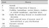
Every child that has ingested a corrosive substance should have a thorough follow-up. The majority of corrosive ingestions may be asymptomatic at presentation. But, absence of oral burns does not exclude ingestion and esophageal/gastric damage and the consequent need for an endoscopic evaluation [1]. Potential mucosal injury and the risk for stricture development should be suspected in a similar way for acidic and alkali ingestion. However, alkali ingestion, especially lye, is associated with more severe esophageal lesions and severe gastric lesions can occur in acidic ingestion. Endoscopy is guided by the presence of symptoms. If symptoms are present, the timing should be within the first 24 hours after ingestion [1]. It is recommended that every child with a suspected caustic ingestion and symptoms/signs (e.g., any oral lesions, vomiting, drooling, dysphagia, hematemesis, dyspnea, abdominal pain, etc.) should undergo an endoscopy. In case the ingestion of a corrosive is suspected, endoscopy is withheld if the child is asymptomatic and that adequate follow-up is assured. Esophageal lesions after corrosive ingestion are described according to the Zargar classification (Table 1) [1011]. Patients with low-grade lesions at endoscopy (grade 0 to IIa) who have in addition a normal physical examination and who can eat and drink normally can be discharged [1213].
DEXAMETHASON
A decreased incidence of grade III burns and stricture formation with early corticosteroid and anti-biotic use compared with controls has been suggested [14]. There is some evidence of benefit for the administration of intravenous dexamethasone at high dose (1 g/1.73 m2 per day) for a period of three 3 days. This may prevent the development of esophageal strictures. The evidence is limited to IIb esophagitis after corrosive ingestion, but it can be debated if a similar management would not be appropriate if there is severe esophagitis after ingestion of corrosives.
There is some recent evidence that intralesional steroid injection may be an effective adjunct to dilatation in children with short-segment strictures [15].
DILATION PROCEDURES
Anesthesiology and surgical assistance should be available during esophageal dilation procedures in children—the latter in case of complications [16].
Esophageal dilation should only be performed only when symptoms occur. Strictures shorter than 5 cm in length appeared to have a significantly better outcome. Wire-guided polyvinyl bougie dilators (Savary Gilliard) and “through-the-scope balloons” are the most frequently used material to dilate benign esophageal strictures. According to a retrospective study from 2001 comparing 125 balloon dilations versus 88 bougie dilations in children with benign esophageal strictures fluoroscopically guided balloon dilatation is safer and has fewer technical failures than surgical bouginage [16]. These findings were confirmed by another retrospective study in patients with esophageal atresia, showing that balloon dilation was more effective and less traumatic than bougienage [17]. However, bougie dilation is also safe and effective. Most centres will prefer balloon dilations over bougies if financially possible. But the experience of the centre with a given technique may be more important. Balloon dilation can be performed under direct endoscopic or fluoroscopic view. The size of the balloon catheter can vary from 4 to 22 mm. There is a heterogeneity in literature regarding the duration of balloon inflation which varies from 20 to 120 seconds [18].
Data on the best timing of esophageal dilation are scarce. Two retrospective studies in children post esophageal atresia compared routine esophageal dilation every 3 weeks starting 3 weeks post-surgery versus when symptoms developed. No difference in outcome and complications were found between both groups but significantly fewer dilations were needed in the on-demand dilation group [1920]. Although there is no evidence regarding a number of practical aspects, there is consensus to apply in most situations the rule of three: dilate maximal up to three times the diameter of stenosis, with an average of three dilations and a minimal period of three weeks between two dilation sessions.
MYTOMYCIN, STENTING AND OTHERS
There is no standard treatment for refractory stenosis. A temporary stent placement or application of topical mytomycin-C following dilation for refractory esophageal stenosis in children is proposed.
Mytomycin-C is anthracycline derived from Streptomyces anti-fibrotic agent that inhibits fibroblast proliferation and decreases scar formation. Mytomycin-C is a cytostatic agent; therefore, dysplasia of healthy tissues after application should be considered as a theoretical risk of a severe adverse effect, which as up to now not reported [21]. Mytomycin-C has been used in ophthalmology (glaucoma), ear-nose-thoroat Medicine (laryngeal and tracheal stenosis), anal, vesical and vaginal strictures. Data are needed on the effect of antifibrotic mitomycin-C used topically to prevent postingestion fibrosis. Local application of mytomycin-C is a therapeutic option for the treatment of refractory esophageal strictures in children. Cotton pledgets soaked in a solution (0.1 mg/mL) of mytomycin-C have been applied endoscopically directly onto the mucosa postdilation with some success, reducing the number of esophageal dilations sessions needed. Questions that still need to be answered regarding mytomycin-C are the role of selection of patients, when to administer it (after more than three 3 dilations?), does the technique of application matter, what about the long term outcome (which surveillance is needed, is there a risk for the development of Barrett esophagus, esophageal cancer).
Currently, self-expandable plastic stents and self-expanding metal stents mostly made from nitinol (alloy of nickel and titanium), dominate the market because of their removability or because of their ability to conform to anatomical angulations. With the development of these removable, fully covered, self-expandable metal stents, the use of esophageal stents in children has expanded in particular in case of refractory stenosis. Most patients will experience nausea or chest pain in the days following stent placement. Complete clinical response following stent removal with no recurrence of dysphagia or need for subsequent dilations was reported. There is a need for better standardization of the duration of stenting as according to literature time intervals of 1 to 24 weeks are reported. Stent migration is the most frequent complication [1].
A recent uncontrolled study in 10 children with intractable esophageal strictures due to caustic ingestion reported symptom resolution using stricture dilation preceded by intralesional triamcinolone injection was reported to be successful but failure was as well reported.
ESOPHAGEAL REPLACEMENT
Replacement of the esophagus in children can be required as the ultimate treatment of refractory stenosis. The new esophagus should allow normal oral feeding, with little or no gastroesophageal reflux, and be able to work well for the lifetime of the patient. For over a century, many substitutes have been used, such as segments of colon, the entire stomach, gastric tubes, or parts of the small bowel, but none are perfect or function like a normal esophagus. The long-term outcome of colon interposition after esophagectomy in children shows that this technique has a high morbidity: 85% digestive symptoms, 58% abnormal lung function, 50% feeding difficulties, and failure to thrive in most of the patients [11].
CONCLUSION
Symptomatic esophageal strictures should be dilated, either using balloons or bouginage. Administration of high dosis corticoids could of interest in some conditions. In case of refractory stricture, mytomycin-C and/or stenting can be useful. Every effort should be made to minimis ethe need for surgery and esophageal replacement.
 XML Download