

 PDF
PDF ePub
ePub Citation
 Print
Print


INTRODUCTION
Urinary tract infection (UTI) is a common febrile illness in children, especially in infancy. Acute pyelonephritis (APN) in particular is of concern because it can easily lead to bacteremia and sepsis in children, and cause renal scarring as a complication [1]. APN is suspected in children with UTI who exhibit signs of systemic inflammatory response, hence the need for diagnostic tests such as renal scintigraphy with 99mTc-dimercaptosuccinic acid (DMSA).
APN, like many other disease entities, can cause liver injury via sepsis. In patients with sepsis, proinflammatory cytokines derived from Kupffer cells are responsible for hepatocellular dysfunction [234]. Liver injury in APN patients has been studied in adults [5]. In addition, a study on children with UTI and elevated alanine aminotransferases (ALT) has been reported. However, it did not show direct correlation of elevated ALT with sepsis [6].
The aim of our study was to investigate the association between elevated ALT and urosepsis in children with APN.
MATERIALS AND METHODS
Children diagnosed with APN at Gachon University Gil Medical Center (Incheon, Korea) between January 2005 and December 2014 were included in our study. The research process was approved by Gachon University Gil Medical Center Institutional Review Board (GCIRB2015-65).
In our study diagnosis of APN was defined as having a positive urine culture and a positive DMSA. The results of urine culture and DMSA were collected via a retrospective chart review of children who were clinically diagnosed with UTI during their stay. Urine samples for urinalysis and urine culture were collected using sterile urine bags, urine catheterization, or suprapubic aspiration. The exact proportion of urine samples collected via each method is unknown, although sterile urine bags were used in most cases. DMSA was performed when a working diagnosis of UTI was made.
Clinical data and laboratory test results were recorded for all children diagnosed with APN. This data include age, gender, duration of fever prior to admission, types of antibiotics used, systolic blood pressure, and laboratory tests such as ALT, total bilirubin, C-reactive protein (CRP), creatinine, hemoglobin, white blood cells (WBC) count, platelet count, and blood culture results, if performed. For the laboratory test results, data from the day of admission before the initiation of treatment were used. Results of voiding cystourethrogram were also recorded if available.
Hypotension was defined as a systolic blood pressure of less than (2 × age in years+65) mmHg, in accordance with an analysis by Hague and Zaritsky [7]. ALT levels over 50 IU/mL were defined as elevated ALT. Cut-off values of total bilirubin and CRP were 1.2 mg/dL and 0.5 mg/dL, respectively. For creatinine, the cut-off values were the upper reference limits calculated by Pottel et al. [8]. Patients with hemoglobin values under the 3rd percentile of Korean children were diagnosed as anemic [9] and a cut-off value of 9.5 g/dL was used for infants under the age of 6 months. Urosepsis was defined as a positive blood culture of the same agent found in the urine culture with at least two of the signs of systemic inflammatory response syndrome [10]. Vesicoureteral reflux (VUR) of grades I and II were grouped as low grade and III and over as high grade.
We compared those with elevated ALT and those with normal ALT according to the following variables: age, gender, duration of fever prior to admission, presence of hypotension, CRP, creatinine, presence of anemia, WBC count, platelet count, blood culture result, and grades of VUR. In addition, the correlation between elevated ALT and positive blood culture was analyzed in detail.
Exclusion criteria
Patients were excluded from the study if any of the following were true: the DMSA scan was performed as a follow-up study; there was any history of hospitalization and administration of parenteral antibiotics within a month of the diagnosis of UTI; there was any history of liver disease; or another etiology for ALT elevation was identified during their hospitalization for APN. A few patients whose laboratory data was unavailable for a variety of reasons were excluded from analysis.
Statistical analysis
For univariate analysis, either a chi-square (χ2) test or an independent-samples t-test was used. The χ2 test was used for categorical variables. For continuous variables, if different cut-off values were used for different ages, the variables were categorized according to normal or abnormal values, and analyzed as categorical variables using a χ2 test. For other continuous variables, independent t-tests were used to compare the means between normal and elevated ALT groups. For analysis with blood culture results, all variables were categorized according to either normal or abnormal values for logistic regression analysis. All statistical analyses were performed using SPSS ver. 12.0 (SPSS Inc., Chicago, IL, USA).
RESULTS
Demography of children with APN
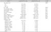
The total number of children diagnosed as APN was 996. The number of patients excluded from this study by each criteria is as follows: 87 were excluded because the DMSA scan was a follow-up study; 18 were excluded due to prior use of antibiotics; three were excluded due to prior history of liver disease; one patient was excluded because another etiology for hepatitis was identified; and four patients were excluded because laboratory data were unavailable. A total of 883 children with APN were included in this study, 514 were boys and 369 were girls. The male to female ratio for all APN patients was 1.4:1. The number of children in each of the age groups was 320 for 0 to 3 months of age, 378 for 4 to 12 months of age, and 185 for over 12 months of age. The proportion of boys in ages 0 to 8 months was greater than 50%, especially during the earlier months when the proportion of boys was greater than 80%. After 8 months of age, the proportion of boys declined rapidly. This difference in proportions was statistically significant when comparing each of the age groups described above (77.2% in ages 0 to 3 months, 55.6% in ages 4 to 12 months, and 30.8% in ages greater than 12 months, p=0.000).
Elevated ALT and its association with demographic and clinical variables
Of the 883 children with APN, 81 children (9.2%) had elevated ALT. The maximum level of ALT was 774 IU/L, and the median elevated ALT was 95 IU/L. In the analysis of demographic characteristics between the children with normal and elevated ALT levels, differences were observed between the number of children in each age group, sex, and blood culture results (Table 1).
Urosepsis and its association with demographic and clinical variables
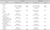
To confirm the correlation between elevated ALT and positive blood culture, we decided that multivariate analysis of variables related to positive blood culture was required. In the multivariate analysis of the variables, age, elevated ALT, elevated CRP, and elevated creatinine showed statistical significance (p<0.050) (Table 2).
The duration of intravenous antibiotic treatment for the children with positive blood culture was longer than that of the children with negative blood culture, 8.2±2.38 days and 6.8±2.14 days, respectively (p=0.000).
Total bilirubin and hypotension
Of the 883 children with APN, 58 had elevated total bilirubin levels (7 children with elevated ALT and 51 children with normal ALT), and 81% of these children were under the age of 3 months. Hypotension was observed in only three children, one with elevated ALT and two without. None of these children had a systemic blood pressure of less than 60 mmHg.
DISCUSSION
The results of our study demonstrate a correlation between elevated ALT levels and increased prevalence of sepsis in children with APN. Roughly 9% of children with APN had ALT elevation, and among these children, 17.6% had a positive blood culture, double the rate in children without ALT elevation, which is 8.2%.
Younger age and elevated serum levels of CRP and creatinine had been found to be associated with urosepsis among children with UTI or APN. Among infants and children with UTI, patients with a positive blood culture were younger [111]. UTI has been studied most often among young infants since they were the most prone to be implicated, and as high as 85% of serious bacterial infections among infants younger than 3 months of age has been identified as UTI [12]. We found that age younger than 3 months was significantly related to increased prevalence of urosepsis among children with APN. Elevated CRP has been associated with increased risk for sepsis [13]. Our study also showed difference of blood culture positivity between children with elevated and normal CRP levels, with adjusted odds ratio of 8.13. Our study also showed difference of blood culture positivity between children with elevated and normal creatinine levels, with adjusted odds ratio of 1.78. This finding is in accordance with a prior study analyzing factors associated with bacteremia in young infants with UTI which found association of increased blood creatinine values with bacteremia [14]. Therefore elevated ALT can be another parameter for anticipating increased prevalence of urosepsis in children with APN, complementary to age younger than 3 months, elevated CRP, and elevated creatinine.
Three main mechanisms have been proposed for the liver injury during sepsis. The liver plays a central role when a sepsis occurs. Kupffer cells are the main scavengers of bacteria and endotoxins, preventing them from entering the systemic circulation [4]. However, Kupffer cells have been found to be responsible for the increase in proinflammatory cytokines and decrease in hepatocellular function. Decrease number of Kupffer cells have shown beneficial effect on hepatocellular function and decreased levels of the cytokines [3]. During sepsis, liver parenchymal cells are involved in the immune response, and acute phase proteins produced by hepatocytes enhances both host defense and protective functions. However, the acute phase proteins response markedly contributes to the procoagulant state [4]. Lastly, neutrophils are proposed to be the main effector cells that cause septic liver injury [15]. Activated neutrophils injure hepatocytes by production of oxygen-derived radicals and protease that may injure hepatocytes [16].
Liver enzyme elevation among patients with UTI or APN has been studied in adults and children. In the study of liver enzyme elevation among adult patients with APN by Campos et al. [5], arterial hypotension and older age seemed to be associated with increased prevalence of urosepsis. In the study among children [6], however, younger age was the only meaningful factor that showed difference between patients with liver enzyme elevation and those without. These studies did not consider presence of urosepsis as a factor in their analysis. Whether the effect of arterial hypotension and older age in adults and younger age in children on elevated liver enzymes is actually caused by the presence of urosepsis remains to be studied.
UTI during the first three months of life is more common in boys, and the sex ratio reverses after the first year [17]. The results of our study are in concordance with the known demography, with boys constituting more than 80% of children under age of 3 months, and dropping with increasing age.
Oddly, in univariate analysis of our study, fever durations of two or more days showed negative correlation with positive blood culture. A prospective cohort study in children of 3 to 36 months of age by Teach and Fleisher reported similar results [18]. Another study on fever and bacteremia in children concluded that a high fever of greater than or equal to 40.5℃ was specific for bacteremia [19]. It would be logical to suppose that parents with younger children or children with high fevers would be more inclined to visit a hospital and consent to a urinalysis for detection of UTI. Although we did not collect data on the degree of fever, an analysis of our data showed that children who had a fever for less than two days were younger than those who had a fever for two or more days (21.9±1.0 months and 25.2±1.2 months, respectively, p=0.001). A logistic regression analysis of fever duration and age showed that fever duration had no statistically significant correlation with blood culture positive rate, which confirmed the possibility of age being the confounding variable for the fever duration (adjusted odds ratio 0.62, 95% confidence interval 0.35-1.09, p=0.094).
Systemic viral, bacterial, and protozoan infections invading the liver may cause cholestasis [20]. This effect occurs predominantly in Gram-negative bacilli, such as Escherichia coli, Kelbsiella pneumoniae, Pseudomonas aeruginosa, and Proteus species. Proposed mechanisms are increased bilirubin load due to hemolysis that can occur in bacterial infections, and decreased bilirubin uptake, intrahepatic processing, and canalicular excretion [21]. In our study, 58 children had elevated total bilirubin levels among 883 children with APN, but the majority (81%) was under the age of 3 months. Neonates with UTI may present with jaundice only [22], and sepsis is more likely to manifest with jaundice in infants and children than in adults [21]. In our study, no association was found between elevated total bilirubin and elevated ALT. More accurate analysis of the relationship between elevated total bilirubin with the adjusted normal values for age and the rate of sepsis would be valuable.
Prior studies on UTI and elevated aminotransferases included both aspartate aminotransferase (AST) and ALT elevation, and the prevalence of increased aminotransferase levels in UTI patients was approximately 20% [56]. If elevated AST was included in our analysis as well, approximately 15% of the children with APN had elevated aminotransferases.
One of the limitations of our analysis is that blood cultures were not performed in 154 children (19.2%) among 802 with normal ALT and in 13 children (16.0%) among 81 with elevated ALT, but the proportions of the children without blood cultures do not differ much between the two groups, and we do not think it has affected the results of our study.
In conclusion, our study demonstrates the association between elevated ALT and increased prevalence of urosepsis, in addition to elevated CRP, elevated creatinine, and age younger than 3 months in children with APN.
 XML Download