

 PDF
PDF ePub
ePub Citation
 Print
Print


INTRODUCTION
Alport syndrome is a glomerulopathy due to abnormal type 4 collagen in the glomerular basement membrane, and predisposes affected individuals to develop glomerulosclerosis. Affected individuals present with hematuria, renal failure, hearing loss, dyslipidemia, and retinal flecks. Diagnosis can be made by skin biopsy, renal biopsy, or genetic testing. Management includes angiotensin blockade therapy in the setting of overt proteinuria, and kidney transplant if/when the disease progresses to end stage renal disease [1]. In Wilson disease (WD), copper accumulates in the liver, kidneys, brain, corneas, and other organs due to mutations in ATP7B. The diagnosis of WD may involve testing for serum ceruloplasmin, urinary excretion of copper, hepatic copper concentration, neurologic or psychiatric abnormalities and screening for ATP7B mutations [23]. In patients with renal disease that causes glomerular damage and non-selective proteinuria, the urinary copper excretion may be difficult to interpret since there are losses of heavy metals associated with proteins in their urine [45]. Little is known about levels of urinary copper and zinc excretion in children with proteinuric kidney diseases. We report on the case of a child diagnosed at an early age with Alport syndrome whom later developed abnormal liver enzymes, low serum ceruloplasmin and elevated urine heavy metals, findings that may be associated with glumerolopathy and medications. The concomitant diagnosis of WD was established, enabling targeted therapy to prevent progression of the liver disease.
CASE REPORT
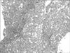
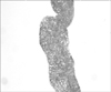
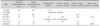
A 17-year-old male was diagnosed with Alport syndrome at the age of 5 years due to the presence of hematuria, proteinuria, and a positive family history in his mother, maternal uncle, and brother. He did not have hearing loss or eye involvement. There was no family history of liver disease. Angiotensin blockade therapy was started. Routine laboratory monitoring revealed persistent elevation of transaminases (alanine aminotransferase 108-120 IU/L, aspartate aminotransferase 64-66 IU/L) for 2 years. Initially, this elevation was thought to be due to medication hepatotoxicity. However, liver enzymes remained persistently elevated after medication discontinuation. Additionally, he had an elevated cholesterol (256 mg/dL) and triglycerides (270 mg/dL) and an abdominal ultrasound with increased echogenicity of the liver. Further workup due to chronic abnormal liver enzymes showed a low serum ceruloplasmin (<10 mg/dL) and albumin (3.5 g/dL) but a normal total serum protein (6.4 g/dL). Urinary 24-hour excretion of copper was 159 mcg. Based on these results, a percutaneous liver biopsy was performed. Histological analysis showed diffuse bridging fibrosis, microvesicular and macrovesicular steatosis, and quantitative copper analysis demonstrated a high concentration of copper (1,260 mcg/gr dry weight liver), suggesting the diagnosis of WD (Fig. 1 and 2). Further confirmation was obtained with genetic testing showing the presence of two heterozygous WD mutations of ATP7B, V1262F and M645R. The patient was started on a chelating agent, trientine, and liver enzymes improved (Table 1). While on trientene, the 24-hour urinary excretion of zinc was high at 4,987 mcg, even though he was not on zinc supplementation at the time. He was transitioned 7 years after the diagnosis of WD to zinc maintenance monotherapy as his liver enzymes were near normal and 24-hour urine copper excretion was decreasing. After 4 months on zinc, the liver enzymes normalized and 24-hour urine copper decreased appropriately to 91 mcg.
DISCUSSION
The diagnosis of WD and its ongoing monitoring is challenging when associated with an underlying kidney disease such as Alport syndrome. The frequency of abnormal liver enzymes in patients with Alport syndrome is unknown and the differential diagnosis can be broad. Elevated transaminases may initially be attributed to dyslipidemia or drug induced hepatotoxicity. Additionally, dyslipidemia may worsen in the setting of nephrotic syndrome. Angiotensin-converting enzyme-inhibitors, including Enalapril, Captopril, and Lisinopril, frequently used in the treatment of Alport syndrome, may infrequently have hepatotoxic effects [678]. However the differential diagnosis for the abnormal liver tests should also include hepatitis B, hepatitis C, alpha-1 antitrypsin, autoimmune hepatitis, nonalcoholic steatohepatitis and WD.
Initial laboratory results in the patient presented in our case report included a low serum ceruloplasmin. However, in Alport syndrome, proteinuria alone might lead to a low serum ceruloplasmin since this protein is among the serum proteins inappropriately filtered by the damaged glomerulus. Furthermore, Alport syndrome can also lead to increased urinary loss of heavy metals such as zinc and copper due to be presence of proteinuria [459], which may also complicate the diagnosis and monitoring of the disease. Trientene treatment may also contribute to increased urinary zinc loss. Zinc deficiency has not been reported in patients with WD on chelation therapy, likely due to the ubiquitous presence of dietary zinc. Urine macroglobulin was not measured which could assist in differentiating the etiology of the urine zinc loss between proteinuria and chelation therapy.
Based on the Leipzig criteria for establishing a diagnosis of WD, elevated urine copper >2 times the upper limit of normal will give 2 points, and low ceruloplasmin below 10 ng/dL will give an additional 2 points, summing 4 points, which is considered as a high likelihood of WD diagnosis [10]. In the patient described and in other patients with non-selective proteinuria, a low serum ceruloplasmin and increased urine copper could be attributed to the glomerulopathy rather than WD. We suggest that in patients with proteinuria and abnormal liver tests or other symptomatology (neurological or psychiatric), additional work up, including a liver biopsy and genetic testing, should be considered.
Little has been published about urinary zinc excretion in patients with WD not on zinc therapy. In our patient, urinary zinc excretion was high even before he was transitioned from trientine to zinc. This was similar to a recent report showing high zinc excretion in WD patients on trientine taken once daily [11]. Urine zinc may therefore be elevated due to chelation with trientene, however in our patient zinc excretion due to the leaky glomerulus and proteinuria could not be excluded. Therefore it may be difficult to monitor compliance with zinc therapy for maintenance as well unless baseline values are known for the patient off of all therapy [459]. On zinc monotherapy for his WD, the patient's liver enzymes have normalized and urinary copper excretion remains decreased, suggesting effective treatment.
 XML Download