

 PDF
PDF ePub
ePub Citation
 Print
Print


INTRODUCTION
Helicobacter pylori causes a chronic, persistent infection that may lead to chronic gastritis, atrophic gastritis, intestinal metaplasia, and even gastric adenocarcinoma [12]. Primary H. pylori infection occurs during early childhood, with most adults in developing countries eventually becoming carriers [34]. Although the stomach is considered a sterile organ because of the acidic conditions (pH<4) [5], H. pylori can survive in the human stomach by activating a cytoplasmic urease, which converts urea into carbon dioxide and ammonia and thereby increases the pH of the environment [6]. The urease test used to diagnose H. pylori infection is based on this activity of the microbe: the urease test uses phenol red, which changes from yellow to pink or red as the pH increases [7]. In children, the diagnosis of H. pylori infection is usually based on the findings of histological examination and the urease test [8].
A previous study identified the sampling site of gastric biopsy examination, histopathologic findings, and the H. pylori load as factors that influence the results of the urease test [9]. Further, several conditions can lead to false-negative results in the urease test, the two most common of which are the recent use of proton-pump inhibitors (PPIs) and the presence of intestinal metaplasia [7]. Both these conditions are associated with low gastric acidity. Thus, the pH of gastric juice may be related to the results of the urease test. In the present study, we assessed the pH of gastric juice in children aged 0-15 years and investigated the correlation between gastric pH and the results of the urease test and histopathologic examination.
MATERIALS AND METHODS
Study population
The institutional review board approved the research protocols in the present retrospective study (GNUHIRB-2015-07-016), and 562 children with upper abdominal pain who underwent endoscopy of the upper digestive tract and gastric juice aspiration via endoscopy at the Department of Pediatrics, Gyeongsang National University Hospital (Jinju, Korea), were enrolled. Endoscopy was performed after overnight fasting, and the indication of endoscopy was upper abdominal pain. No participant was taking PPIs before endoscopy, and no active bleeding was observed during endoscopy. The study population was stratified into 3 age groups: 0-4 years (n=75), 5-9 years (n=244), and 10-14 years (n=243).
Measurement of gastric pH
The pH of gastric juice was measured using a Metter Toledo Delta 320 pH meter (Metter-Toledo International Inc., Columbus, OH, USA) immediately after it was thawed. To ensure accuracy, the pH was adjusted using 3 buffer systems (pH 4.00, 7.00, and 10.01). Hypochlorhydria was defined as a gastric juice pH of ≥4.0 [5].
Urease test
A clinical diagnosis of H. pylori infection was obtained by conducting the urease test and histopathological analysis on gastric antral biopsy samples. Urease tests were performed in the endoscopy room according to a previous method [9]. Briefly, three biopsy specimens from the antrum were incubated in a 2% urea broth (20 g/L urea, 0.04 g/L phenol red, 0.2 g/L KH2PO4, 0.5 g/L NaCl; pH 6.8), and if color change occurred in the following 24 hours, the biopsy was deemed to be urease test positive.
Histopathologic findings
Histopathological examination was performed using biopsy specimens that were fixed in 10% buffered formalin overnight, processed for paraffin embedding, cut into 4- to 5-µm thick sections, and stained with hematoxylin-eosin. The histological results were interpreted using the Updated Sydney System [10]. For this, the degrees of chronic gastritis, active gastritis, and H. pylori infiltration were classified as normal, mild, moderate, or marked.
Statistical analysis
Data were analyzed using IBM SPSS Statistics ver. 21.0 for Windows (IBM Co., Armonk, NY, USA). Variation in urease-test positivity, histopathologic findings, and gastric pH depending on age group was evaluated. Statistically significant differences in urease-test positivity and gastric pH among the 3 age groups and in urease-test positivity and the degrees of chronic and active gastritis and H. pylori infiltration between those with normal gastric pH and hypochlorhydria were determined by the χ2 test and generalized linear models. Nonparametric tests were used to analyze the mean gastric juice pH in different age groups. p-values of <0.05 were considered statistically significant.
RESULTS
Gastric juice pH
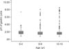
The median pH of the gastric juice was 1.77 in the 0-4 years group, 1.59 in the 5-9 years group, and 1.55 in the 10-15 years group (Fig. 1). No differences were found in the range of gastric pH among the 3 age groups (p=0.655). The proportion of individuals with hypochlorhydria was 1.3% (1/75) in the 0-4 years group, 6.1% (15/244) in the 5-9 years group, and 8.2% (20/243) in the 10-15 years group (p=0.101; Table 1).
Comparison of urease-test positivity and histopathologic findings between subjects with hypochlorhydria and normal gastric pH
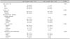
Table 1 shows a comparison of urease-test positivity and histopathologic findings between subjects with normal gastric pH and hypochlorhydria.
The rate of urease-test positivity was 31.7% among subjects with normal gastric pH and 77.8% among those with hypochlorhydria (p<0.001). Urease-test positivity showed a positive correlation with gastric acidity or pH (R=0.237, p<0.001).
Subjects with hypochlorhydria had moderate to severe degrees of chronic and active gastritis while those with normal gastric pH did not (p<0.001 for both, Table 1). H. pylori was detected in 38.9% of the subjects with hypochlorhydria, and this proportion was higher than the proportion of subjects with normal gastric pH in whom H. pylori was detected (20.9%).
Urease-test positivity and histopathologic findings
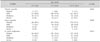
The rate of urease-test positivity was 36.0% in the 0-4 years group, 32.0% in the 5-9 years group, and 37.0% in the 10-15 years group. This parameter did not differ significantly among the 3 age groups (p=0.487). The proportions of individuals with moderate and severe chronic gastritis (p=0.003), active gastritis (p=0.046), and H. pylori infiltration (p=0.021) increased with age (Table 2).
DISCUSSION
In the present study, the range of gastric juice pH was similar in the 3 age groups, and the median gastric juice pH was <2.0 in all age groups. A previous study reported that gastric acidity increased with age and reached adult levels by age 14 years [11]. It found that the ranges of gastric pH while fasting were 3-4 in neonates, 1.5-3 in infants, 1-3 in preschoolers, 1-2 in school-going children, and 0.5-2 in adolescents and adults [11].
In this study, 93.6% of the subjects had normal gastric pH, while 6.4% had hypochlorhydria (pH > 4.0). This proportion of subjects with hypochlorhydria was lower than that in a study on Bangladeshi children aged 2-5 years, in which 70.0% of 30 children with H. pylori infection and 43.3% of 30 children without H. pylori infection had hypochlorhydria [12]. These differences in the proportions of hypochlorhydria in children might be related to differences in H. pylori infection rates in the general population and socioeconomic conditions between the countries under study. In the present study, the range of urease-test positivity was 32.0-37.0% in children aged 0-15 years, but in the Bangladeshi study, the prevalence of H. pylori infection was about 60% in children aged 0-5 years [13]. The proportion of subjects with hypochlorhydria in the present study increased with age, from 1.3% in the 0-4 years group to 8.2% in the 10-15 years group, although without statistical significance.
In a previous study, adult individuals with acute H. pylori infection were found to have transient hypochlorhydria [14]. Further, among male Japanese patients, H. pylori showed a stronger inhibitory effect on acid secretion [15]. Nonetheless, the relation between hypochlorhydria and H. pylori infection in children remains controversial. Park et al. [16] reported that H. pylori infection was significantly more frequent in children with hypochlorhydria (pH > 4) than in those with normal gastric pH (≤4). Sarker et al. reported that hypochlorhydria was observed in 70.0% of children with H. pylori infection [12] and that gastric acid output improved after treatment of the infection [17]. However, Nagita et al. [11] reported no significant correlation between the histologic density of H. pylori and gastric juice pH.
In normal healthy adults, age has an independent positive effect on acid secretion, while H. pylori infection has an independent negative effect [18]. The proportion of hypochlorhydria was found to increase with age in the present study. This finding may be related to the higher degree of chronic gastritis and active gastritis and higher urease-test positivity observed in the older subjects than the younger ones.
Hypochlorhydria in adult patients was found to result from H. pylori-associated corpus atrophy [19]. H. pylori infection occurs in early childhood, and chronic H. pylori infection induces atrophy of the gastric mucosa [20]. However, the incidence of atrophic gastritis and intestinal metaplasia is lower in children than in adults [2122]. In fact, no subject in the present study had atrophic gastritis or intestinal metaplasia. A previous study showed that although there was no difference in the proportion of moderate and severe degrees of chronic and acute gastritis between the antrum and body in children, the positivity rate in the urease test was higher for gastric body samples (49.4%) than antrum samples (85.1%) [9]. Another pediatric study found that the pH of gastric juice was higher in the urease-test positive group (2.52±1.45) than the negative group (1.80±0.52) [11]. Further, the pH was significantly correlated with the degree of chronic gastritis and was significantly higher in urease-test positive subjects [11]. In our study as well, we compared urease-test positivity and histopathologic findings between children with normal gastric acidity and those with hypochlorhydria. The rate of urease-test positivity was higher in the latter than the former. Additionally, moderate to severe degrees of active gastritis and chronic gastritis were observed in children with hypochlorhydria than in those with normal gastric acidity, and the presence of H. pylori was more frequently observed in children with hypochlorhydria than in those without hypochlorhydria, although without statistical significance. These results collectively suggest that hypochlorhydria in children is related to H. pylori infection.
Subjects with H. pylori-related body gastritis were previously found to have hypochlorhydria and a low density of H. pylori colonization [20]. In the present study, moderate and severe degrees of H. pylori infiltration were observed in subjects with normal gastric pH even though the presence of H. pylori was higher in subjects with hypochlorhydria (38.9%) than those with normal gastric pH (20.9%). We did not evaluate the histopathologic findings of the gastric body and did not know the exact duration of H. pylori infection. Further investigation is needed to better understand the association between the degree of H. pylori infiltration and hypochlorhydria.
Chronic H. pylori-related gastritis with hypochlorhydria was previously found to be related to false negative results in the urea breath test [20], and as mentioned in the introduction, PPI use is known to produce false negative results in the urease test [7]. Although hypochlorhydria is observed in acute H. pylori infection and chronic atrophic gastritis of the gastric body and with the use of PPIs, the results of the urease test may be different: positive in acute H. pylori infection and negative in chronic atrophic gastritis and use of PPIs. In the present study, we were not able to determine the cause of hypochlorhydria in children with negative urease test results and no H. pylori infection.
The present study has some limitations. First, it was a retrospective study. Further, we could not investigate the exact reason for hypochlorhydria in the children who did not take PPIs and did not have H. pylori infection. Second, we did not measure the acid output. Finally, we did not evaluate the pH of gastric juice in healthy children.
In summary, the pH of gastric juice while fasting seems to be normal in children regardless of age. Urease-test positivity is related to hypochlorhydria in children who do not take PPIs. Our results suggest that hypochlorhydria is related to H. pylori infection in children, although further studies are needed for prove the pathogenesis of hypochlorhydria in children and the clinical course of H. pylori-infected children with hypochlorhydria.
 XML Download