

 PDF
PDF ePub
ePub Citation
 Print
Print


INTRODUCTION
Progressive familial intrahepatic cholestasis type 3 (PFIC3) is an autosomal recessive disorder of cholestasis of hepatocellular origin. The onset of PFIC3 is typically in infancy or in childhood. PFIC3 is caused by a defect in the ABCB4 located on chromosome 7 that encodes a class III multidrug resistance (MDR3) P-glycoprotein. This protein functions as a phospholipid translocator involved in biliary phospholipid (phosphatidylcholine) excretion, and is predominantly expressed in the canalicular membrane of the hepatocyte [12]. Altered MDR3 function causes continuous exposure of biliary epithelium to hydrophobic bile salts with subsequent cholangitis, and also increases lithogenicity of bile with resultant cholelithiasis, cholestasis and biliary cirrhosis. The exact prevalence of PFIC3 remains unknown, but the estimated incidence varies between 1/150,000 and 1/300,000 [3].
Prior reports showed increased hepatic copper in patients with PFIC3 that presented with liver disease at ages 2-11 years [45]. Here we report on an older patient that presented with biochemical abnormalities in copper metabolism at age 16 that led to an initial consideration of a diagnosis of Wilson disease (WD), resulting in a delayed diagnosis of PFIC3. The diagnosis of PFIC3 was later confirmed by molecular studies that identified novel mutations in the ABCB4 gene. We present an analysis of her liver tests, bile acids, liver histology and immunohistochemistry (IHC) and parameters of copper metabolism before and following therapy with ursodeoxycholic acid (UDCA).
CASE REPORT
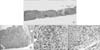
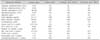
The patient presented with fatigue at the age of 15 years and was found to have splenomegaly and mild thrombocytopenia. Esophageal varices were seen on screening endoscopy. Viral studies and serology for autoimmune hepatitis were negative and testing for WD was performed. Twenty four hours urine copper was 342 µg and slit lamp examination showed absence of Kayser-Fleischer (K-F) rings. Ceruloplasmin level was normal, 35 mg/dL, and no mutations in the full sequence analysis of ATP7B, the gene for WD, were detected. Liver biopsy was small and fragmented and showed bridging fibrosis with a suspicion of cirrhosis. The interpretation was limited due to its small size and fragmentation, but other histologic changes seen included a mild chronic portal inflammatory infiltrate with mild interface activity, moderate bile duct proliferation and neutrophilic pericholangitis (Fig. 1). Liver copper quantitation showed a markedly elevated hepatic copper content of 1,471 µg/g, suggesting a diagnosis of WD. Electron microscopic findings included electron dense lysosomes, microvesicular steatosis and focal degeneration of biliary epithelium associated with mild bile stasis. She was started on zinc therapy for presumed WD, but was switched to trientine due to gastrointestinal side effects from zinc. Nadolol was initiated for primary prophylaxis for esophageal bleeding. Though treated with trientine for over 12 months, her transaminase levels did not improve and 24 h urine copper determination remained elevated at 200 µg (normal range, 15-60 µg). Lab testing showed aspartate aminotransferase 141 U/L, alanine aminotransferase 114 U/L, alkaline phosphatase 596 U/L, gamma glutamyltransferase (GGT) 734 U/L, albumin 4.2 g/dL, and conjugated bilirubin 1.67 mg/dL. Ceruloplasmin level was 38 mg/dL (normal range, 18-46 mg/dL), total serum copper 1.51 µg/dL (normal range, 0.75-1.45 µg/dL). In view of a normal ceruloplasmin, absent K-F rings and lack of ATB7B mutations alternate diagnostic possibilities were considered. Further evaluation for another possible etiology of her liver disease included total and primary bile acid levels that were elevated (Table 1). She was started on a trial of UDCA and ABCB4 gene sequencing was performed. The patient was found to have compound heterozygosity for novel mutations in the ABCB4 gene and had inherited one mutation from each parent (father: c.984T>G [p.Y328*], mother: c.3218G>A [p.C1073Y]), consistent with a diagnosis of PFIC3. Trientine was stopped, but UDCA was continued. With further UDCA treatment, serum transaminases decreased but did not normalize (Table 1). Another liver biopsy performed after 4 months of UDCA therapy showed histologic findings similar to the previous biopsy, and confirmed the presence of cirrhosis (Fig. 2A) on trichrome and reticulin stains. In addition, subtle changes of cholate stasis in the form of pale cytoplasm of hepatocytes was seen at the periphery of the cirrhotic nodules (Fig. 2B). There was accumulation of brownish granular pigment in the periseptal hepatocytes (Fig. 2C), suggestive of copper, which was also confirmed with a rhodanine stain (Fig. 2D). These findings suggested chronic cholestasis and a biliary type cirrhosis consistent with PFIC3. Quantitative copper analysis showed hepatic copper content decreased to 135 µg/g dry weight liver. Immunostains for CD10, bile salt export pump and MDR3 showed normal canalicular expression of these proteins (Fig. 3). At the time of biopsy, the hepatic venous portal vein pressure gradient was measured as 11 mmHg while the patient was on nadolol. After 14 months of UDCA treatment, liver indices remained mildly abnormal but synthetic function is stable. Esophageal varices were still seen on endoscopy 6 and 12 months later, and banding was performed.
DISCUSSION
Etiologies for cirrhosis presenting in late adolescence and early adulthood includes late onset of all childhood metabolic or inherited liver disorders and many disorders that typically present in adults. In those patients without clear causes for their liver disease, molecular genetic testing for some of the inherited metabolic disorders has proven to be a useful diagnostic tool.
In our patient, biochemical testing led to an initial concern for a disorder of copper metabolism, specifically WD, due to elevated urine and hepatic copper above typical diagnostic levels. Current guidelines for the diagnosis of WD utilizes a scoring system developed by experts at the 8th International Meeting on Wilson's disease in Leipzig in 2001 that includes graded values for clinical, biochemical and molecular testing. This patient met criteria for a diagnosis of WD based on elevated urine copper excretion and hepatic copper content, giving a total score of 4 (a score above 3 is considered diagnostic for WD) [6]. However, corneal K-F rings were absent, ceruloplasmin was normal and full sequence analysis of ATP7B failed to detect mutations. Furthermore, a treatment trial of zinc and then the copper chelator trientine did not improve her liver tests, and therefore an alternative diagnoses of a cholestatic disorder (where copper accumulation would be secondarily) was considered. In support of a cholestatic disorder were elevated serum bile acids and GGT, and biochemical response to a trial of UDCA. Sequence analysis for mutations of ABCB4 revealed compound heterozygosity for novel mutations of ABCB4. These mutations included a nonsense mutation inherited from her father and predicted to cause early termination of protein synthesis, and a novel variant of uncertain significance on the other allele inherited from mother indicating these mutations were not biallelic.
Indeed our data show that PFIC3 can lead to a false positive result for the diagnostic scoring system for WD. Two recent reports [45] showed elevated hepatic copper concentrations in some PFIC3 patients with chronic cholestasis, that were 248, 860, and 863 µg/g dry weight of liver, near or above the typical 250 µg/g found in most patients with WD. Our patients had an even higher copper concentration which could be a consequence of the more advanced age and prolonged cholestasis compared to the younger patients previously reported. Published data was not detailed enough to calculate Leipzig scores for diagnosis of WD in the prior case reports. However we would speculate similar elevations in urine copper would have been present given the elevated liver copper. This would have given diagnostic scores for WD of either 3 or 4 for these patients.
Therefore response to therapy and histology must be carefully considered in cases where pathognomonic findings of ATP7B mutations, a low ceruloplasmin and K-F rings are absent. In this patient molecular testing helped refute the diagnosis of WD and establish that she had PFIC3.
Chronic cholestasis is the likely cause for the increased hepatic copper in our patient. Following treatment with UDCA, liver tests corresponding to cholestatic change (GGT, alkaline phosphatase) were reduced and a follow up determination of hepatic copper was markedly reduced as well. We cannot exclude that her brief treatment with zinc then trientine helped with the initial reduction of hepatic copper; however, we did note that elevated urine copper persisted on trientine so it was unlikely that stores were depleted prior to UDCA treatment. After UDCA treatment, an elevated 24 hour urine copper prior to treatment decreased to the normal range in parallel with the reduction of hepatic copper. In the prior reports of patients with PFIC3 and increased hepatic copper, no data was presented on their response to UDCA therapy with respect to copper metabolism.
The histologic findings in our patient included mild chronic hepatitis like changes, mild macrovesicular steatosis, few glycogenated nuclei, bile ductular proliferation with neutrophilic cholangitis, patchy areas of cholate stasis, rare hepatocytes with Mallory-Denk bodies, and cirrhosis. While some of the findings (mild chronic hepatitis, steatosis, glycogenated nuclei and Mallory-Denk bodies) are seen in cases of WD, none of them are specific and the overall pattern in this patient suggested a biliary cirrhosis more consistent with PFIC3. However we speculate the initial findings of dense lysosomal deposits are common to both untreated WD and PFIC3 due to aggregates of copper and copper binding proteins such as metallothioneins [7].
Expression of MDR3 detected by IHC is highly variable. While some patients with early onset PFIC3 disease and homozygous mutations show absent or markedly decreased MDR3 expression on the canalicular surface of hepatocytes, others, particularly with heterozygous mutations may show normal MDR3 by IHC. This patient had two unique mutations of ABCB4. One of the mutations, c.984T >G (p.Y328*), has not been previously reported in the literature. This is predicted to be a nonsense mutation that results in a premature stop codon (p.Y328*) and thus is categorized as a deleterious mutation. The other mutation, c.3218G >A (p.C1073Y), is a novel variant not previously reported in the literature. The functional effect of this mutation is uncertain but is likely deleterious based on our patients' disease state. Normal cannalicular MDR3 expression by IHC, suggests the protein had retained antigenicity despite loss of function. This has been reported in prior studies showing retained MDR3 expression in some cases with phenotypic PFIC3 with clinical disease. Therefore IHC evidence for MDR3 expression should not be taken as an evidence against a diagnosis of PFIC3 disease [89].
Long term outcomes of UDCA treated patients with PFIC3 is unknown and previous reports include follow up only up to 4 years [10]. Interestingly, Davit-Spraul et al. [11] reported that the threshold predicting a positive response to UDCA therapy in PFIC was represented by a percentage of biliary phospholipids of around 7%. This means that PFIC3 cases with milder mutations causing partial protein dysfunction also respond the UDCA treatment better. We believe that PFIC3 cases presenting later in life are therefore more likely to have milder mutations affecting MDR3 function and UDCA responsiveness.
Jacquemin et al. [10] observed a decrease in serum bile acid concentration in PFIC patients who responded to UDCA therapy (where response was defined by decreased transaminases) reflecting an improvement of biliary bile acid secretion. Serum total bile acid concentrations increased under UDCA therapy in patients who failed to respond. Although we found a significant decline in transaminase, GGT and alkaline phosphatase levels in our patient after UDCA treatment, we didn't observe a significant decline in serum bile acid concentrations. This may be due to a shorter time of follow up when compared to the study mentioned above where the follow up time interval ranged from 2-4 years.
In their case report, Lai et al. [12] presented a pediatric patient with small duct sclerosing cholangitis who later developed cholangiocellular carcinoma. However, in their report ABCB4 genetic testing of the patient was reported as sent but the result was not available at the time of submission. The patient indeed had two different missense variants in the ABCB4 gene that are associated with PFIC3 (personal communication to Dr. Joanne Lai, Ronen Arnon and Benjamin Shneider). Cholestasis, cholangitis, biliary cirrhosis and hepatocellular failure [13] are well described in the course of PFIC3 however cholangiocellular carcinoma must now also be considered. Screening and ongoing surveillance is likely indicated, but we lack data on the true incidence of malignant transformation of the biliary epithelum in this disorder. In our patient, CA 19-9 was elevated and MRI/MRCP did not show evidence of malignancy. Over time on UDCA, CA 19-9 began to decrease as well.
In summary, the diagnosis of PFIC3 should be considered in young adults with features of chronic cholestasis and/or biliary cirrhosis. If this disorder is not recognized early, prolonged cholestasis can cause elevated hepatic copper and increased urine copper excretion that overlap with current diagnostic criteria for WD. Failure of adequate response to treatment for WD in such a patient should raise suspicion of alternative diagnoses. As illustrated by this case, molecular diagnostics are very useful for establishing a diagnosis of PFIC3, and can be used for the screening of primary relatives of the patient. UDCA ameliorates cholestasis in PFIC3, and also helps mediate a reduction in hepatic copper content in response to treatment. Ongoing surveillance for cholangiocellular carcinoma should be considered as well.


 XML Download