

 PDF
PDF ePub
ePub Citation
 Print
Print


INTRODUCTION
Alimentary tract duplications are rare congenital malformations of the gastrointestinal tract, which may be cystic or tubular and occur anywhere from the mouth to the anus [1]. More than 80% of cases present before the age of 2 years as an acute abdomen or bowel obstruction [23]. The commonest site for duplication is the ileum, while colonic duplication is rare, accounting for 6-13% of all gastrointestinal duplications, commonly located in the cecum [14]. These lesions, if encountered incidentally, should be surgically managed to prevent any future complications. Although perineal canal or H-type congenital fistula is a relatively common anomaly in Asian countries, it's association with Y-shaped tubular colonic duplication is rare [5]. We report a case of colorectal duplication in a two month old female child passing stools from normal anal opening and a fistulous opening in the vestibule.
CASE REPORT
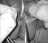
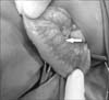
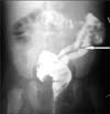
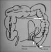
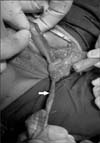
A two-month-old female child presented with the complaints of passing stools from normal anal opening and a fistulous opening in the vestibule. A provisional diagnosis of perineal canal was made. Abdominal sonography, echocardiography and spinal radiography were normal, ruling out associated urinary tract, cardiac and vertebral anomalies. Examination under anaesthesia revealed a normal anal opening. The vestibule had 3 openings, which were a normal urethral and vaginal opening and a rectovestibular fistula (Fig. 1) with no communication with the normal anorectum. Considering this to be a complex anomaly, a diverting stoma was planned. On exploration of the abdomen for the stoma, tubular duplication of the sigmoid colon was noted (Fig. 2). A high sigmoid loop colostomy was done which resulted in four stomal lumens. Later, for better delineation of the anatomy of the anomaly, a proximal and distal colostomogram was performed (Fig. 3). It revealed duplicated colon with separate lumens from the transverse colon downwards with two anal openings. The duplicated colonic segment towards the mesenteric side terminated as a rectovestibular fistula (Fig. 4). At 6 months of age, the patient was taken up for definitive surgical management. An abdominoperineal approach was used, in which initially the colostomy was mobilized. For the distal duplicated colonic segment, the mucosal cuff of the colon leading to the vestibular fistula was dissected like a Soave's endorectal pullthrough from the distal stoma to the vestibular fistula and excised (Fig. 5). The remaining muscular cuff was plicated and closed.
The proximal duplicated colonic segment shared the vascular supply with the native colon, hence resection was not possible. The duplicated segment extended proximally till the ascending colon and common wall comprised of two layers of mucosa. Therefore, the intervening mucosal septum between the duplicated colon, upto the non-duplicated ascending colon was divided (Fig. 6). A colo-colic anastomosis between the proximal unified colon with the distal native colon leading to the normal anus was done. In view of the significant disparity in the proximal and distal colonic luminar diameter, a covering ileostomy was also done. The postoperative course was uneventful and the ileostomy was closed after 3 months. At follow up of 6 months post-surgery, the opening in the vestibule had closed, with no bowel complaints.
DISCUSSION
Duplications of the colon are uncommon malformations, which are tubular or cystic in nature. Early aberration in the formation of the primitive hindgut is hypothesized to cause a split or twinning process of the anlage, which results in terminal gut duplication with or without duplication of the genitourinary organs [6]. Tubular colonic duplications are double-barreled or Y-shaped. They possess a double muscular layer and epithelium similar to the rest of the colon [17]. Either both lumina may be unobstructed and function normally as two perineal ani, or terminate distally blindly as imperforate anus of one or both lumina. In some cases, the ventral colon may end as a rectourinary, rectovaginal or vestibular fistula [7]. The presenting features are constipation, vomiting, volvulus, perforation, and, most commonly, intestinal obstruction due to compression of the normal bowel by the blind end of the duplication [8]. Associated anomalies include genitourinary duplications, skeletal anomalies bladder exstrophy, malrotation of gut, omphalocele, Meckel's diverticulum [178]. In absence of other associated malformations or an ectopic opening, tubular duplications of the colon remain unnoticed, until it's complications warrant surgical intervention.
Most authors recommend that once the diagnosis is made, an elective surgical procedure in an optimal state of the patient should be performed to avoid complications. The recommended surgical procedure is excision of the duplicated colon. Although malignant changes have been reported in adults [9], colorectal duplications are essentially benign lesions and radical surgical excision is not required. Need for surgery when asymptomatic is debatable [8].
In most cases, resection of the duplicated colon may not be possible because of common blood supply with a single mesentery to the two colons [8]. In our case, separation of the two proximal colonic segments was impossible because in addition to the common blood supply, the intervening wall comprised of only mucosa. Also, excision of common wall resulted in gross disparity between proximal and distal loops.
The options to manage unresectable proximal duplicated colons include dividing the septum to convert it into a common colonic channel; long side-to-side anastomosis of adjacent duplicated bowel; transection of the rectum over the peritoneal reflection and anastomosis of the proximal end of the neighboring colon to the main colon, excision of the distal common septum with creation of a common channel should prevent accumulation of feces in the false lumen or performing mucosectomy on one limb of the proximal duplication [10111213].
Excision of the distal duplicated segment is even more difficult, as although, all ectopic openings require surgical intervention, it can compromise the continence mechanism of the normal sited anus. Stephens and Smith [14] presented a series of double perineal ani and opined that, provided that there is no neurogenic element, and both recta appear to lie within the same puborectalis muscle sling and within a single external sphincter, it may be best to accept the 2 ani permanently. Attempt to excise one rectum from the other may jeopardize normal continence.
Hence, for the distal segment, if the colon proper ends as a fistula, posterior sagittal anorectoplasty approach helps to prevent compromise of continence mechanism [15]. Management options for the distal duplicated segment include fistula closure with excision of distal few centimeters of septum to create a common distal channel; and mucosal stripping of the duplicated colon like a soave's procedure [151617].
Also, in cases in whom hindgut duplication is discovered unexpectedly at initial laparotomy for stoma formation for anorectal malformations, sigmoidostomy has proven to be well tolerated as it does not compromise subsequent reconstruction, and facilitates easier radiological evaluation of the complex anatomy [15]. To prevent unexpected findings on surgical exploration, radiological evaluation is recommended. Contrast enema and a fistulogram performed concomitantly, preferably with contrast media of differing densities, helps to delineate both colons. Occasionally, upper gastrointestinal contrast follow-through studies may help to locate the proximal extent of duplication [1018].
In our case, the child was asymptomatic for the proximal colonic duplication and the duplicated colon could not be resected without resection of almost the entire colon in view of the shared vascular supply. Hence, dividing the septum between the two lumens of the proximal colonic segment with mucosectomy and closure of fistula treated the problem without hindering continence.
In conclusion, tubular colorectal duplication should be considered as one of the differential diagnosis with perineal canal in cases of vestibular fistula along with a normal anus. Resection of the duplicated colon may not be possible due to shared blood supply between the duplicated segments. Sigmoidostomy at initial laparotomy is useful for future reconstruction and radiographic evaluation. Endorectal mucosal resection of the duplicated segment effectively manages the condition without compromise of the continence mechanism.
 XML Download