

 PDF
PDF ePub
ePub Citation
 Print
Print


INTRODUCTION
Guillain-Barre syndrome (GBS) refers to an acute demyelinating polyneuropathy with progressive ascending motor weakness and decreased deep tendon reflex [1], which is known to be accompanied by autonomic dysfunction in 2/3 of patients [2]. The most common symptom of dysautonomia is cardiac dysfunction, while ileus, urinary retention, altered sweating, and mild hypotension are also found. The authors experienced gastrointestinal autonomic neuropathy that was accompanied by vomiting and constipation in a pediatric patient who was being treated for GBS. In addition, cyclic vomiting syndrome (CVS)-like symptoms appeared after recovery from the autonomic neuropathy, and they improved after follow-up treatment for a few years.
CASE REPORT
An 8-year-old boy who was previously healthy was treated for fever, vomiting, and diarrhea symptoms in a private clinic. He was transferred to our hospital because of convulsions. A generalized tonic seizure that lasted for about 10 min was observed. In a blood test that was performed in our hospital, the level of serum sodium was 115 mmol/L, which indicated a syndrome of inappropriate antidiuretic hormone secretion (SIADH). No abnormalities were observed with brain magnetic resonance imaging (MRI), electroencephalography, or abdominal computed tomography. Thereafter, the serum sodium value was normalized, and no more convulsions were observed.
On the 3rd day of hospitalization, the patient's oxygen saturation dropped to 90% during sleep, and fine crackles and wheezing were auscultated in both lung fields. A chest x-ray identified infiltration in both lung fields. A blood test found an increase of the mycoplasma IgM antibody titer to 10.0 (reference range: <0.9 index), and the respiratory symptoms improved during treatment.
The boy began to complain of pains in both legs about a week after hospitalization, and he would not walk and flopped down, even when being helped, on the 13th day of hospitalization. Because a loss of the deep tendon reflex and peripheral neuropathy were observed in an electromyogram and nerve conduction studies, he was diagnosed with GBS. Non-specific arachnoiditis (radiculitis) was observed in spine MRI, which corresponded with the GBS. The boy was treated with intravenous immunoglobulin, and he underwent rehabilitation. Thereafter, his muscle weakness symptoms gradually improved for several weeks.
About a month after his admission, symptoms of voiding difficulty, dizziness during voiding and defecation, and orthostatic hypotension appeared. In addition, because vomiting and constipation were also observed, the boy had difficulty of ingesting food. The symptoms waxed and waned for 2 weeks. During the follow-up examination after discharge, his vomiting was observed to be worse, and he had lost 2 kg. Thus, he was hospitalized again in the gastrointestinal division 1 week after discharge. At the time of the hospitalization, he vomited whenever he ate, and it was non-bilious. He could hardly ingest anything orally due to his rejection of food. The pediatric patient had no abnormal medical history or surgical history and no recent vaccinations or travel history. In addition, there was no abnormal family history.
His body weight was 25 kg (25th-50th percentile), and his height was 134 cm (90th-95th percentile). His vital signs were as follows: 36.7℃ for body temperature, 90 counts/min for heart rate, 25 breaths/min for breathing rate, and 100/60 mmHg for blood pressure. There was no orthostatic change. In the abdominal examination, his bowel sounds were normal, and there was no tenderness, rebound tenderness, or hepatosplenomegaly. There was no anemia in the sclera and no abnormal heart sounds. The oral mucosa was dry. The blood test results were 4,500/mm3 for leukocytes, 12.9 g/dL for hemoglobin, 293,000/mm3 for platelets, 7.6 g/dL for serum total protein, and 4.4 g/dL for albumin. Serum electrolytes, blood urea nitrogen, creatinine, liver enzymes, and total cholesterol were unremarkable. The blood gas analysis results were pH 7.45, 31 mmHg for pCO2, and 22 mmol/L for HCO3, urine ketones were 3+, and a thyroid function test was normal. An abdominal x-ray showed no abnormalities other than constipation, and an abdominal ultrasound was normal. Peristalsis was normal without achalasia on an esophagogram. An esophagogastroduodenoscopy identified reduced antral motility. The gastric emptying half-time of solid food was delayed to 285 min in a gastric emptying scan.
Gastrointestinal motility modulating drugs, such as low-dose erythromycin, were not effective. Vomiting was exacerbated, even during conservative treatment. No improvement was observed in a follow-up gastric emptying scan. There was no progress with the oral diet, and nutrition was therefore supplied through a nasojejunal tube. Liquid food and PediaSure were supplied through the tube, and it was replaced about every 3 weeks. Meanwhile, oral ingestion was attempted, but it failed. Low-dose erythromycin and Forlax were continued, and his body weight was maintained.
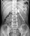
Because the pediatric patient had difficulty with oral ingestion due to insertion of the tube through the nose and experienced severe psychological stress at school, the nasojejunal tube was removed about 6 months after its insertion. Percutaneous endoscopic gastrostomy (PEG) was performed, and a tube was inserted into the jejunum through the gastrostomy. Thus, nutritional support was started through the gastrojejunal tube (Fig. 1). After gastrojejunal tube feeding for 3 months, the patient's general symptoms and gastrointestinal symptoms rapidly improved, and his oral ingestion increased. The PEG was then removed following a request from the parents. The other autonomic symptoms (voiding difficulty, dizziness during voiding and defecation, and orthostatic hypotension) also gradually decreased, and they were completely improved in the end, despite slight relapses for about 6 months.
Although the patient's health was maintained well with the oral feeding after tube removal, vomiting attacks appeared intermittently, which resulted in hospitalization. Thus, the pediatric patient was repeatedly discharged after the parenteral nutrition allowed him to improve enough to be able to undergo oral ingestion. The hospitalization interval was approximately once every few months or twice a month, and each hospitalization period was 5-10 days. During the vomiting episodes, the pediatric patient exhibited very severe vomiting symptoms, and he required fluid treatment due to his inability to ingest food. There was no accompanying abdominal pain or headache, and he did not have a migraine history or a family history of migraine. There were no prodromal symptoms, and the daily vomiting starting time varied from morning to night. Although it sometimes occurred after overeating or with cold symptoms that were accompanied by fever, there were many instances without abnormal causative factors. In addition, no abnormalities were found in additional examinations, including a blood test and upper gastrointestinal series.
Because of the CVS-like symptoms, low-dose amitriptyline was prescribed, but the child's guardian refused to administer it to the patient. Because his symptoms did not improve, the pediatric patient was taken to another hospital by the guardian for another examination. He took propranolol for a few months for CVS prevention, and it had no effect. His body weight again decreased. The pediatric patient returned to our hospital, and he started treatment with low-dose amitriptyline, which resulted in improvement. The dose was increased due to continuous recurrence. Currently, the patient is taking a dose of 30 mg, and he has been maintained well for 6 months without recurrence. After PEG removal, he had 30 months of outpatient follow-up examination. His present growth status is normal (height, 90th-95th percentile; body weight, 50th-75th percentile).
DISCUSSION
Dysautonomia, which is a common complication of GBS [3], can result in disorders in both the sympathetic nervous system and the parasympathetic nervous system. Various symptoms, including cardiovascular dysfunction, dyshidrosis, digestive disorders, and dysuria, can appear [4]. In the present case, dysuria and orthostatic hypotension were observed. Orthostatic hypotension is due to malfunction of the sympathetic vasomotor fibers. In addition, it is possible that the cardiovascular function was reduced and that the sympathetic response was weakened while the pediatric patient had been lying down, which resulted in a temporary exacerbation of the orthostatic hypotension [4]. The patient also exhibited gastroenteritis, SIADH, and mycoplasma pneumonia before the typical GBS symptoms appeared. Although the co-occurrence of SIADH and GBS has been reported in a few studies [56], the underlying mechanism is not clear. The only hypotheses include downward osmotic resetting and enhanced tubular sensitivity to antidiuretic hormone. A prospective study reported that hyponatremia was found in 48% of the patients [6], and motor dysfunction preceded the hyponatremia in most cases. To the best of the authors' knowledge, there have been only 3 cases in which hyponatremia preceded motor dysfunction as in the present pediatric patient [578]. Slight pulmonary infection or gastrointestinal infection precedes in 60% of patients with GBS 1-3 weeks before the appearance of the neurological manifestations [1]. A Campylobacter jejuni infection is the most common preceding factor. In addition, viruses, including cytomegalovirus, Epstein-Barr virus, and human immunodeficiency virus; bacteria, such as Mycoplasma pneumoniae; exposure to thrombolytics; and lymphoma are possible preceding factors [1]. Although the acute gastroenteritis symptoms appeared first in this pediatric patient, the specific causative organism could not be isolated through a stool culture or blood test. He had the mycoplasma infection but because the symptoms appeared after the gastroenteritis and SIADH, it is doubtful that mycoplasma infection was a preceding factor of GBS.
Autonomic dysfunctions, including resting tachycardia, orthostatic hypotension, and hypertensive surge, have been observed in 2/3 of patients with severe GBS [9]. Despite the reports on adynamic ileus in about 15% (17 of 114) of patients with GBS worldwide [10], there have been no reports on gastrointestinal complications in patients with GBS in Korea. There is a hypothesis that GI dysautonomia could be related to immune-mediated inflammation in GBS [10], and this would mean that an immune attack breaks the balance between the sympathetic and parasympathetic tones. This hypothesis has been supported by the findings of mononuclear inflammatory cell infiltration and demyelination of somatic, parasympathetic, and sympathetic fibers in autopsies of patients with GBS [11]. Multiple reports claim that autonomic dysfunction is related to the more severe motor deficit and respiratory failure symptoms [1213], while other studies have claimed that there is no relevance [914]. Thus, the relationship between autonomic dysfunction and motor disturbance is not yet clear. However, because it has been reported that clear dysautonomia mostly occurs in severe patients who require mechanical ventilation [15], the relationship between dysautonomia and prognosis should be investigated further.
In this pediatric patient, gastroparesis was the major symptom among the autonomic symptoms. The other autonomic symptoms improved after only about 6 months, whereas the gastroparesis lasted for about 9 months before improving. In a follow-up gastric emptying scan, the gastric emptying half-time of solid food improved to 50 min.
In general, when a nasogastric feeding tube is used for more than 3 months, PEG is performed. However, because the guardian did not want it, the PEG was delayed. After the PEG, the patient's life quality greatly improved as the discomforts in the nose and neck disappeared and the psychological stress diminished.
CVS refers to a disease involving cyclic vomiting symptoms without specific causes in the nervous or digestive system [16]. According to the Rome III, the definition of CVS is the state that accompanies severe nausea that lasts for several hours to several days and that involves 2 or more episodes of continuous vomiting or nausea symptoms and a healthy period that lasts several weeks to several months between the episodes [16]. The pediatric patient in the present report was maintained well without gastrointestinal symptoms for 3 months after removal of the PEG, had repeated vomiting episodes, and exhibited a healthy state without symptoms between the episodes, which lead to the suspicion of CVS. However, even though CVS has no underlying preceding diseases and because this pediatric patient had a history of gastroparesis after GBS, his symptoms were described as a CVS-like disorder. Because this pediatric patient had a history of gastroparesis and his gastric emptying time increased in each periodic vomiting symptom, CVS was not suspected in the beginning, but low-dose amitriptyline was prescribed to prevent CVS. Because studies have reported that some patients with CVS have gastric emptying delay [1718], it can be speculated that the gastric emptying delaying in the present patient was related to CVS.
Amitriptyline, which is a type of tricyclic antidepressant and which is used for prevention therapy against CVS, interferes with the reabsorption of norepinephrine and serotonin and increases neurotransmitter concentrations in synapses [19]. The pediatric patient started with a dose of 0.5 mg/kg/day, and it was increased to 1 mg/kg/day. He took it once before he went to sleep, and his compliance was therefore satisfactory.
According to a report on the natural progress of patients with CVS in Korea [20], the CVS tends to improve 3-4 years after the first incidence, which suggests that CVS might be cured by natural progress, and the patients slowly improved during their growth. The pediatric patient had no differences in the intervals between the vomiting episodes or in their severities, and the intervals between episodes became longer with the administration of amitriptyline. The present state of the patient has been maintained without symptoms along with the increased dose, and, thus, it is thought to have a preventative effect.
About 40-80% of patients with CVS have trigger factors, including excessive psychologic stress, emotionally excited stress, physical exhaustion, motion sickness, infection, overeating, and specific foods (cheese and chocolate), and the CVS tended to be exacerbated by overeating or a high-fat diet in the present case due to gastroparesis. According to the guidelines of NASPGAN (North American Society for Pediatric Gastroenterology, Hepatology and Nutrition), amitriptyline is recommended as the primary medicine for children who are 5 years old and up. Previously, our center has also reported a superior effect of low-dose amitriptyline for preventing recurrence in patients with CVS [18].
In this case report, the authors describe a rare case of a 8-year-old boy with gastrointestinal autonomic neuropathy that accompanied GBS, and this is the first report of such a patient in Korea. The patient's symptoms progressed to a CVS-like disorder after improvement of the gastrointestinal autonomic neuropathy, and his symptoms were improved by treatment with low-dose amitriptyline.
 XML Download